Loading
PDBj
メニューPDBj@FacebookPDBj@TwitterPDBj@YouTubewwPDB FoundationwwPDB
RCSB PDBPDBeBMRBAdv. SearchSearch help
Search by PDB author
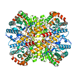
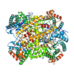
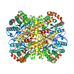
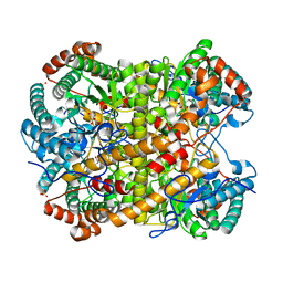
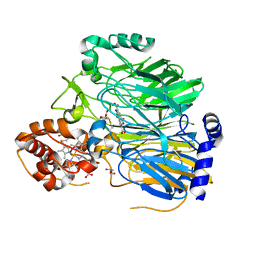
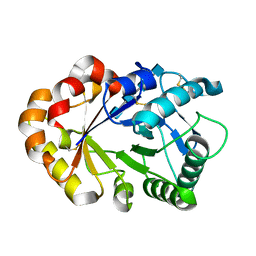
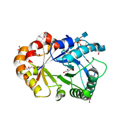
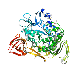
6F74
DownloadVisualize
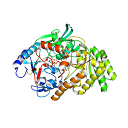
BU of 6f74 by Molmil
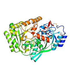
Crystal structure of VAO-type flavoprotein MtVAO713 from Myceliophthora thermophila C1
分子名称: 2-acetamido-2-deoxy-beta-D-glucopyranose, 2-acetamido-2-deoxy-beta-D-glucopyranose-(1-4)-2-acetamido-2-deoxy-beta-D-glucopyranose, Alcohol oxidase, ...
著者Rozeboom, H.J, Fraaije, M.W.
登録日2017-12-07
公開日2018-01-10
最終更新日2024-05-01
実験手法X-RAY DIFFRACTION (2.2 Å)
主引用文献Characterization of Two VAO-Type Flavoprotein Oxidases from Myceliophthora thermophila.
Molecules, 23, 2018
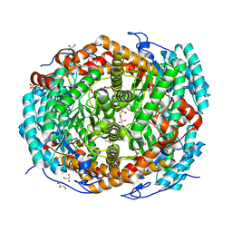
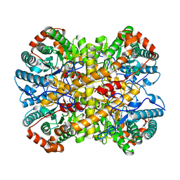
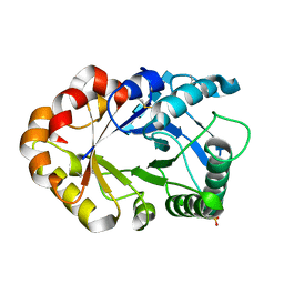
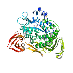
6F73
DownloadVisualize
BU of 6f73 by Molmil
Crystal structure of VAO-type flavoprotein MtVAO615 at pH 5.0 from Myceliophthora thermophila C1
分子名称: 2-acetamido-2-deoxy-beta-D-glucopyranose, 2-acetamido-2-deoxy-beta-D-glucopyranose-(1-4)-2-acetamido-2-deoxy-beta-D-glucopyranose, FLAVIN-ADENINE DINUCLEOTIDE, ...
著者Rozeboom, H.J, Fraaije, M.W.
登録日2017-12-07
公開日2018-01-10
最終更新日2024-01-17
実験手法X-RAY DIFFRACTION (2.22 Å)
主引用文献Characterization of Two VAO-Type Flavoprotein Oxidases from Myceliophthora thermophila.
Molecules, 23, 2018
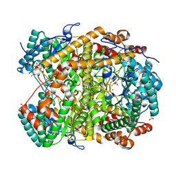
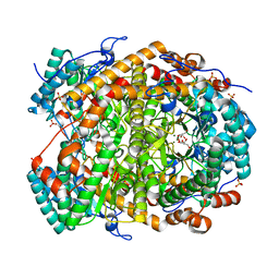
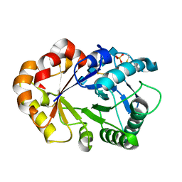
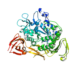
6F72
DownloadVisualize
BU of 6f72 by Molmil
Crystal structure of VAO-type flavoprotein MtVAO615 at pH 7.5 from Myceliophthora thermophila C1
分子名称: 2-acetamido-2-deoxy-beta-D-glucopyranose, 2-acetamido-2-deoxy-beta-D-glucopyranose-(1-4)-2-acetamido-2-deoxy-beta-D-glucopyranose, FLAVIN-ADENINE DINUCLEOTIDE, ...
著者Rozeboom, H.J, Fraaije, M.W.
登録日2017-12-07
公開日2018-01-10
最終更新日2024-05-01
実験手法X-RAY DIFFRACTION (2 Å)
主引用文献Characterization of Two VAO-Type Flavoprotein Oxidases from Myceliophthora thermophila.
Molecules, 23, 2018
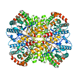
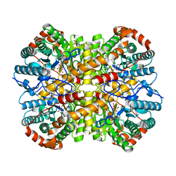
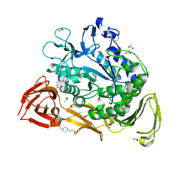
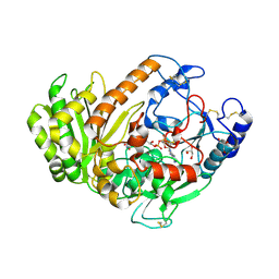
6G4C
DownloadVisualize
BU of 6g4c by Molmil
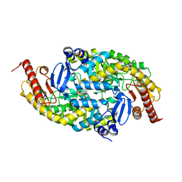
Crystal structure of the omega transaminase from Pseudomonas jessenii in the apo form, crystallized from ammonium phosphate
分子名称: Aspartate aminotransferase family protein, PHOSPHATE ION
著者Rozeboom, H.J, Janssen, D.B.
登録日2018-03-27
公開日2019-04-10
最終更新日2024-05-01
実験手法X-RAY DIFFRACTION (1.87 Å)
主引用文献Biochemical properties of a Pseudomonas aminotransferase involved in caprolactam metabolism.
Febs J., 286, 2019
6G4F
DownloadVisualize
BU of 6g4f by Molmil
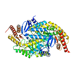
Crystal structure of the omega TRANSAMINASE FROM PSEUDOMONAS Jessenii in complex with PMP
分子名称: 4'-DEOXY-4'-AMINOPYRIDOXAL-5'-PHOSPHATE, Aspartate aminotransferase family protein, GLYCEROL, ...
著者Rozeboom, H.J, Janssen, D.B.
登録日2018-03-27
公開日2019-04-10
最終更新日2024-05-01
実験手法X-RAY DIFFRACTION (2.5 Å)
主引用文献Biochemical properties of a Pseudomonas aminotransferase involved in caprolactam metabolism.
Febs J., 286, 2019
5NH8
DownloadVisualize
BU of 5nh8 by Molmil
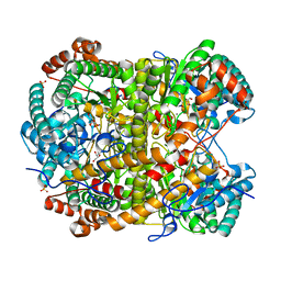
Crystal structure of xylose isomerase from Piromyces E2 in complex with two Ca2+ ions and xylose
分子名称: CALCIUM ION, D-xylose, SULFATE ION, ...
著者Rozeboom, H.J, Janssen, D.B.
登録日2017-03-21
公開日2017-11-01
最終更新日2024-05-01
実験手法X-RAY DIFFRACTION (1.86 Å)
主引用文献Metal Dependence of the Xylose Isomerase from Piromyces sp. E2 Explored by Activity Profiling and Protein Crystallography.
Biochemistry, 56, 2017
5NHM
DownloadVisualize
BU of 5nhm by Molmil
Crystal structure of apo xylose isomerase from Piromyces E2
分子名称: ACETIC ACID, GLYCEROL, SULFATE ION, ...
著者Rozeboom, H.J, Janssen, D.B.
登録日2017-03-21
公開日2017-11-01
最終更新日2024-05-01
実験手法X-RAY DIFFRACTION (1.67 Å)
主引用文献Metal Dependence of the Xylose Isomerase from Piromyces sp. E2 Explored by Activity Profiling and Protein Crystallography.
Biochemistry, 56, 2017
5NH5
DownloadVisualize
BU of 5nh5 by Molmil
Crystal structure of native xylose isomerase from Piromyces E2
分子名称: CALCIUM ION, FE (II) ION, GLYCEROL, ...
著者Rozeboom, H.J, Janssen, D.B.
登録日2017-03-21
公開日2017-11-01
最終更新日2024-01-17
実験手法X-RAY DIFFRACTION (1.8 Å)
主引用文献Metal Dependence of the Xylose Isomerase from Piromyces sp. E2 Explored by Activity Profiling and Protein Crystallography.
Biochemistry, 56, 2017
5NHE
DownloadVisualize
BU of 5nhe by Molmil
Crystal structure of xylose isomerase from Piromyces E2 in complex with two Cd2+ ions and xylose
分子名称: CADMIUM ION, D-xylose, SULFATE ION, ...
著者Rozeboom, H.J, Janssen, D.B.
登録日2017-03-21
公開日2017-11-01
最終更新日2024-05-01
実験手法X-RAY DIFFRACTION (1.86 Å)
主引用文献Metal Dependence of the Xylose Isomerase from Piromyces sp. E2 Explored by Activity Profiling and Protein Crystallography.
Biochemistry, 56, 2017
5NHB
DownloadVisualize
BU of 5nhb by Molmil
Crystal structure of xylose isomerase from Piromyces E2 in complex with two Fe2+ ions
分子名称: FE (II) ION, SULFATE ION, Xylose isomerase
著者Rozeboom, H.J, Janssen, D.B.
登録日2017-03-21
公開日2017-11-01
最終更新日2024-05-01
実験手法X-RAY DIFFRACTION (2.4 Å)
主引用文献Metal Dependence of the Xylose Isomerase from Piromyces sp. E2 Explored by Activity Profiling and Protein Crystallography.
Biochemistry, 56, 2017
5NHA
DownloadVisualize
BU of 5nha by Molmil
Crystal structure of xylose isomerase from Piromyces sp. E2 in complex with two Mn2+ ions and sorbitol
分子名称: MANGANESE (II) ION, SULFATE ION, Xylose isomerase, ...
著者Rozeboom, H.J, Janssen, D.B.
登録日2017-03-21
公開日2017-11-01
最終更新日2024-05-01
実験手法X-RAY DIFFRACTION (1.8 Å)
主引用文献Metal Dependence of the Xylose Isomerase from Piromyces sp. E2 Explored by Activity Profiling and Protein Crystallography.
Biochemistry, 56, 2017
5NH9
DownloadVisualize
BU of 5nh9 by Molmil
Crystal structure of xylose isomerase from Piromyces E2 in complex with two Mn2+ ions and xylose
分子名称: D-xylose, MANGANESE (II) ION, SULFATE ION, ...
著者Rozeboom, H.J, Janssen, D.B.
登録日2017-03-21
公開日2017-11-01
最終更新日2024-05-01
実験手法X-RAY DIFFRACTION (2.08 Å)
主引用文献Metal Dependence of the Xylose Isomerase from Piromyces sp. E2 Explored by Activity Profiling and Protein Crystallography.
Biochemistry, 56, 2017
5NH7
DownloadVisualize
BU of 5nh7 by Molmil
Crystal structure of xylose isomerase from Piromyces E2 in complex with two Mg2+ ions and xylose
分子名称: D-xylose, MAGNESIUM ION, SULFATE ION, ...
著者Rozeboom, H.J, Janssen, D.B.
登録日2017-03-21
公開日2017-11-01
最終更新日2024-05-01
実験手法X-RAY DIFFRACTION (1.9 Å)
主引用文献Metal Dependence of the Xylose Isomerase from Piromyces sp. E2 Explored by Activity Profiling and Protein Crystallography.
Biochemistry, 56, 2017
5NHC
DownloadVisualize
BU of 5nhc by Molmil
Crystal structure of xylose isomerase from Piromyces E2 in complex with two Co2+ ions and xylulose
分子名称: 4-HYDROXYPROLINE, COBALT (II) ION, D-XYLULOSE, ...
著者Rozeboom, H.J, Janssen, D.B.
登録日2017-03-21
公開日2017-11-01
最終更新日2024-05-01
実験手法X-RAY DIFFRACTION (1.93 Å)
主引用文献Metal Dependence of the Xylose Isomerase from Piromyces sp. E2 Explored by Activity Profiling and Protein Crystallography.
Biochemistry, 56, 2017
5NH4
DownloadVisualize
BU of 5nh4 by Molmil
Crystal structure of xylose isomerase from Piromyces E2 in complex with one Mg2+ ions and glycerol
分子名称: GLYCEROL, MAGNESIUM ION, SULFATE ION, ...
著者Rozeboom, H.J, Janssen, D.B.
登録日2017-03-21
公開日2017-11-01
最終更新日2024-01-17
実験手法X-RAY DIFFRACTION (1.8 Å)
主引用文献Metal Dependence of the Xylose Isomerase from Piromyces sp. E2 Explored by Activity Profiling and Protein Crystallography.
Biochemistry, 56, 2017
5NHD
DownloadVisualize
BU of 5nhd by Molmil
Crystal structure of xylose isomerase from Piromyces E2 in complex with 2 Ni2+ ions and xylose
分子名称: D-xylose, NICKEL (II) ION, SULFATE ION, ...
著者Rozeboom, H.J, Janssen, D.B.
登録日2017-03-21
公開日2017-11-01
最終更新日2024-05-01
実験手法X-RAY DIFFRACTION (1.8 Å)
主引用文献Metal Dependence of the Xylose Isomerase from Piromyces sp. E2 Explored by Activity Profiling and Protein Crystallography.
Biochemistry, 56, 2017
1KB0
DownloadVisualize
BU of 1kb0 by Molmil
Crystal Structure of Quinohemoprotein Alcohol Dehydrogenase from Comamonas testosteroni
分子名称: CALCIUM ION, GLYCEROL, HEME C, ...
著者Rozeboom, H.J, Oubrie, A.
登録日2001-11-05
公開日2001-12-28
最終更新日2023-08-16
実験手法X-RAY DIFFRACTION (1.44 Å)
主引用文献Crystal structure of quinohemoprotein alcohol dehydrogenase from Comamonas testosteroni: structural basis for substrate oxidation and electron transfer.
J.Biol.Chem., 277, 2002
1KQZ
DownloadVisualize
BU of 1kqz by Molmil
Hevamine Mutant D125A/E127A/Y183F in Complex with Tetra-NAG
分子名称: 2-acetamido-2-deoxy-beta-D-glucopyranose-(1-4)-2-acetamido-2-deoxy-beta-D-glucopyranose-(1-4)-2-acetamido-2-deoxy-beta-D-glucopyranose-(1-4)-2-acetamido-2-deoxy-beta-D-glucopyranose, Hevamine A
著者Rozeboom, H.J, Dijkstra, B.W.
登録日2002-01-08
公開日2002-01-23
最終更新日2023-08-16
実験手法X-RAY DIFFRACTION (1.92 Å)
主引用文献Expression and Characterization of Active Site Mutants of Hevamine, a Chitinase from the Rubber Tree Hevea brasiliensis.
Eur.J.Biochem., 269, 2002
1KQY
DownloadVisualize
BU of 1kqy by Molmil
Hevamine Mutant D125A/E127A/Y183F in Complex with Penta-NAG
分子名称: 2-acetamido-2-deoxy-beta-D-glucopyranose-(1-4)-2-acetamido-2-deoxy-beta-D-glucopyranose-(1-4)-2-acetamido-2-deoxy-beta-D-glucopyranose-(1-4)-2-acetamido-2-deoxy-beta-D-glucopyranose-(1-4)-2-acetamido-2-deoxy-beta-D-glucopyranose, GLYCEROL, Hevamine A, ...
著者Rozeboom, H.J, Dijkstra, B.W.
登録日2002-01-08
公開日2002-01-23
最終更新日2023-08-16
実験手法X-RAY DIFFRACTION (1.92 Å)
主引用文献Expression and characterization of active site mutants of hevamine, a chitinase from the rubber tree Hevea brasiliensis.
Eur.J.Biochem., 269, 2002
1KR0
DownloadVisualize
BU of 1kr0 by Molmil
Hevamine Mutant D125A/Y183F in Complex with Tetra-NAG
分子名称: 2-acetamido-2-deoxy-beta-D-glucopyranose-(1-4)-2-acetamido-2-deoxy-beta-D-glucopyranose-(1-4)-2-acetamido-2-deoxy-beta-D-glucopyranose-(1-4)-2-acetamido-2-deoxy-beta-D-glucopyranose, Hevamine A, SULFATE ION
著者Rozeboom, H.J, Dijkstra, B.W.
登録日2002-01-08
公開日2002-01-23
最終更新日2023-08-16
実験手法X-RAY DIFFRACTION (1.92 Å)
主引用文献Expression and Characterization of Active Site Mutants of Hevamine, a Chitinase from the Rubber Tree Hevea brasiliensis.
Eur.J.Biochem., 269, 2002
1KR1
DownloadVisualize
BU of 1kr1 by Molmil
Hevamine Mutant D125A/E127A in Complex with Tetra-NAG
分子名称: 2-acetamido-2-deoxy-beta-D-glucopyranose-(1-4)-2-acetamido-2-deoxy-beta-D-glucopyranose-(1-4)-2-acetamido-2-deoxy-beta-D-glucopyranose-(1-4)-2-acetamido-2-deoxy-beta-D-glucopyranose, Hevamine A, SULFATE ION
著者Rozeboom, H.J, Dijkstra, B.W.
登録日2002-01-08
公開日2002-01-23
最終更新日2023-08-16
実験手法X-RAY DIFFRACTION (2 Å)
主引用文献Expression and Characterization of Active Site Mutants of Hevamine, a Chitinase from the Rubber Tree Hevea brasiliensis.
Eur.J.Biochem., 269, 2002
1PEZ
DownloadVisualize
BU of 1pez by Molmil
Bacillus circulans strain 251 mutant A230V
分子名称: (4S)-2-METHYL-2,4-PENTANEDIOL, 4-(2-HYDROXYETHYL)-1-PIPERAZINE ETHANESULFONIC ACID, ACETIC ACID, ...
著者Rozeboom, H.J, Dijkstra, B.W.
登録日2003-05-23
公開日2003-10-28
最終更新日2023-08-16
実験手法X-RAY DIFFRACTION (2.32 Å)
主引用文献Conversion of Cyclodextrin Glycosyltransferase into a Starch Hydrolase by Directed Evolution: The Role of Alanine 230 in Acceptor Subsite +1
Biochemistry, 42, 2003
1PJ9
DownloadVisualize
BU of 1pj9 by Molmil
Bacillus circulans strain 251 loop mutant 183-195
分子名称: (4S)-2-METHYL-2,4-PENTANEDIOL, ACETIC ACID, CALCIUM ION, ...
著者Rozeboom, H.J, Dijkstra, B.W.
登録日2003-06-02
公開日2004-02-03
最終更新日2023-08-16
実験手法X-RAY DIFFRACTION (2 Å)
主引用文献Improved thermostability of bacillus circulans cyclodextrin glycosyltransferase by the introduction of a salt bridge
PROTEINS: STRUCT.,FUNCT.,GENET., 54, 2004
1KCK
DownloadVisualize
BU of 1kck by Molmil
Bacillus circulans strain 251 Cyclodextrin glycosyl transferase mutant N193G
分子名称: 1-AMINO-2,3-DIHYDROXY-5-HYDROXYMETHYL CYCLOHEX-5-ENE, CALCIUM ION, CYCLODEXTRIN GLYCOSYLTRANSFERASE, ...
著者Rozeboom, H.J, Uitdehaag, J.C.M, Dijkstra, B.W.
登録日2001-11-09
公開日2002-01-16
最終更新日2023-08-16
実験手法X-RAY DIFFRACTION (2.43 Å)
主引用文献The remote substrate binding subsite -6 in cyclodextrin-glycosyltransferase controls the transferase activity of the enzyme via an induced-fit mechanism.
J.Biol.Chem., 277, 2002
1KCL
DownloadVisualize
BU of 1kcl by Molmil
Bacillus ciruclans strain 251 Cyclodextrin glycosyl transferase mutant G179L
分子名称: (4S)-2-METHYL-2,4-PENTANEDIOL, CALCIUM ION, Cyclodextrin glycosyltransferase, ...
著者Rozeboom, H.J, Uitdehaag, J.C.M, Dijkstra, B.W.
登録日2001-11-09
公開日2002-01-16
最終更新日2023-08-16
実験手法X-RAY DIFFRACTION (1.94 Å)
主引用文献The remote substrate binding subsite -6 in cyclodextrin-glycosyltransferase controls the transferase activity of the enzyme via an induced-fit mechanism.
J.Biol.Chem., 277, 2002

219869

件を2024-05-15に公開中

PDB statisticsPDBj update infoContact PDBjnumon