Loading
PDBj
MenuPDBj@FacebookPDBj@TwitterPDBj@YouTubewwPDB FoundationwwPDB
RCSB PDBPDBeBMRBAdv. SearchSearch help
Search by PDB author
1OG3
DownloadVisualize
BU of 1og3 by Molmil
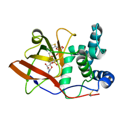
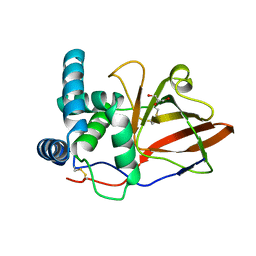
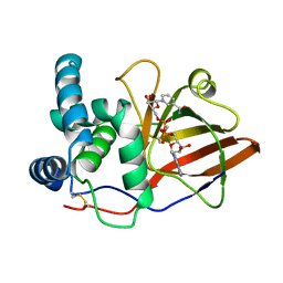
Crystal structure of the eukaryotic mono-ADP-ribosyltransferase ART2.2 mutant E189I in complex with NAD
Descriptor: NICOTINAMIDE-ADENINE-DINUCLEOTIDE, T-CELL ECTO-ADP-RIBOSYLTRANSFERASE 2
Authors:Ritter, H, Koch-Nolte, F, Marquez, V.E, Schulz, G.E.
Deposit date:2003-04-24
Release date:2003-08-28
Last modified:2024-10-23
Method:X-RAY DIFFRACTION (2.6 Å)
Cite:Substrate Binding and Catalysis of Ecto-Adp-Ribosyltransferase 2.2 From Rat
Biochemistry, 42, 2003
1OG1
DownloadVisualize
BU of 1og1 by Molmil
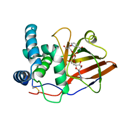
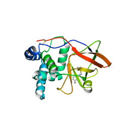
CRYSTAL STRUCTURE OF THE EUCARYOTIC MONO-ADP-RIBOSYLTRANSFERASE ART2.2 IN COMPLEX WITH TAD
Descriptor: BETA-METHYLENE-THIAZOLE-4-CARBOXYAMIDE-ADENINE DINUCLEOTIDE, T-CELL ECTO-ADP-RIBOSYLTRANSFERASE 2
Authors:Ritter, H, Koch-Nolte, F, Marquez, V.E, Schulz, G.E.
Deposit date:2003-04-23
Release date:2003-08-28
Last modified:2024-10-16
Method:X-RAY DIFFRACTION (2 Å)
Cite:Substrate Binding and Catalysis of Ecto-Adp-Ribosyltransferase 2.2 From Rat
Biochemistry, 42, 2003
1W27
DownloadVisualize
BU of 1w27 by Molmil
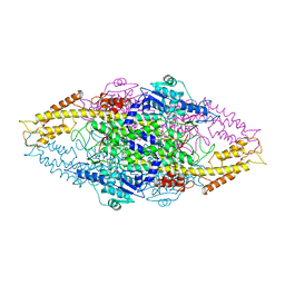
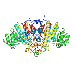
Phenylalanine ammonia-lyase (PAL) from Petroselinum crispum
Descriptor: 2,3-DIHYDROXY-1,4-DITHIOBUTANE, PHENYLALANINE AMMONIA-LYASE 1
Authors:Ritter, H, Schulz, G.E.
Deposit date:2004-06-29
Release date:2004-11-25
Last modified:2023-11-15
Method:X-RAY DIFFRACTION (1.7 Å)
Cite:Structural Basis for the Entrance Into the Phenylpropanoid Metabolism Catalyzed by Phenylalanine Ammonia-Lyase
Plant Cell, 16, 2004
1OG4
DownloadVisualize
BU of 1og4 by Molmil
Crystal Structure of the Eucaryotic Mono-ADP-Ribosyltransferase ART2.2 Mutant E189A in Complex with NADH
Descriptor: 1,4-DIHYDRONICOTINAMIDE ADENINE DINUCLEOTIDE, T-CELL ECTO-ADP-RIBOSYLTRANSFERASE 2
Authors:Ritter, H, Koch-Nolte, F, Marquez, V.E, Schulz, G.E.
Deposit date:2003-04-24
Release date:2003-08-28
Last modified:2023-12-13
Method:X-RAY DIFFRACTION (2.6 Å)
Cite:Substrate Binding and Catalysis of Ecto-Adp-Ribosyltransferase 2.2 From Rat
Biochemistry, 42, 2003
1GY0
DownloadVisualize
BU of 1gy0 by Molmil
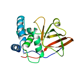
crystal structure of the eucaryotic mono-ADP-ribosyltransferase ART2.2; CRYSTAL FORM C (P3121)
Descriptor: T-CELL ECTO-ADP-RIBOSYLTRANSFERASE 2
Authors:Mueller-Dieckmann, C, Schulz, G.E.
Deposit date:2002-04-15
Release date:2002-09-26
Last modified:2024-10-23
Method:X-RAY DIFFRACTION (2.08 Å)
Cite:Structure of the Ecto-Adp-Ribosyl Transferase Art2.2 From Rat
J.Mol.Biol., 322, 2002
1GXY
DownloadVisualize
BU of 1gxy by Molmil
crystal structure of the eucaryotic mono-ADP-ribosyltransferase ART2.2; CRYSTAL FORM A (P21)
Descriptor: GLYCEROL, T-CELL ECTO-ADP-RIBOSYLTRANSFERASE 2
Authors:Mueller-Dieckmann, C, Schulz, G.E.
Deposit date:2002-04-15
Release date:2002-09-26
Last modified:2019-07-24
Method:X-RAY DIFFRACTION (1.71 Å)
Cite:Structure of the Ecto-Adp-Ribosyl Transferase Art2.2 From Rat
J.Mol.Biol., 322, 2002
1GXZ
DownloadVisualize
BU of 1gxz by Molmil
crystal structure of the eukaryotic mono-ADP-ribosyltransferase ART2.2; Crystal form B (P212121)
Descriptor: 5-BROMONICOTINAMIDE, T-CELL ECTO-ADP-RIBOSYLTRANSFERASE 2
Authors:Mueller-Dieckmann, C, Schulz, G.E.
Deposit date:2002-04-15
Release date:2002-09-26
Method:X-RAY DIFFRACTION (2.1 Å)
Cite:Structure of the Ecto-Adp-Ribosyl Transferase Art2.2 From Rat
J.Mol.Biol., 322, 2002
3BDG
DownloadVisualize
BU of 3bdg by Molmil
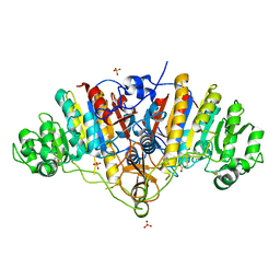
Crystal structure of wild-type/T155V mixed dimer of E. coli alkaline phosphatase
Descriptor: Alkaline phosphatase, SULFATE ION
Authors:Grigg, J.C, Murphy, M.E.
Deposit date:2007-11-14
Release date:2008-11-25
Last modified:2024-10-30
Method:X-RAY DIFFRACTION (1.4 Å)
Cite:The Active-Site Trimetallic Cluster of Alkaline Phosphatase is Lost Upon Isosteric Mutation at the Mg2+-Coordinating Residue Threonine-155
To be Published
3BDF
DownloadVisualize
BU of 3bdf by Molmil
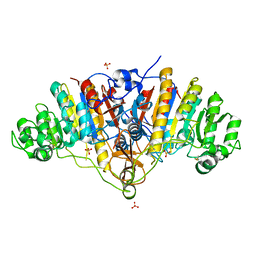
Crystal structure of metal-free E. coli alkaline phosphatase (T155V)
Descriptor: Alkaline phosphatase, SULFATE ION
Authors:Grigg, J.C, Murphy, M.E.
Deposit date:2007-11-14
Release date:2008-11-25
Last modified:2024-10-30
Method:X-RAY DIFFRACTION (1.4 Å)
Cite:The Active-Site Trimetallic Cluster of Alkaline Phosphatase is Lost Upon Isosteric Mutation at the Mg2+-Coordinating Residue Threonine-155
To be Published
3BDH
DownloadVisualize
BU of 3bdh by Molmil
Crystal structure of zinc-deficient wild-type E. coli alkaline phosphatase
Descriptor: Alkaline phosphatase, MAGNESIUM ION
Authors:Grigg, J.C, Murphy, M.E.
Deposit date:2007-11-14
Release date:2008-11-18
Last modified:2023-08-30
Method:X-RAY DIFFRACTION (1.85 Å)
Cite:The Active-Site Trimetallic Cluster of Alkaline Phosphatase is Lost Upon Isosteric Mutation at the Mg2+-Coordinating Residue Threonine-155
To be Published

226707

PDB entries from 2024-10-30

PDB statisticsPDBj update infoContact PDBjnumon