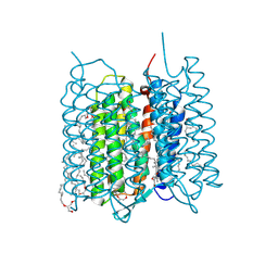
4V8K
 
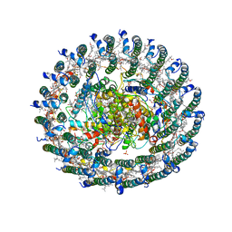 | | Crystal structure of the LH1-RC complex from Thermochromatium tepidum in P21 form | | 分子名称: | BACTERIOCHLOROPHYLL A, BACTERIOPHEOPHYTIN A, CALCIUM ION, ... | | 著者 | Niwa, S, Takeda, K, Wang-Otomo, Z.-Y, Miki, K. | | 登録日 | 2013-11-22 | | 公開日 | 2014-07-09 | | 最終更新日 | 2024-03-20 | | 実験手法 | X-RAY DIFFRACTION (3.006 Å) | | 主引用文献 | Structure of the LH1-RC complex from Thermochromatium tepidum at 3.0 angstrom
Nature, 508, 2014
|
|
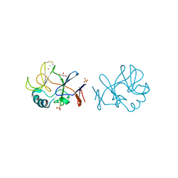
6IED
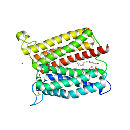 | | Crystal structure of heme A synthase from Bacillus subtilis | | 分子名称: | (2R)-2,3-dihydroxypropyl (9Z)-octadec-9-enoate, COPPER (II) ION, Heme A synthase, ... | | 著者 | Niwa, S, Takeda, K, Kosugi, M, Tsutsumi, E, Miki, K. | | 登録日 | 2018-09-13 | | 公開日 | 2018-11-21 | | 最終更新日 | 2023-11-22 | | 実験手法 | X-RAY DIFFRACTION (3 Å) | | 主引用文献 | Crystal structure of heme A synthase fromBacillus subtilis.
Proc. Natl. Acad. Sci. U.S.A., 115, 2018
|
|
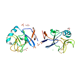
6A2J
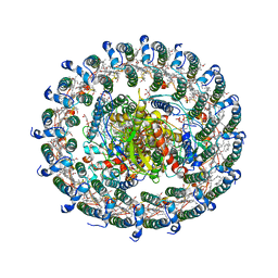
 | | Crystal structure of heme A synthase from Bacillus subtilis | | 分子名称: | (2R)-2,3-dihydroxypropyl (9Z)-octadec-9-enoate, COPPER (II) ION, Heme A synthase, ... | | 著者 | Niwa, S, Takeda, K, Kosugi, M, Tsutsumi, E, Miki, K. | | 登録日 | 2018-06-12 | | 公開日 | 2018-11-21 | | 最終更新日 | 2018-12-12 | | 実験手法 | X-RAY DIFFRACTION (2.2 Å) | | 主引用文献 | Crystal structure of heme A synthase fromBacillus subtilis.
Proc. Natl. Acad. Sci. U.S.A., 115, 2018
|
|
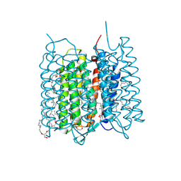
3WMM
 | | Crystal structure of the LH1-RC complex from Thermochromatium tepidum in C2 form | | 分子名称: | (1R)-2-{[(S)-{[(2S)-2,3-dihydroxypropyl]oxy}(hydroxy)phosphoryl]oxy}-1-[(hexadecanoyloxy)methyl]ethyl (9Z)-octadec-9-enoate, BACTERIOCHLOROPHYLL A, BACTERIOPHEOPHYTIN A, ... | | 著者 | Niwa, S, Takeda, K, Wang-Otomo, Z.-Y, Miki, K. | | 登録日 | 2013-11-22 | | 公開日 | 2014-04-09 | | 最終更新日 | 2024-03-20 | | 実験手法 | X-RAY DIFFRACTION (3.008 Å) | | 主引用文献 | Structure of the LH1-RC complex from Thermochromatium tepidum at 3.0 angstrom
Nature, 508, 2014
|
|
5DHE
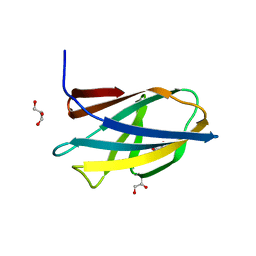 | | Crystal structure of ChBD3 from Thermococcus kodakarensis KOD1 | | 分子名称: | Chitinase, GLYCEROL | | 著者 | Niwa, S, Hibi, M, Takeda, K, Miki, K. | | 登録日 | 2015-08-30 | | 公開日 | 2016-02-10 | | 最終更新日 | 2024-03-20 | | 実験手法 | X-RAY DIFFRACTION (1.6 Å) | | 主引用文献 | Crystal structures of chitin binding domains of chitinase from Thermococcus kodakarensis KOD1
Febs Lett., 590, 2016
|
|
5WQR
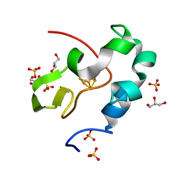 | | High resolution structure of high-potential iron-sulfur protein in the reduced state | | 分子名称: | GLYCEROL, High-potential iron-sulfur protein, IRON/SULFUR CLUSTER, ... | | 著者 | Ohno, H, Takeda, K, Niwa, S, Tsujinaka, T, Hanazono, Y, Hirano, Y, Miki, K. | | 登録日 | 2016-11-28 | | 公開日 | 2017-06-07 | | 最終更新日 | 2023-11-08 | | 実験手法 | X-RAY DIFFRACTION (0.8 Å) | | 主引用文献 | Crystallographic characterization of the high-potential iron-sulfur protein in the oxidized state at 0.8 angstrom resolution
PLoS ONE, 12, 2017
|
|
5WQQ
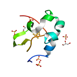 | | High resolution structure of high-potential iron-sulfur protein in the oxidized state | | 分子名称: | GLYCEROL, High-potential iron-sulfur protein, IRON/SULFUR CLUSTER, ... | | 著者 | Ohno, H, Takeda, K, Niwa, S, Tsujinaka, T, Hanazono, Y, Hirano, Y, Miki, K. | | 登録日 | 2016-11-28 | | 公開日 | 2017-06-07 | | 最終更新日 | 2023-11-08 | | 実験手法 | X-RAY DIFFRACTION (0.8 Å) | | 主引用文献 | Crystallographic characterization of the high-potential iron-sulfur protein in the oxidized state at 0.8 angstrom resolution
PLoS ONE, 12, 2017
|
|
5DHD
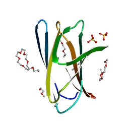 | | Crystal structure of ChBD2 from Thermococcus kodakarensis KOD1 | | 分子名称: | 3,6,9,12,15,18,21,24,27,30,33,36,39-TRIDECAOXAHENTETRACONTANE-1,41-DIOL, Chitinase, SULFATE ION | | 著者 | Hibi, M, Niwa, S, Takeda, K, Miki, K. | | 登録日 | 2015-08-30 | | 公開日 | 2016-02-10 | | 最終更新日 | 2023-11-08 | | 実験手法 | X-RAY DIFFRACTION (1.27 Å) | | 主引用文献 | Crystal structures of chitin binding domains of chitinase from Thermococcus kodakarensis KOD1
Febs Lett., 590, 2016
|
|
7XJE
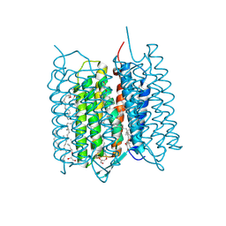 | | Crystal structure of bacteriorhodopsin in the K state refined against the extrapolated dataset | | 分子名称: | 2,3-DI-PHYTANYL-GLYCEROL, Bacteriorhodopsin, RETINAL | | 著者 | Taguchi, S, Niwa, S, Takeda, K. | | 登録日 | 2022-04-16 | | 公開日 | 2023-03-01 | | 最終更新日 | 2024-04-03 | | 実験手法 | X-RAY DIFFRACTION (1.33 Å) | | 主引用文献 | Detailed analysis of distorted retinal and its interaction with surrounding residues in the K intermediate of bacteriorhodopsin
Commun Biol, 6, 2023
|
|
7XJC
 | | Crystal structure of bacteriorhodopsin in the ground and K states after green laser irradiation | | 分子名称: | 2,10,23-TRIMETHYL-TETRACOSANE, 2,3-DI-PHYTANYL-GLYCEROL, Bacteriorhodopsin, ... | | 著者 | Taguchi, S, Niwa, S, Takeda, K. | | 登録日 | 2022-04-16 | | 公開日 | 2023-03-01 | | 最終更新日 | 2023-11-29 | | 実験手法 | X-RAY DIFFRACTION (1.33 Å) | | 主引用文献 | Detailed analysis of distorted retinal and its interaction with surrounding residues in the K intermediate of bacteriorhodopsin
Commun Biol, 6, 2023
|
|
7XJD
 | | Crystal structure of bacteriorhodopsin in the ground state by red laser irradiation | | 分子名称: | 2,10,23-TRIMETHYL-TETRACOSANE, 2,3-DI-PHYTANYL-GLYCEROL, Bacteriorhodopsin, ... | | 著者 | Taguchi, S, Niwa, S, Takeda, K. | | 登録日 | 2022-04-16 | | 公開日 | 2023-03-22 | | 実験手法 | X-RAY DIFFRACTION (1.33 Å) | | 主引用文献 | Detailed analysis of distorted retinal and its interaction with surrounding residues in the K intermediate of bacteriorhodopsin.
Commun Biol, 6, 2023
|
|
7YR9
 | | Crystal structure of the immature form of TtPetA | | 分子名称: | CHLORIDE ION, GLYCEROL, SULFATE ION, ... | | 著者 | Tsutsumi, E, Niwa, S, Takeda, K. | | 登録日 | 2022-08-09 | | 公開日 | 2023-09-20 | | 実験手法 | X-RAY DIFFRACTION (1.7 Å) | | 主引用文献 | Structure of a putative immature form of a Rieske-type iron-sulfur protein in complex with zinc chloride.
Commun Chem, 6, 2023
|
|
7YRA
 | | Crystal structure of [2Fe-2S]-TtPetA | | 分子名称: | CHLORIDE ION, FE2/S2 (INORGANIC) CLUSTER, GLYCEROL, ... | | 著者 | Tsutsumi, E, Niwa, S, Takeda, K. | | 登録日 | 2022-08-09 | | 公開日 | 2023-09-20 | | 実験手法 | X-RAY DIFFRACTION (1.79 Å) | | 主引用文献 | Structure of a putative immature form of a Rieske-type iron-sulfur protein in complex with zinc chloride.
Commun Chem, 6, 2023
|
|
5X1D
 | |
5X1A
 | |
5X1C
 | |
7X4N
 | | Crystal Structure of C. elegans kinesin-4 KLP-12 complexed with tubulin and DARPin | | 分子名称: | DARPin, GUANOSINE-5'-DIPHOSPHATE, GUANOSINE-5'-TRIPHOSPHATE, ... | | 著者 | Taguchi, S, Imasaki, T, Saijo-Hamano, Y, Sakai, N, Nitta, R. | | 登録日 | 2022-03-03 | | 公開日 | 2022-09-21 | | 最終更新日 | 2023-11-29 | | 実験手法 | X-RAY DIFFRACTION (2.88 Å) | | 主引用文献 | Structural model of microtubule dynamics inhibition by kinesin-4 from the crystal structure of KLP-12 -tubulin complex.
Elife, 11, 2022
|
|
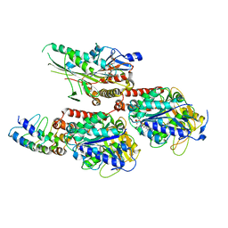
2Z5C
 | | Crystal Structure of a Novel Chaperone Complex for Yeast 20S Proteasome Assembly | | 分子名称: | Proteasome component PUP2, Protein YPL144W, Uncharacterized protein YLR021W | | 著者 | Yashiroda, H, Mizushima, T, Okamoto, K, Kameyama, T, Hayashi, H, Kishimoto, T, Kasahara, M, Kurimoto, E, Sakata, E, Suzuki, A, Hirano, Y, Murata, S, Kato, K, Yamane, T, Tanaka, K. | | 登録日 | 2007-07-03 | | 公開日 | 2008-01-22 | | 最終更新日 | 2023-11-01 | | 実験手法 | X-RAY DIFFRACTION (2.9 Å) | | 主引用文献 | Crystal structure of a chaperone complex that contributes to the assembly of yeast 20S proteasomes
Nat.Struct.Mol.Biol., 15, 2008
|
|
2Z5B
 | | Crystal Structure of a Novel Chaperone Complex for Yeast 20S Proteasome Assembly | | 分子名称: | Protein YPL144W, Uncharacterized protein YLR021W | | 著者 | Yashiroda, H, Mizushima, T, Okamoto, K, Kameyama, T, Hayashi, H, Kishimoto, T, Kasahara, M, Kurimoto, E, Sakata, E, Suzuki, A, Hirano, Y, Murata, S, Kato, K, Yamane, T, Tanaka, K. | | 登録日 | 2007-07-03 | | 公開日 | 2008-01-22 | | 最終更新日 | 2024-03-13 | | 実験手法 | X-RAY DIFFRACTION (1.96 Å) | | 主引用文献 | Crystal structure of a chaperone complex that contributes to the assembly of yeast 20S proteasomes
Nat.Struct.Mol.Biol., 15, 2008
|
|
2Z5E
 | | Crystal Structure of Proteasome Assembling Chaperone 3 | | 分子名称: | Proteasome Assembling Chaperone 3 | | 著者 | Okamoto, K, Kurimoto, E, Sakata, E, Suzuki, A, Yamane, T, Hirano, Y, Murata, S, Tanaka, K, Kato, K. | | 登録日 | 2007-07-06 | | 公開日 | 2008-02-19 | | 最終更新日 | 2024-03-13 | | 実験手法 | X-RAY DIFFRACTION (2 Å) | | 主引用文献 | Crystal structure of a chaperone complex that contributes to the assembly of yeast 20S proteasomes
Nat.Struct.Mol.Biol., 15, 2008
|
|


