Loading
PDBj
MenuPDBj@FacebookPDBj@TwitterPDBj@YouTubewwPDB FoundationwwPDB
RCSB PDBPDBeBMRBAdv. SearchSearch help
Search by PDB author
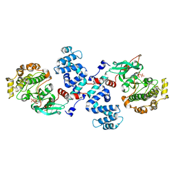
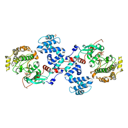
3C50
DownloadVisualize
BU of 3c50 by Molmil
Crystal Structure of G protein coupled receptor kinase 1 bound to ADP and magnesium chloride at 2.6A
Descriptor: 2-(N-MORPHOLINO)-ETHANESULFONIC ACID, ADENOSINE-5'-DIPHOSPHATE, CHLORIDE ION, ...
Authors:Singh, P, Tesmer, J.J.G.
Deposit date:2008-01-30
Release date:2008-03-11
Last modified:2023-08-30
Method:X-RAY DIFFRACTION (2.6 Å)
Cite:Structures of rhodopsin kinase in different ligand states reveal key elements involved in G protein-coupled receptor kinase activation.
J.Biol.Chem., 283, 2008
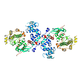
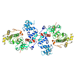
3C4Y
DownloadVisualize
BU of 3c4y by Molmil
Crystal Structure of Apo form of G protein coupled receptor kinase 1 at 7.51A
Descriptor: Rhodopsin kinase
Authors:Singh, P, Tesmer, J.J.G.
Deposit date:2008-01-30
Release date:2008-03-11
Last modified:2023-08-30
Method:X-RAY DIFFRACTION (7.509 Å)
Cite:Structures of rhodopsin kinase in different ligand states reveal key elements involved in G protein-coupled receptor kinase activation.
J.Biol.Chem., 283, 2008
3C51
DownloadVisualize
BU of 3c51 by Molmil
Crystal structure of G protein coupled receptor kinase 1 bound to ADP and magnesium chloride at 3.55A
Descriptor: ADENOSINE-5'-DIPHOSPHATE, BROMIDE ION, MAGNESIUM ION, ...
Authors:Singh, P, Tesmer, J.J.G.
Deposit date:2008-01-30
Release date:2008-03-11
Last modified:2023-08-30
Method:X-RAY DIFFRACTION (3.55 Å)
Cite:Structures of rhodopsin kinase in different ligand states reveal key elements involved in G protein-coupled receptor kinase activation.
J.Biol.Chem., 283, 2008
3C4X
DownloadVisualize
BU of 3c4x by Molmil
Crystal Structure of G protein coupled receptor kinase 1 bound to ATP and magnesium chloride at 2.9A
Descriptor: ADENOSINE-5'-TRIPHOSPHATE, CHLORIDE ION, MAGNESIUM ION, ...
Authors:Singh, P, Tesmer, J.J.G.
Deposit date:2008-01-30
Release date:2008-03-11
Last modified:2023-08-30
Method:X-RAY DIFFRACTION (2.9 Å)
Cite:Structures of rhodopsin kinase in different ligand states reveal key elements involved in G protein-coupled receptor kinase activation.
J.Biol.Chem., 283, 2008
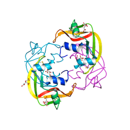
4OXF
DownloadVisualize
BU of 4oxf by Molmil
Structure of ECP in complex with citrate ions at 1.50 Angstroms
Descriptor: CITRIC ACID, Eosinophil cationic protein, FE (III) ION
Authors:Blanco, J.A, Boix, E, Moussaoui, M, Salazar, V.A.
Deposit date:2014-02-05
Release date:2015-03-04
Last modified:2023-12-27
Method:X-RAY DIFFRACTION (1.5 Å)
Cite:Structure of ECP in complex with citrate ions at 1.50 Angstroms
To be published
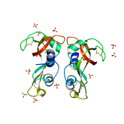
4X08
DownloadVisualize
BU of 4x08 by Molmil
Structure of H128N/ECP mutant in complex with sulphate anions at 1.34 Angstroms.
Descriptor: Eosinophil cationic protein, SULFATE ION
Authors:Blanco, J.A, Garcia, J.M, Salazar, V.A, Sanchez, D, Moussauoi, M, Boix, E.
Deposit date:2014-11-21
Release date:2015-10-07
Last modified:2024-01-10
Method:X-RAY DIFFRACTION (1.34 Å)
Cite:Structure of H128N/ECP mutant in complex with sulphate anions at 1.34 Angstroms.
To Be Published
<12


218853

PDB entries from 2024-04-24

PDB statisticsPDBj update infoContact PDBjnumon