7PUD
 
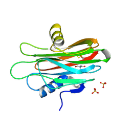
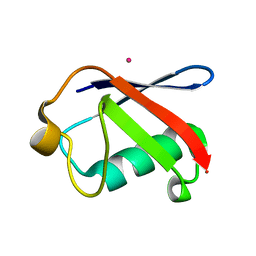
 | | Bryoporin - actinoporin from moss Physcomitrium patens | | Descriptor: | (4S)-2-METHYL-2,4-PENTANEDIOL, Bryoporin, SULFATE ION | | Authors: | Solinc, G, Anderluh, G, Podobnik, M. | | Deposit date: | 2021-09-29 | | Release date: | 2022-09-28 | | Last modified: | 2024-01-31 | | Method: | X-RAY DIFFRACTION (1.25 Å) | | Cite: | Pore-forming moss protein bryoporin is structurally and mechanistically related to actinoporins from evolutionarily distant cnidarians.
J.Biol.Chem., 298, 2022
|
|
9XRW
 | | Probing Positions 3 and 8: Insights into a 960 nm emissive DNA-Stabilized Silver Nanocluster | | Descriptor: | CHLORIDE ION, DNA (5'-D(*CP*CP*GP*CP*GP*CP*GP*TP*GP*CP*CP*GP*CP*GP*AP*A)-3'), SILVER ION, ... | | Authors: | Romolini, G, Kanazawa, H, Cerretani, C, Huang, Z, Kondo, J, Vosch, T. | | Deposit date: | 2025-11-20 | | Release date: | 2026-01-28 | | Last modified: | 2026-02-18 | | Method: | X-RAY DIFFRACTION (1.9 Å) | | Cite: | Insights into the crystal packing interactions of a 960-nm-emissive DNA-stabilized silver nanocluster.
Chem.Commun.(Camb.), 62, 2026
|
|
8BXL
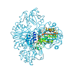
 | | Patulin Synthase from Penicillium expansum | | Descriptor: | 2-acetamido-2-deoxy-beta-D-glucopyranose, 2-acetamido-2-deoxy-beta-D-glucopyranose-(1-4)-2-acetamido-2-deoxy-beta-D-glucopyranose, DI(HYDROXYETHYL)ETHER, ... | | Authors: | Tjallinks, G, Boverio, A, Rozeboom, H.J, Fraaije, M.W. | | Deposit date: | 2022-12-09 | | Release date: | 2023-09-06 | | Last modified: | 2024-10-23 | | Method: | X-RAY DIFFRACTION (2.4 Å) | | Cite: | Structure elucidation and characterization of patulin synthase, insights into the formation of a fungal mycotoxin.
Febs J., 290, 2023
|
|
9H8Z
 | | FAD-dependent monooxygenase sorC with sorbicillin bound | | Descriptor: | (2~{Z})-1-[3,5-dimethyl-2,4-bis(oxidanyl)phenyl]hexa-2,4-dien-1-one, FAD-dependent monooxygenase sorC, FLAVIN-ADENINE DINUCLEOTIDE | | Authors: | Tjallinks, G, Mattevi, A. | | Deposit date: | 2024-10-29 | | Release date: | 2025-03-19 | | Last modified: | 2025-04-02 | | Method: | X-RAY DIFFRACTION (1.71 Å) | | Cite: | Structural and Mechanistic Characterization of the Flavin-Dependent Monooxygenase and Oxidase Involved in Sorbicillinoid Biosynthesis.
Acs Chem.Biol., 20, 2025
|
|
9H8U
 | | FAD-dependent oxidase sorD with sorbicillin bound | | Descriptor: | (2~{Z})-1-[3,5-dimethyl-2,4-bis(oxidanyl)phenyl]hexa-2,4-dien-1-one, 2-acetamido-2-deoxy-beta-D-glucopyranose, FAD-linked oxidoreductase sorD, ... | | Authors: | Tjallinks, G, Mattevi, A. | | Deposit date: | 2024-10-29 | | Release date: | 2025-03-19 | | Last modified: | 2025-04-02 | | Method: | X-RAY DIFFRACTION (3 Å) | | Cite: | Structural and Mechanistic Characterization of the Flavin-Dependent Monooxygenase and Oxidase Involved in Sorbicillinoid Biosynthesis.
Acs Chem.Biol., 20, 2025
|
|
9H92
 | | FAD-dependent oxidase sorD | | Descriptor: | 2-acetamido-2-deoxy-beta-D-glucopyranose, 2-acetamido-2-deoxy-beta-D-glucopyranose-(1-4)-2-acetamido-2-deoxy-beta-D-glucopyranose, FAD-linked oxidoreductase sorD, ... | | Authors: | Tjallinks, G, Mattevi, A. | | Deposit date: | 2024-10-29 | | Release date: | 2025-03-19 | | Last modified: | 2025-04-02 | | Method: | X-RAY DIFFRACTION (1.55 Å) | | Cite: | Structural and Mechanistic Characterization of the Flavin-Dependent Monooxygenase and Oxidase Involved in Sorbicillinoid Biosynthesis.
Acs Chem.Biol., 20, 2025
|
|
9H8M
 | | FAD-dependent monooxygenase sorC | | Descriptor: | DI(HYDROXYETHYL)ETHER, FAD-dependent monooxygenase sorC, FLAVIN-ADENINE DINUCLEOTIDE | | Authors: | Tjallinks, G, Mattevi, A. | | Deposit date: | 2024-10-29 | | Release date: | 2025-03-19 | | Last modified: | 2025-04-02 | | Method: | X-RAY DIFFRACTION (1.38 Å) | | Cite: | Structural and Mechanistic Characterization of the Flavin-Dependent Monooxygenase and Oxidase Involved in Sorbicillinoid Biosynthesis.
Acs Chem.Biol., 20, 2025
|
|
1NBO
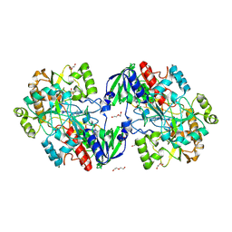
 | | The dual coenzyme specificity of photosynthetic glyceraldehyde-3-phosphate dehydrogenase interpreted by the crystal structure of A4 isoform complexed with NAD | | Descriptor: | NICOTINAMIDE-ADENINE-DINUCLEOTIDE, SULFATE ION, glyceraldehyde-3-phosphate dehydrogenase A | | Authors: | Falini, G, Fermani, S, Ripamonti, A, Sabatino, P, Sparla, F, Pupillo, P, Trost, P. | | Deposit date: | 2002-12-03 | | Release date: | 2003-05-13 | | Last modified: | 2024-11-20 | | Method: | X-RAY DIFFRACTION (2.6 Å) | | Cite: | Dual Coenzyme Specificity of Photosynthetic Glyceraldehyde-3-phosphate
Dehydrogenase Interpreted by the Crystal Structure of A(4) Isoform
Complexed with NAD
Biochemistry, 42, 2003
|
|
9EYM
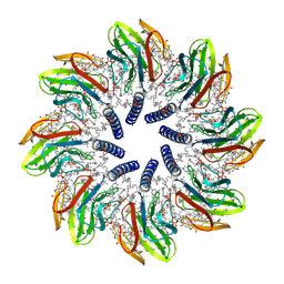
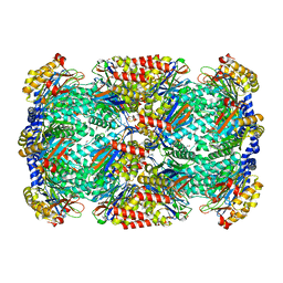
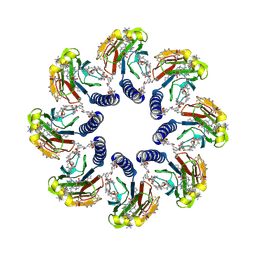 | | The structure of solubilized octameric pore of actinoporin Fav prepared on DOPC:sphingomyelin membranes | | Descriptor: | Actinoporin, Sphingomyelin C18 | | Authors: | Solinc, G, Srnko, M, Svigel, T, Anderluh, G, Podobnik, M. | | Deposit date: | 2024-04-09 | | Release date: | 2025-04-23 | | Last modified: | 2025-11-05 | | Method: | ELECTRON MICROSCOPY (2.6 Å) | | Cite: | Cryo-EM structures of a protein pore reveal a cluster of cholesterol molecules and diverse roles of membrane lipids.
Nat Commun, 16, 2025
|
|
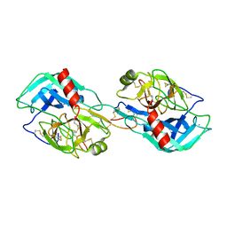
9EYL
 | | dN53 deletion variant of monomeric Fav - actinoporin from Orbicella faveolata | | Descriptor: | SULFATE ION, TRIS-HYDROXYMETHYL-METHYL-AMMONIUM, dN53_Fav | | Authors: | Solinc, G, Svigelj, T, Anderluh, G, Podobnik, M. | | Deposit date: | 2024-04-09 | | Release date: | 2025-04-23 | | Last modified: | 2025-11-05 | | Method: | X-RAY DIFFRACTION (1.5 Å) | | Cite: | Cryo-EM structures of a protein pore reveal a cluster of cholesterol molecules and diverse roles of membrane lipids.
Nat Commun, 16, 2025
|
|
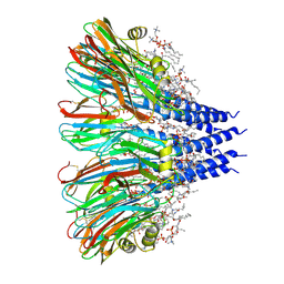
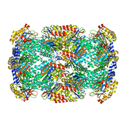
9EYO
 | | The structure of solubilized octameric pore of actinoporin Fav prepared on POPG, cholesterol, sphingomyelin membranes | | Descriptor: | Actinoporin, CHOLESTEROL, Sphingomyelin C18 | | Authors: | Solinc, G, Srnko, M, Anderluh, G, Podobnik, M. | | Deposit date: | 2024-04-09 | | Release date: | 2025-04-23 | | Last modified: | 2025-11-05 | | Method: | ELECTRON MICROSCOPY (2.86 Å) | | Cite: | Cryo-EM structures of a protein pore reveal a cluster of cholesterol molecules and diverse roles of membrane lipids.
Nat Commun, 16, 2025
|
|
9EYN
 | | The structure of solubilized octameric pore of actinoporin Fav prepared on DOPC, cholesterol, sphingomyelin membranes | | Descriptor: | Actinoporin, CHOLESTEROL, Sphingomyelin C18 | | Authors: | Solinc, G, Srnko, M, Svigel, T, Anderluh, G, Podobnik, M. | | Deposit date: | 2024-04-09 | | Release date: | 2025-04-23 | | Last modified: | 2025-11-05 | | Method: | ELECTRON MICROSCOPY (3.06 Å) | | Cite: | Cryo-EM structures of a protein pore reveal a cluster of cholesterol molecules and diverse roles of membrane lipids.
Nat Commun, 16, 2025
|
|
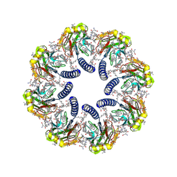
9EYQ
 | | The structure of nonameric pore of RN1 variant of actinoporin Fav | | Descriptor: | Actinoporin, Sphingomyelin C18 | | Authors: | Solinc, G, Srnko, M, Anderluh, G, Crnkovic, A, Podobnik, M. | | Deposit date: | 2024-04-09 | | Release date: | 2025-04-23 | | Method: | ELECTRON MICROSCOPY (3.28 Å) | | Cite: | The structure of nonameric pore of RN1 variant of actinoporin Fav
To Be Published
|
|
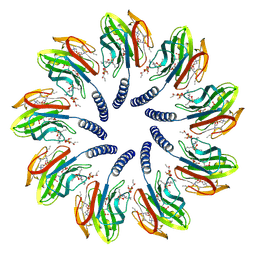
9EYP
 | | The structure of octameric pore of RN1 variant of actinoporin Fav | | Descriptor: | Actinoporin, Sphingomyelin C18 | | Authors: | Solinc, G, Srnko, M, Anderluh, G, Crnkovic, A, Podobnik, M. | | Deposit date: | 2024-04-09 | | Release date: | 2025-04-23 | | Method: | ELECTRON MICROSCOPY (3.05 Å) | | Cite: | The structure of octameric pore of RN1 variant of actinoporin Fav
To Be Published
|
|
9R43
 | | FAD-dependent oxidoreductase McrA | | Descriptor: | 4-(2-HYDROXYETHYL)-1-PIPERAZINE ETHANESULFONIC ACID, ACETATE ION, DI(HYDROXYETHYL)ETHER, ... | | Authors: | Tjallinks, G, Mattevi, A. | | Deposit date: | 2025-05-07 | | Release date: | 2025-12-10 | | Method: | X-RAY DIFFRACTION (2.1 Å) | | Cite: | Characterization of the Mitomycin C Resistance Protein McrA
Biorxiv, 2025
|
|
3EHV
 | |
3EFU
 | |
3EEC
 | | X-ray structure of human ubiquitin Cd(II) adduct | | Descriptor: | CADMIUM ION, Ubiquitin | | Authors: | Falini, G, Fermani, S, Tosi, G, Arnesano, F, Natile, G. | | Deposit date: | 2008-09-04 | | Release date: | 2009-03-10 | | Last modified: | 2023-11-01 | | Method: | X-RAY DIFFRACTION (3 Å) | | Cite: | Structural probing of Zn(II), Cd(II) and Hg(II) binding to human ubiquitin.
Chem.Commun.(Camb.), 45, 2008
|
|
3H6I
 | |
3H6F
 | |
3MYW
 | | The Bowman-Birk type inhibitor from mung bean in ternary complex with porcine trypsin | | Descriptor: | Bowman-Birk type trypsin inhibitor, CALCIUM ION, Trypsin | | Authors: | Engh, R.A, Bode, W, Huber, R, Lin, G, Chi, C. | | Deposit date: | 2010-05-11 | | Release date: | 2010-12-29 | | Last modified: | 2024-11-06 | | Method: | X-RAY DIFFRACTION (2.5 Å) | | Cite: | The 0.25-nm X-ray structure of the Bowman-Birk-type inhibitor from mung bean in ternary complex with porcine trypsin.
Eur.J.Biochem., 212, 1993
|
|
5XKN
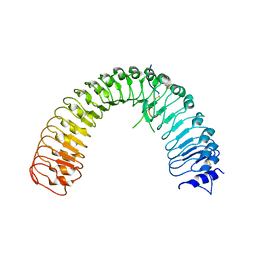 | | Crystal structure of plant receptor ERL2 in complexe with EPFL4 | | Descriptor: | EPIDERMAL PATTERNING FACTOR-like protein 4, LRR receptor-like serine/threonine-protein kinase ERL2 | | Authors: | Chai, J, Lin, G. | | Deposit date: | 2017-05-08 | | Release date: | 2019-01-23 | | Last modified: | 2024-10-23 | | Method: | X-RAY DIFFRACTION (3.651 Å) | | Cite: | A receptor-like protein acts as a specificity switch for the regulation of stomatal development.
Genes Dev., 31, 2017
|
|
5XJO
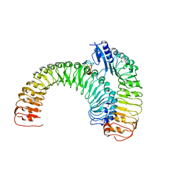 | | Plant receptor ERL1-TMM in complex with peptide EPF1 | | Descriptor: | LRR receptor-like serine/threonine-protein kinase ERL1, Protein EPIDERMAL PATTERNING FACTOR 1, Protein TOO MANY MOUTHS | | Authors: | Chai, J, Lin, G, Zhang, L, Han, Z, Shpak, E.D, Yang, X. | | Deposit date: | 2017-05-03 | | Release date: | 2019-01-23 | | Last modified: | 2024-11-06 | | Method: | X-RAY DIFFRACTION (2.626 Å) | | Cite: | A receptor-like protein acts as a specificity switch for the regulation of stomatal development.
Genes Dev., 31, 2017
|
|
5XJX
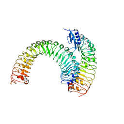 | | Pre-formed plant receptor ERL1-TMM complex | | Descriptor: | LRR receptor-like serine/threonine-protein kinase ERL1, Protein TOO MANY MOUTHS | | Authors: | Chai, J, Lin, G, Zhang, L, Han, Z, Yang, X, Liu, W, Qi, Y, Chang, J, Li, E. | | Deposit date: | 2017-05-04 | | Release date: | 2019-01-23 | | Last modified: | 2024-11-13 | | Method: | X-RAY DIFFRACTION (3.055 Å) | | Cite: | A receptor-like protein acts as a specificity switch for the regulation of stomatal development.
Genes Dev., 31, 2017
|
|
5XKJ
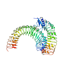 | | Crystal structure of plant receptor ERL1-TMM in complexe with EPF2 | | Descriptor: | LRR receptor-like serine/threonine-protein kinase ERL1, Protein EPIDERMAL PATTERNING FACTOR 2, Protein TOO MANY MOUTHS | | Authors: | Chai, J, Lin, G. | | Deposit date: | 2017-05-07 | | Release date: | 2019-01-23 | | Last modified: | 2024-10-30 | | Method: | X-RAY DIFFRACTION (3.475 Å) | | Cite: | A receptor-like protein acts as a specificity switch for the regulation of stomatal development.
Genes Dev., 31, 2017
|
|


