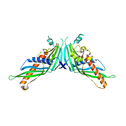
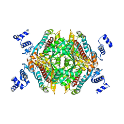
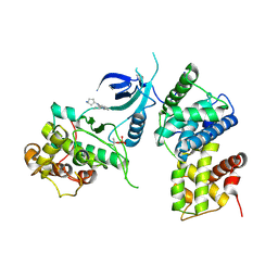
2J3K
 
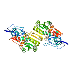 | | Crystal structure of Arabidopsis thaliana Double Bond Reductase (AT5G16970)-Ternary Complex II | | 分子名称: | (2E,4R)-4-HYDROXYNON-2-ENAL, NADP NICOTINAMIDE-ADENINE-DINUCLEOTIDE PHOSPHATE, NADPH-dependent oxidoreductase 2-alkenal reductase | | 著者 | Youn, B, Kim, S.J, Moinuddin, S.G, Lee, C, Bedgar, D.L, Harper, A.R, Davin, L.B, Lewis, N.G, Kang, C. | | 登録日 | 2006-08-22 | | 公開日 | 2006-10-05 | | 最終更新日 | 2024-05-08 | | 実験手法 | X-RAY DIFFRACTION (2.8 Å) | | 主引用文献 | Mechanistic and structural studies of apoform, binary, and ternary complexes of the Arabidopsis alkenal double bond reductase At5g16970.
J. Biol. Chem., 281, 2006
|
|
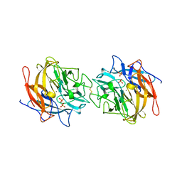
2J3J
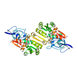 | | Crystal structure of Arabidopsis thaliana Double Bond Reductase (AT5G16970)-Ternary Complex I | | 分子名称: | 4'-HYDROXYCINNAMIC ACID, NADP NICOTINAMIDE-ADENINE-DINUCLEOTIDE PHOSPHATE, NADPH-dependent oxidoreductase 2-alkenal reductase | | 著者 | Youn, B, Kim, S.J, Moinuddin, S.G, Lee, C, Bedgar, D.L, Harper, A.R, Davin, L.B, Lewis, N.G, Kang, C. | | 登録日 | 2006-08-21 | | 公開日 | 2006-10-05 | | 最終更新日 | 2024-05-08 | | 実験手法 | X-RAY DIFFRACTION (2.8 Å) | | 主引用文献 | Mechanistic and structural studies of apoform, binary, and ternary complexes of the Arabidopsis alkenal double bond reductase At5g16970.
J. Biol. Chem., 281, 2006
|
|
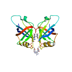
2J3I
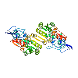 | | Crystal structure of Arabidopsis thaliana Double Bond Reductase (AT5G16970)-Binary Complex | | 分子名称: | NADP NICOTINAMIDE-ADENINE-DINUCLEOTIDE PHOSPHATE, NADP-DEPENDENT OXIDOREDUCTASE P1 | | 著者 | Youn, B, Kim, S.J, Moinuddin, S.G, Lee, C, Bedgar, D.L, Harper, A.R, Davin, L.B, Lewis, N.G, Kang, C. | | 登録日 | 2006-08-21 | | 公開日 | 2006-10-05 | | 最終更新日 | 2011-07-13 | | 実験手法 | X-RAY DIFFRACTION (2.8 Å) | | 主引用文献 | Mechanistic and Structural Studies of Apoform, Binary, and Ternary Complexes of the Arabidopsis Alkenal Double Bond Reductase at5G16970.
J.Biol.Chem., 281, 2006
|
|
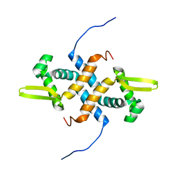
5GYD
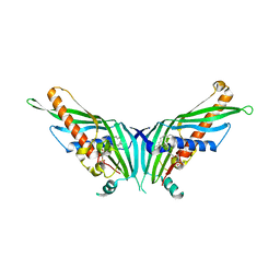 | | Crystal Structure of Mdm12 | | 分子名称: | 1,2-dioleoyl-sn-glycero-3-phosphoethanolamine, Mitochondrial distribution and morphology protein 12 | | 著者 | Jeong, H, Park, J, Lee, C. | | 登録日 | 2016-09-22 | | 公開日 | 2016-11-16 | | 最終更新日 | 2024-03-20 | | 実験手法 | X-RAY DIFFRACTION (3.106 Å) | | 主引用文献 | Crystal structure of Mdm12 reveals the architecture and dynamic organization of the ERMES complex
EMBO Rep., 17, 2016
|
|
5GYK
 | | Crystal Structure of Mdm12-deletion mutant | | 分子名称: | 1,2-dioleoyl-sn-glycero-3-phosphoethanolamine, Mitochondrial distribution and morphology protein 12 | | 著者 | Jeong, H, Park, J, Lee, C. | | 登録日 | 2016-09-22 | | 公開日 | 2016-11-16 | | 最終更新日 | 2023-11-08 | | 実験手法 | X-RAY DIFFRACTION (3.596 Å) | | 主引用文献 | Crystal structure of Mdm12 reveals the architecture and dynamic organization of the ERMES complex
EMBO Rep., 17, 2016
|
|
4MLG
 | | Structure of RS223-Beta-xylosidase | | 分子名称: | Beta-xylosidase, CALCIUM ION, SULFATE ION | | 著者 | Jordan, D, Braker, J, Wagschal, K, Lee, C, Dubrovska, I, Anderson, S, Wawrzak, Z. | | 登録日 | 2013-09-06 | | 公開日 | 2014-09-17 | | 最終更新日 | 2023-09-20 | | 実験手法 | X-RAY DIFFRACTION (2.7 Å) | | 主引用文献 | Structure of RS223-Beta-xylosidase
To be Published
|
|
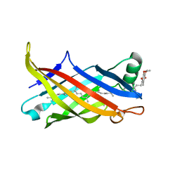
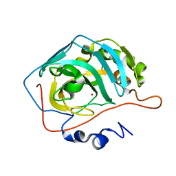
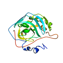
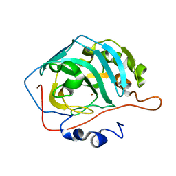
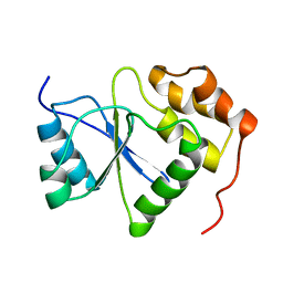
2KCC
 | | Solution Structure of biotinoyl domain from human acetyl-CoA carboxylase 2 | | 分子名称: | Acetyl-CoA carboxylase 2 | | 著者 | Lee, C, Cheong, H, Ryu, K, Lee, J, Lee, W, Jeon, Y, Cheong, C. | | 登録日 | 2008-12-19 | | 公開日 | 2009-02-17 | | 最終更新日 | 2023-09-27 | | 実験手法 | SOLUTION NMR | | 主引用文献 | Biotinoyl domain of human acetyl-CoA carboxylase: Structural insights into the carboxyl transfer mechanism.
Proteins, 72, 2008
|
|
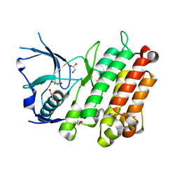
6L2U
 | | Soluble methane monooxygenase reductase FAD-binding domain from Methylosinus sporium. | | 分子名称: | FLAVIN-ADENINE DINUCLEOTIDE, Methane monooxygenase | | 著者 | Park, J.H, Ha, S.C, Rao, Z, Yoo, H, Yoon, C, Kim, S.Y, Kim, D.S, Lee, S.J. | | 登録日 | 2019-10-07 | | 公開日 | 2021-03-03 | | 最終更新日 | 2021-12-08 | | 実験手法 | X-RAY DIFFRACTION (1.5 Å) | | 主引用文献 | Elucidation of the electron transfer environment in the MMOR FAD-binding domain from Methylosinus sporium 5.
Dalton Trans, 50, 2021
|
|
7BWL
 | | Structure of antibiotic sequester from Pseudomonas aerurinosa | | 分子名称: | UPF0312 protein PA0423, Ubiquinone-8 | | 著者 | Hong, M. | | 登録日 | 2020-04-14 | | 公開日 | 2020-12-02 | | 最終更新日 | 2023-11-29 | | 実験手法 | X-RAY DIFFRACTION (2.25 Å) | | 主引用文献 | Crystal structure of the Pseudomonas aeruginosa PA0423 protein and its functional implication in antibiotic sequestration.
Biochem.Biophys.Res.Commun., 528, 2020
|
|
3L9P
 | |
6X84
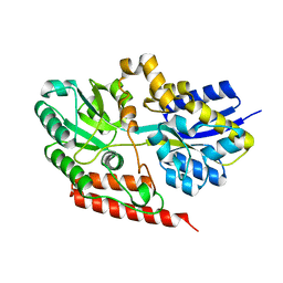 | |
5ZQH
 | | Crystal structure of Streptococcus transcriptional regulator | | 分子名称: | PadR family transcriptional regulator | | 著者 | Kim, M, Hong, M. | | 登録日 | 2018-04-19 | | 公開日 | 2019-05-01 | | 最終更新日 | 2023-11-22 | | 実験手法 | X-RAY DIFFRACTION (2.4 Å) | | 主引用文献 | Structure-based functional analysis of a PadR transcription factor from Streptococcus pneumoniae and characteristic features in the PadR subfamily-2.
Biochem.Biophys.Res.Commun., 532, 2020
|
|
2L14
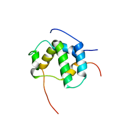 | | Structure of CBP nuclear coactivator binding domain in complex with p53 TAD | | 分子名称: | CREB-binding protein, Cellular tumor antigen p53 | | 著者 | Lee, C, Martinez-Yamout, M.A, Dyson, H.J, Wright, P.E. | | 登録日 | 2010-07-22 | | 公開日 | 2010-11-03 | | 最終更新日 | 2024-05-01 | | 実験手法 | SOLUTION NMR | | 主引用文献 | Structure of the p53 transactivation domain in complex with the nuclear receptor coactivator binding domain of CREB binding protein.
Biochemistry, 49, 2010
|
|
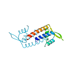
2LKP
 | | solution structure of apo-NmtR | | 分子名称: | HTH-type transcriptional regulator NmtR | | 著者 | Lee, C, Giedroc, D. | | 登録日 | 2011-10-18 | | 公開日 | 2012-04-18 | | 最終更新日 | 2023-06-14 | | 実験手法 | SOLUTION NMR | | 主引用文献 | Solution Structure of Mycobacterium tuberculosis NmtR in the Apo State: Insights into Ni(II)-Mediated Allostery.
Biochemistry, 51, 2012
|
|
2L6I
 | |
2Q1L
 | | Design and Synthesis of Pyrrole-based, Hepatoselective HMG-CoA Reductase Inhibitors | | 分子名称: | (3R,5R)-7-[5-(ANILINOCARBONYL)-3,4-BIS(4-FLUOROPHENYL)-1-ISOPROPYL-1H-PYRROL-2-YL]-3,5-DIHYDROXYHEPTANOIC ACID, 3-hydroxy-3-methylglutaryl-coenzyme A reductase | | 著者 | Pavlovsky, A, Pfefferkorn, J.A, Harris, M.S, Finzel, B.C. | | 登録日 | 2007-05-24 | | 公開日 | 2007-07-17 | | 最終更新日 | 2024-02-21 | | 実験手法 | X-RAY DIFFRACTION (2.05 Å) | | 主引用文献 | Design and synthesis of hepatoselective, pyrrole-based HMG-CoA reductase inhibitors.
Bioorg.Med.Chem.Lett., 17, 2007
|
|
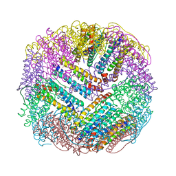
8K5R
 | | CDK9/cyclin T1 in complex with KB-0742 | | 分子名称: | (1S,3S)-N3-(5-pentan-3-ylpyrazolo[1,5-a]pyrimidin-7-yl)cyclopentane-1,3-diamine, Cyclin-T1, Cyclin-dependent kinase 9 | | 著者 | Zhou, M, Li, H, Gao, H, Trotter, B.W, Freeman, D. | | 登録日 | 2023-07-24 | | 公開日 | 2023-12-06 | | 最終更新日 | 2023-12-27 | | 実験手法 | X-RAY DIFFRACTION (3.751 Å) | | 主引用文献 | Discovery of KB-0742, a Potent, Selective, Orally Bioavailable Small Molecule Inhibitor of CDK9 for MYC-Dependent Cancers.
J.Med.Chem., 66, 2023
|
|
8SD8
 | |
8SD6
 | |
8SD9
 | |
8SD7
 | |
8SD1
 | |
8SF1
 | |
1V3A
 | |
4XGS
 | |


