Loading
PDBj
MenuPDBj@FacebookPDBj@TwitterPDBj@YouTubewwPDB FoundationwwPDB
RCSB PDBPDBeBMRBAdv. SearchSearch help
Search by PDB author
1LSL
DownloadVisualize
BU of 1lsl by Molmil
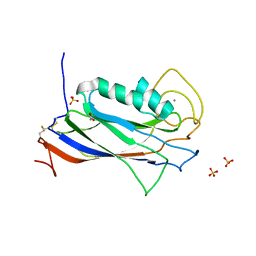
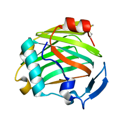
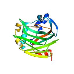
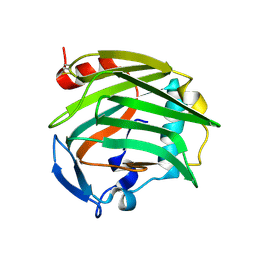
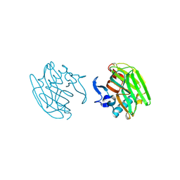
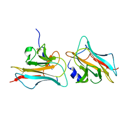
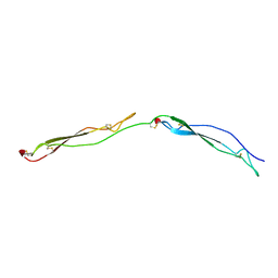
Crystal Structure of the Thrombospondin-1 Type 1 Repeats
Descriptor: Thrombospondin 1, alpha-L-fucopyranose, beta-L-fucopyranose
Authors:Tan, K, Duquette, M, Liu, J, Dong, Y, Zhang, R, Joachimiak, A, Lawler, J, Wang, J.-H.
Deposit date:2002-05-17
Release date:2002-12-18
Last modified:2024-10-30
Method:X-RAY DIFFRACTION (1.9 Å)
Cite:Crystal structure of the TSP-1 type 1 repeats: a novel layered fold and its biological implication.
J.Cell Biol., 159, 2002
3Q13
DownloadVisualize
BU of 3q13 by Molmil
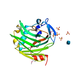
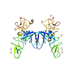
The Structure of the Ca2+-binding, Glycosylated F-spondin Domain of F-spondin, A C2-domain Variant from Extracellular Matrix
Descriptor: ACETATE ION, CALCIUM ION, SULFATE ION, ...
Authors:Tan, K, Lawler, J.
Deposit date:2010-12-16
Release date:2011-06-29
Last modified:2023-09-13
Method:X-RAY DIFFRACTION (1.95 Å)
Cite:The structure of the Ca2+-binding, glycosylated F-spondin domain of F-spondin - A C2-domain variant in an extracellular matrix protein.
Bmc Struct.Biol., 11, 2011
2ERF
DownloadVisualize
BU of 2erf by Molmil
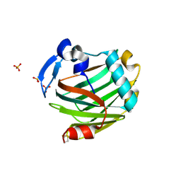
Crystal Structure of the Thrombospondin-1 N-terminal Domain at 1.45A Resolution
Descriptor: Thrombospondin-1
Authors:Tan, K, Wang, J, Lawler, J.
Deposit date:2005-10-24
Release date:2006-01-17
Last modified:2023-08-23
Method:X-RAY DIFFRACTION (1.45 Å)
Cite:The structures of the thrombospondin-1 N-terminal domain and its complex with a synthetic pentameric heparin.
Structure, 14, 2006
2ES3
DownloadVisualize
BU of 2es3 by Molmil
Crystal Structure of Thrombospondin-1 N-terminal Domain in P1 Form at 1.85A Resolution
Descriptor: Thrombospondin-1
Authors:Tan, K, Wang, J, Lawler, J.
Deposit date:2005-10-25
Release date:2006-10-31
Last modified:2024-10-30
Method:X-RAY DIFFRACTION (1.85 Å)
Cite:Heparin-induced cis- and trans-Dimerization Modes of the Thrombospondin-1 N-terminal Domain.
J.Biol.Chem., 283, 2008
1Z78
DownloadVisualize
BU of 1z78 by Molmil
Crystal Structure of the Thrombospondin-1 N-terminal domain
Descriptor: Thrombospondin 1
Authors:Tan, K, Wang, J, Lawler, J.
Deposit date:2005-03-24
Release date:2006-01-17
Last modified:2024-04-03
Method:X-RAY DIFFRACTION (1.8 Å)
Cite:The structures of the thrombospondin-1 N-terminal domain and its complex with a synthetic pentameric heparin.
Structure, 14, 2006
2OUJ
DownloadVisualize
BU of 2ouj by Molmil
The crystal structure of the Thrombospondin-1 N-terminal domain in complex with fractionated Heparin DP8
Descriptor: Thrombospondin-1
Authors:Tan, K, Joachimiak, A, Wang, J, Lawler, J.
Deposit date:2007-02-11
Release date:2008-01-08
Last modified:2023-08-30
Method:X-RAY DIFFRACTION (1.9 Å)
Cite:Heparin-induced cis- and trans-Dimerization Modes of the Thrombospondin-1 N-terminal Domain.
J.Biol.Chem., 283, 2008
2OUH
DownloadVisualize
BU of 2ouh by Molmil
Crystal structure of the Thrombospondin-1 N-terminal domain in complex with fractionated Heparin DP10
Descriptor: SULFATE ION, Thrombospondin-1
Authors:Tan, K, Joachimiak, A, Wang, J, Lawler, J.
Deposit date:2007-02-11
Release date:2008-01-08
Last modified:2024-10-09
Method:X-RAY DIFFRACTION (2.4 Å)
Cite:Heparin-induced cis- and trans-Dimerization Modes of the Thrombospondin-1 N-terminal Domain.
J.Biol.Chem., 283, 2008
1ZA4
DownloadVisualize
BU of 1za4 by Molmil
Crystal Structure of the Thrombospondin-1 N-terminal Domain in Complex with Arixtra
Descriptor: 2-deoxy-6-O-sulfo-2-(sulfoamino)-alpha-D-glucopyranose, SULFATE ION, Thrombospondin 1
Authors:Tan, K, Wang, J.H, Lawler, J.
Deposit date:2005-04-05
Release date:2006-01-24
Last modified:2020-07-29
Method:X-RAY DIFFRACTION (1.9 Å)
Cite:The Structures of the Thrombospondin-1 N-terminal Domain and its Complex with a Synthetic Pentameric Heparin
Structure, 14, 2006
3FBY
DownloadVisualize
BU of 3fby by Molmil
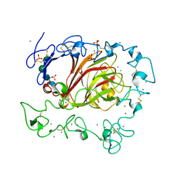
The crystal structure of the signature domain of cartilage oligomeric matrix protein.
Descriptor: CALCIUM ION, Cartilage oligomeric matrix protein, SULFATE ION, ...
Authors:Tan, K, Lawler, J.
Deposit date:2008-11-20
Release date:2009-06-23
Last modified:2023-09-06
Method:X-RAY DIFFRACTION (3.15 Å)
Cite:The crystal structure of the signature domain of cartilage oligomeric matrix protein: implications for collagen, glycosaminoglycan and integrin binding.
Faseb J., 23, 2009
3COO
DownloadVisualize
BU of 3coo by Molmil
The crystal structure of Reelin-N domain of F-spondin
Descriptor: Spondin-1
Authors:Tan, K, Lawler, J, Wang, J.H.
Deposit date:2008-03-28
Release date:2008-08-26
Last modified:2024-10-09
Method:X-RAY DIFFRACTION (2 Å)
Cite:The crystal structure of the heparin-binding reelin-N domain of f-spondin.
J.Mol.Biol., 381, 2008
3SP8
DownloadVisualize
BU of 3sp8 by Molmil
Crystal structure of NK2 in complex with fractionated Heparin DP10
Descriptor: (4R)-2-METHYLPENTANE-2,4-DIOL, (4S)-2-METHYL-2,4-PENTANEDIOL, 2-(N-MORPHOLINO)-ETHANESULFONIC ACID, ...
Authors:Recacha, R, Mulloy, B, Gherardi, E.
Deposit date:2011-07-01
Release date:2012-07-04
Last modified:2023-09-13
Method:X-RAY DIFFRACTION (1.86 Å)
Cite:Crystal structure of NK2 in complex with fractionated Heparin DP10
TO BE PUBLISHED

226707

PDB entries from 2024-10-30

PDB statisticsPDBj update infoContact PDBjnumon