1A13
 
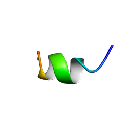 | | G PROTEIN-BOUND CONFORMATION OF MASTOPARAN-X, NMR, 14 STRUCTURES | | 分子名称: | MASTOPARAN-X | | 著者 | Kusunoki, H, Wakamatsu, K, Sato, K, Miyazawa, T, Kohno, T. | | 登録日 | 1997-12-20 | | 公開日 | 1999-01-13 | | 最終更新日 | 2022-02-16 | | 実験手法 | SOLUTION NMR | | 主引用文献 | G protein-bound conformation of mastoparan-X: heteronuclear multidimensional transferred nuclear overhauser effect analysis of peptide uniformly enriched with 13C and 15N.
Biochemistry, 37, 1998
|
|
2EV8
 | |
2RQ1
 | |
2RQ5
 | |
1S35
 | |
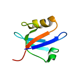
1K1V
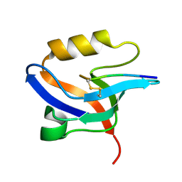
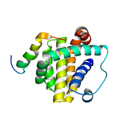
 | | Solution Structure of the DNA-Binding Domain of MafG | | 分子名称: | MafG | | 著者 | Kusunoki, H, Motohashi, H, Katsuoka, F, Morohashi, A, Yamamoto, M, Tanaka, T. | | 登録日 | 2001-09-25 | | 公開日 | 2002-04-10 | | 最終更新日 | 2022-02-23 | | 実験手法 | SOLUTION NMR | | 主引用文献 | Solution structure of the DNA-binding domain of MafG.
Nat.Struct.Biol., 9, 2002
|
|
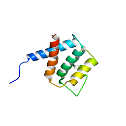
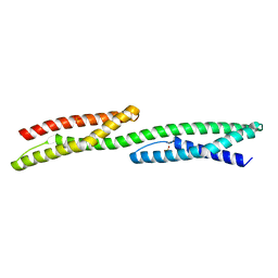
1U5P
 | | Crystal Structure of Repeats 15 and 16 of Chicken Brain Alpha Spectrin | | 分子名称: | PHOSPHATE ION, POTASSIUM ION, Spectrin alpha chain, ... | | 著者 | Kusunoki, H, Minasov, G, MacDonald, R.I, Mondragon, A. | | 登録日 | 2004-07-28 | | 公開日 | 2004-10-19 | | 最終更新日 | 2024-03-13 | | 実験手法 | X-RAY DIFFRACTION (2 Å) | | 主引用文献 | Independent Movement, Dimerization and Stability of Tandem Repeats of Chicken Brain alpha-Spectrin
J.Mol.Biol., 344, 2004
|
|
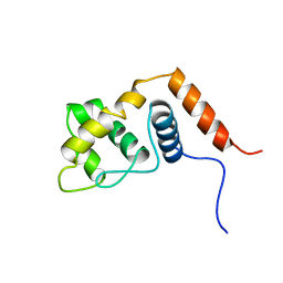
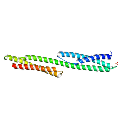
1U4Q
 | | Crystal Structure of Repeats 15, 16 and 17 of Chicken Brain Alpha Spectrin | | 分子名称: | Spectrin alpha chain, brain | | 著者 | Kusunoki, H, Minasov, G, MacDonald, R.I, Mondragon, A. | | 登録日 | 2004-07-26 | | 公開日 | 2004-10-19 | | 最終更新日 | 2024-03-13 | | 実験手法 | X-RAY DIFFRACTION (2.5 Å) | | 主引用文献 | Independent Movement, Dimerization and Stability of Tandem Repeats of Chicken Brain alpha-Spectrin
J.Mol.Biol., 344, 2004
|
|
2EJY
 | |
8WLS
 | |


