Loading
PDBj
MenuPDBj@FacebookPDBj@X(formerly Twitter)PDBj@BlueSkyPDBj@YouTubewwPDB FoundationwwPDBDonate
RCSB PDBPDBeBMRBAdv. SearchSearch help
Search by PDB author
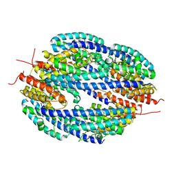
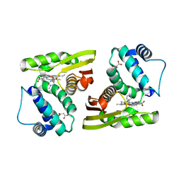
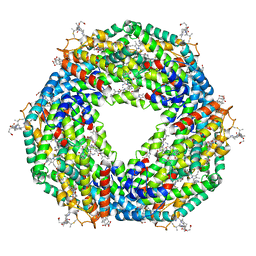
5FVB
DownloadVisualize
BU of 5fvb by Molmil
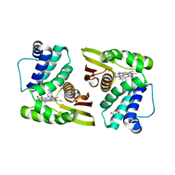
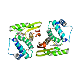
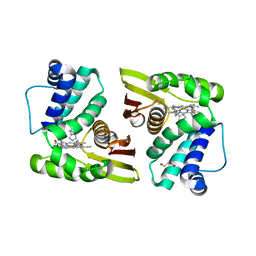
CRYSTAL STRUCTURE OF PHORMIDIUM C-PHYCOERYTHRIN AT PH 5.0
Descriptor: C-PHYCOERYTHRIN ALPHA SUBUNIT, C-PHYCOERYTHRIN BETA SUBUNIT, GLYCEROL, ...
Authors:Kumar, V, Sonani, R.R, Sharma, M, Gupta, G.D, Madamwar, D.
Deposit date:2016-02-05
Release date:2016-06-29
Last modified:2024-01-10
Method:X-RAY DIFFRACTION (1.93 Å)
Cite:Crystal Structure Analysis of C-Phycoerythrin from Marine Cyanobacterium Phormidium Sp. A09Dm.
Photosynth.Res., 129, 2016
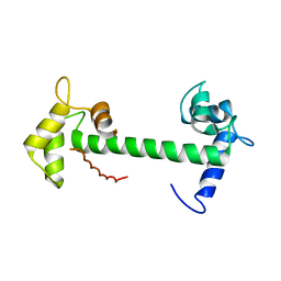
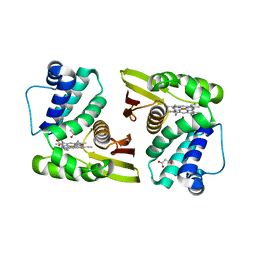
4E53
DownloadVisualize
BU of 4e53 by Molmil
Calmodulin and Nm peptide complex
Descriptor: Calmodulin, Linker, IQ motif of Neuromodulin
Authors:Kumar, V, Sivaraman, J.
Deposit date:2012-03-13
Release date:2013-03-20
Last modified:2024-03-20
Method:X-RAY DIFFRACTION (2.69 Å)
Cite:Structural basis for the interaction of unstructured neuron specific substrates neuromodulin and neurogranin with calmodulin
Sci Rep, 3, 2013
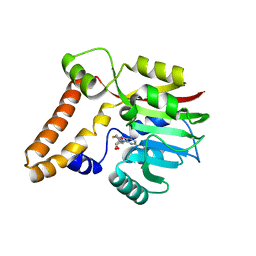
3T0I
DownloadVisualize
BU of 3t0i by Molmil
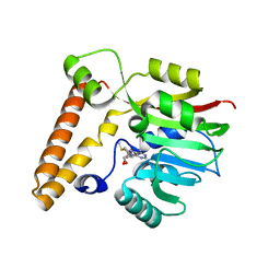
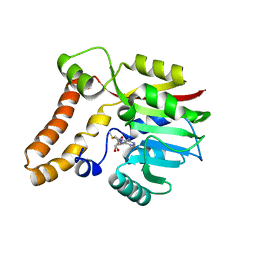
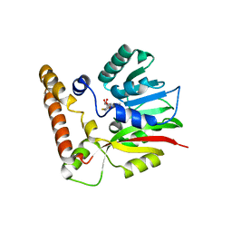
Crystal structure of complex of BT_2972 and AdoHcy, a methyltransferase from Bacteroides thetaiotaomicron
Descriptor: Putative methyltransferase, S-ADENOSYL-L-HOMOCYSTEINE
Authors:Kumar, V, Sivaraman, J.
Deposit date:2011-07-20
Release date:2011-09-07
Last modified:2023-11-01
Method:X-RAY DIFFRACTION (2.4 Å)
Cite:Crystal structure of complex of BT_2972 and AdoHcy,a methyltransferase from Bacteroides thetaiotaomicron
To be published
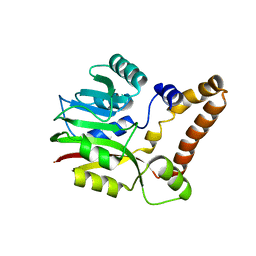
3T7R
DownloadVisualize
BU of 3t7r by Molmil
Crystal structure of apo BVU_3255, a methyltransferase from Bacteroides vulgatus ATCC 8482
Descriptor: 3-[(2Z)-3-methylpent-2-en-1-yl]benzene-1,2-diol, Putative methyltransferase
Authors:Kumar, V, Sivaraman, J.
Deposit date:2011-07-31
Release date:2011-10-12
Last modified:2023-11-01
Method:X-RAY DIFFRACTION (2.9 Å)
Cite:Structural characterization of BVU_3255, a methyltransferase from human intestine antibiotic resistant pathogen Bacteroides vulgatus
J.Struct.Biol., 2011
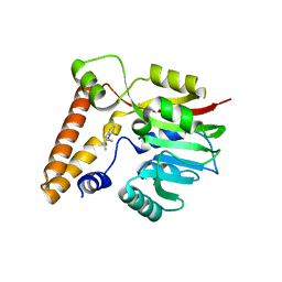
3SVZ
DownloadVisualize
BU of 3svz by Molmil
Crystal structure of apo BT_2972, a methyltransferase from Bacteroides thetaiotaomicron
Descriptor: Putative methyltransferase
Authors:Kumar, V, Sivaraman, J.
Deposit date:2011-07-13
Release date:2011-09-07
Last modified:2023-11-01
Method:X-RAY DIFFRACTION (2.9 Å)
Cite:Crystal structure of apo BT_2972, a methyltransferase from Bacteroides thetaiotaomicron
To be published
3T7S
DownloadVisualize
BU of 3t7s by Molmil
Crystal structure of complex of SAM and BVU_3255, a methyltransferase from Bacteroides vulgatus ATCC 8482
Descriptor: Putative methyltransferase, S-ADENOSYLMETHIONINE
Authors:Kumar, V, Sivaraman, J.
Deposit date:2011-07-31
Release date:2011-10-12
Last modified:2023-11-01
Method:X-RAY DIFFRACTION (2.2 Å)
Cite:Structural characterization of BVU_3255, a methyltransferase from human intestine antibiotic resistant pathogen Bacteroides vulgatus
J.Struct.Biol., 2011
3SXJ
DownloadVisualize
BU of 3sxj by Molmil
Crystal structure of complex of BT_2972 and AdoMet, a methyltransferase from Bacteroides thetaiotaomicron
Descriptor: Putative methyltransferase, S-ADENOSYLMETHIONINE
Authors:Kumar, V, Sivaraman, J.
Deposit date:2011-07-14
Release date:2011-09-07
Last modified:2023-11-01
Method:X-RAY DIFFRACTION (2.5 Å)
Cite:Crystal structure of complex of BT_2972 and AdoMet, a methyltransferase from Bacteroides thetaiotaomicron
To be Published
3T7T
DownloadVisualize
BU of 3t7t by Molmil
Crystal structure of complex of SAH and BVU_3255, a methyltransferase from Bacteroides vulgatus ATCC 8482
Descriptor: Putative methyltransferase, S-ADENOSYL-L-HOMOCYSTEINE
Authors:Kumar, V, Sivaraman, J.
Deposit date:2011-07-31
Release date:2011-10-12
Last modified:2023-11-01
Method:X-RAY DIFFRACTION (2.5 Å)
Cite:Structural characterization of BVU_3255, a methyltransferase from human intestine antibiotic resistant pathogen Bacteroides vulgatus
J.Struct.Biol., 2011
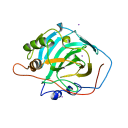
1HCB
DownloadVisualize
BU of 1hcb by Molmil
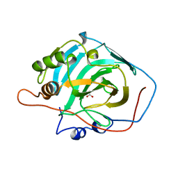
ENZYME-SUBSTRATE INTERACTIONS: STRUCTURE OF HUMAN CARBONIC ANHYDRASE I COMPLEXED WITH BICARBONATE
Descriptor: BICARBONATE ION, CARBONIC ANHYDRASE I, ZINC ION
Authors:Kumar, V, Kannan, K.K.
Deposit date:1994-01-07
Release date:1994-04-30
Last modified:2024-02-07
Method:X-RAY DIFFRACTION (1.6 Å)
Cite:Enzyme-substrate interactions. Structure of human carbonic anhydrase I complexed with bicarbonate.
J.Mol.Biol., 241, 1994
4DG7
DownloadVisualize
BU of 4dg7 by Molmil
Low resolution structure of Drosophila Translin
Descriptor: GM27569p
Authors:Kumar, V, Gupta, G.D.
Deposit date:2012-01-25
Release date:2012-06-06
Last modified:2023-11-29
Method:X-RAY DIFFRACTION (4.195 Å)
Cite:Low-resolution structure of Drosophila translin
FEBS Open Bio, 2, 2012
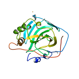
1HUH
DownloadVisualize
BU of 1huh by Molmil
DIFFERENCES IN ANIONIC INHIBITION OF HUMAN CARBONIC ANHYDRASE I REVEALED FROM THE STRUCTURES OF IODIDE AND GOLD CYANIDE INHIBITOR COMPLEXES
Descriptor: CARBONIC ANHYDRASE I, IODIDE ION, ZINC ION
Authors:Kumar, V, Kannan, K.K.
Deposit date:1993-10-28
Release date:1994-04-30
Last modified:2024-02-07
Method:X-RAY DIFFRACTION (2.2 Å)
Cite:Differences in anionic inhibition of human carbonic anhydrase I revealed from the structures of iodide and gold cyanide inhibitor complexes.
Acta Crystallogr.,Sect.D, 50, 1994
1HUG
DownloadVisualize
BU of 1hug by Molmil
Differences in anionic inhibition of Human Carbonic Anhydrase I revealed from the structures of iodide and gold cyanide inhibitor complexes
Descriptor: CARBONIC ANHYDRASE I, GOLD (I) CYANIDE ION, ZINC ION
Authors:Kumar, V, Kannan, K.K.
Deposit date:1993-10-28
Release date:1994-04-30
Last modified:2024-02-07
Method:X-RAY DIFFRACTION (2 Å)
Cite:Differences in anionic inhibition of human carbonic anhydrase I revealed from the structures of iodide and gold cyanide inhibitor complexes.
Acta Crystallogr.,Sect.D, 50, 1994
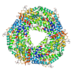
5AQD
DownloadVisualize
BU of 5aqd by Molmil
Crystal structure of Phormidium Phycoerythrin at pH 8.5
Descriptor: GLYCEROL, PHYCOERYTHRIN ALPHA SUBUNIT, PHYCOERYTHRIN BETA SUBUNIT, ...
Authors:Kumar, V, Sharma, M, Sonani, R.R, Gupta, G.D, Madamwar, D.
Deposit date:2015-09-22
Release date:2016-06-29
Last modified:2024-01-10
Method:X-RAY DIFFRACTION (2.121 Å)
Cite:Crystal Structure Analysis of C-Phycoerythrin from Marine Cyanobacterium Phormidium Sp. A09Dm.
Photosynth.Res., 129, 2016
5IXX
DownloadVisualize
BU of 5ixx by Molmil
Crystal structure of the signaling protein complex 4
Descriptor: 1-(1H-imidazol-1-yl)ethan-1-one, Alr2278 protein, MALONATE ION, ...
Authors:Kumar, V, van den Akker, F.
Deposit date:2016-03-23
Release date:2017-08-09
Last modified:2023-09-27
Method:X-RAY DIFFRACTION (2.35 Å)
Cite:Crystal structure of the signaling protein complex 4
To Be Published
5IXR
DownloadVisualize
BU of 5ixr by Molmil
Crystal Structure of C139A nostoc H-NOX domain with imidazole
Descriptor: Alr2278 protein, IMIDAZOLE, MALONATE ION, ...
Authors:Kumar, V, van den Akker, F.
Deposit date:2016-03-23
Release date:2017-08-09
Last modified:2023-09-27
Method:X-RAY DIFFRACTION (2.5 Å)
Cite:Crystal Structure of C139A nostoc H-NOX domain with imidazole
To Be Published
5IXW
DownloadVisualize
BU of 5ixw by Molmil
Crystal structure of the signaling protein complex 3
Descriptor: 1-VINYLIMIDAZOLE, Alr2278 protein, MALONATE ION, ...
Authors:Kumar, V, van den Akker, F.
Deposit date:2016-03-23
Release date:2017-08-09
Last modified:2023-09-27
Method:X-RAY DIFFRACTION (2.46 Å)
Cite:Crystal structure of the signaling protein complex 3
To Be Published
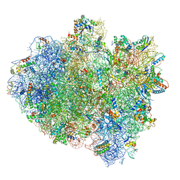
5IMR
DownloadVisualize
BU of 5imr by Molmil
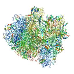
Structure of ribosome bound to cofactor at 5.7 angstrom resolution
Descriptor: 16S ribosomal RNA, 23S ribosomal RNA, 30S ribosomal protein S10, ...
Authors:Kumar, V, Ero, R, Jian, G.K, Ahmed, T, Zhan, Y, Bhushan, S, Gao, Y.G.
Deposit date:2016-03-06
Release date:2016-05-18
Last modified:2024-10-09
Method:ELECTRON MICROSCOPY (5.7 Å)
Cite:Structure of the GTP Form of Elongation Factor 4 (EF4) Bound to the Ribosome
J.Biol.Chem., 291, 2016
5IXV
DownloadVisualize
BU of 5ixv by Molmil
Crystal structure of the signaling protein complex 2
Descriptor: 1-METHYLIMIDAZOLE, Alr2278 protein, MALONATE ION, ...
Authors:Kumar, V, van den Akker, F.
Deposit date:2016-03-23
Release date:2017-08-09
Last modified:2024-11-20
Method:X-RAY DIFFRACTION (2.3 Å)
Cite:Crystal structure of the signaling protein complex 2
To Be Published
5IXZ
DownloadVisualize
BU of 5ixz by Molmil
Crystal structure of the signaling protein complex 5
Descriptor: Alr2278 protein, DIMETHYL SULFOXIDE, MALONATE ION, ...
Authors:Kumar, V, van den Akker, F.
Deposit date:2016-03-23
Release date:2017-11-01
Last modified:2023-09-27
Method:X-RAY DIFFRACTION (2.4 Å)
Cite:Crystal structure of the signaling protein complex 5
To Be Published
5IMQ
DownloadVisualize
BU of 5imq by Molmil
Structure of ribosome bound to cofactor at 3.8 angstrom resolution
Descriptor: 16S ribosomal RNA, 23S ribosomal RNA, 30S ribosomal protein S10, ...
Authors:Kumar, V, Ero, R, Jian, G.K, Ahmed, T, Zhan, Y, Bhushan, S, Gao, Y.G.
Deposit date:2016-03-06
Release date:2016-05-18
Last modified:2025-04-09
Method:ELECTRON MICROSCOPY (3.8 Å)
Cite:Structure of the GTP Form of Elongation Factor 4 (EF4) Bound to the Ribosome
J.Biol.Chem., 291, 2016
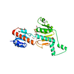
1MX3
DownloadVisualize
BU of 1mx3 by Molmil
Crystal structure of CtBP dehydrogenase core holo form
Descriptor: ACETIC ACID, C-terminal binding protein 1, NICOTINAMIDE-ADENINE-DINUCLEOTIDE
Authors:Kumar, V, Carlson, J.E, Ohgi, K.E, Edwards, T.E, Rose, D.W, Escalante, C.R, Aggarwal, A.K.
Deposit date:2002-10-01
Release date:2002-12-18
Last modified:2024-02-14
Method:X-RAY DIFFRACTION (1.95 Å)
Cite:Transcription Corepressor CtBP Is an NAD+-Regulated Dehydrogenase
Mol.Cell, 10, 2002
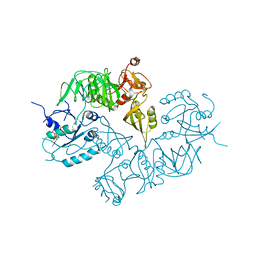
5A9Y
DownloadVisualize
BU of 5a9y by Molmil
Structure of ppGpp BipA
Descriptor: GTP-BINDING PROTEIN, GUANOSINE-5',3'-TETRAPHOSPHATE
Authors:Kumar, V, Chen, Y, Ero, R, Li, Z, Gao, Y.-G.
Deposit date:2015-07-23
Release date:2015-08-26
Last modified:2024-05-01
Method:X-RAY DIFFRACTION (4 Å)
Cite:Structure of Bipa in GTP Form Bound to the Ratcheted Ribosome.
Proc.Natl.Acad.Sci.USA, 112, 2015
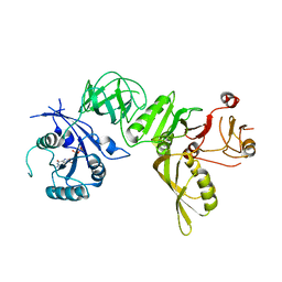
5A9X
DownloadVisualize
BU of 5a9x by Molmil
Structure of GDP bound BipA
Descriptor: GTP-BINDING PROTEIN, GUANOSINE-5'-DIPHOSPHATE
Authors:Kumar, V, Chen, Y, Ero, R, Li, Z, Gao, Y.-G.
Deposit date:2015-07-23
Release date:2015-08-26
Last modified:2024-05-08
Method:X-RAY DIFFRACTION (3.8 Å)
Cite:Structure of Bipa in GTP Form Bound to the Ratcheted Ribosome.
Proc.Natl.Acad.Sci.USA, 112, 2015
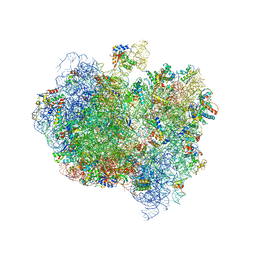
5A9Z
DownloadVisualize
BU of 5a9z by Molmil
Complex of Thermous thermophilus ribosome bound to BipA-GDPCP
Descriptor: 16S ribosomal RNA, 23S ribosomal RNA, 30S ribosomal protein S10, ...
Authors:Kumar, V, Chen, Y, Ahmed, T, Tan, J, Ero, R, Bhushan, S, Gao, Y.-G.
Deposit date:2015-07-23
Release date:2015-10-14
Last modified:2024-10-16
Method:ELECTRON MICROSCOPY (4.7 Å)
Cite:Structure of Bipa in GTP Form Bound to the Ratcheted Ribosome.
Proc.Natl.Acad.Sci.USA, 112, 2015
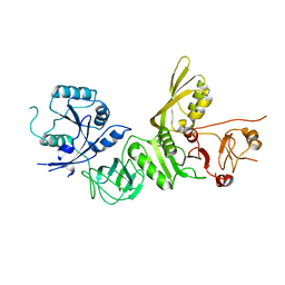
5A9V
DownloadVisualize
BU of 5a9v by Molmil
Structure of apo BipA
Descriptor: GTP-BINDING PROTEIN
Authors:Kumar, V, Chen, Y, Ero, R, Li, Z, Gao, Y.
Deposit date:2015-07-23
Release date:2015-09-02
Last modified:2024-01-10
Method:X-RAY DIFFRACTION (3.31 Å)
Cite:Structure of Bipa in GTP Form Bound to the Ratcheted Ribosome.
Proc.Natl.Acad.Sci.USA, 112, 2015

237423

PDB entries from 2025-06-11

PDB statisticsPDBj update infoContact PDBjnumon