Loading
PDBj
MenuPDBj@FacebookPDBj@TwitterPDBj@YouTubewwPDB FoundationwwPDB
RCSB PDBPDBeBMRBAdv. SearchSearch help
Search by PDB author
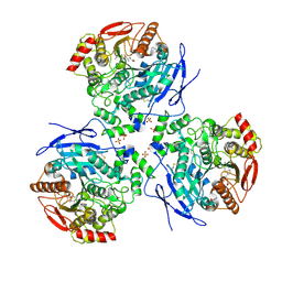
2DQZ
DownloadVisualize
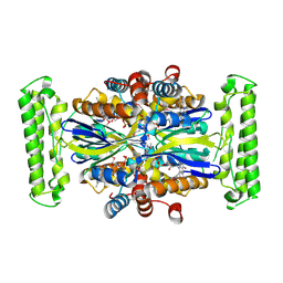
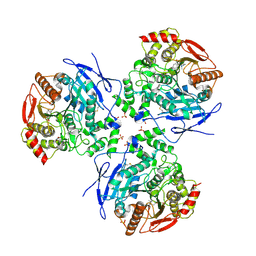
BU of 2dqz by Molmil
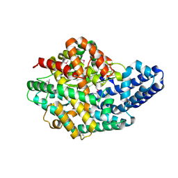
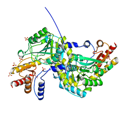
Crystal structure of human carboxylesterase in complex with homatropine, coenzyme A, and palmitate
Descriptor: 2-acetamido-2-deoxy-beta-D-glucopyranose, COENZYME A, FLUORIDE ION, ...
Authors:Bencharit, S, Redinbo, M.R.
Deposit date:2006-06-02
Release date:2006-08-29
Last modified:2023-10-25
Method:X-RAY DIFFRACTION (2.8 Å)
Cite:Multisite promiscuity in the processing of endogenous substrates by human carboxylesterase 1
J.Mol.Biol., 363, 2006
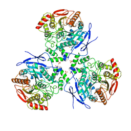
2DQY
DownloadVisualize
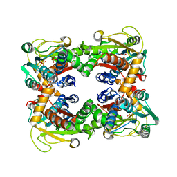
BU of 2dqy by Molmil
Crystal structure of human carboxylesterase in complex with cholate and palmitate
Descriptor: 2-acetamido-2-deoxy-beta-D-glucopyranose, CHOLIC ACID, Liver carboxylesterase 1, ...
Authors:Bencharit, S, Edwards, C.C, Morton, C.L, Howard-Williams, E.L, Potter, P.M, Redinbo, M.R.
Deposit date:2006-06-02
Release date:2006-08-29
Last modified:2023-10-25
Method:X-RAY DIFFRACTION (3 Å)
Cite:Multisite promiscuity in the processing of endogenous substrates by human carboxylesterase 1
J.Mol.Biol., 363, 2006
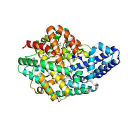
2DR0
DownloadVisualize
BU of 2dr0 by Molmil
Crystal structure of human carboxylesterase in complex with taurocholate
Descriptor: 2-acetamido-2-deoxy-beta-D-glucopyranose, Liver carboxylesterase 1, N-acetyl-alpha-neuraminic acid, ...
Authors:Bencharit, S, Redinbo, M.R.
Deposit date:2006-06-02
Release date:2006-08-29
Last modified:2020-07-29
Method:X-RAY DIFFRACTION (3.2 Å)
Cite:Multisite promiscuity in the processing of endogenous substrates by human carboxylesterase 1
J.Mol.Biol., 363, 2006
2H1J
DownloadVisualize
BU of 2h1j by Molmil
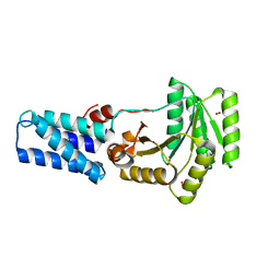
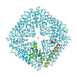
3.1 A X-ray structure of putative Oligoendopeptidase F: Crystals grown by microfluidic seeding
Descriptor: Oligoendopeptidase F, ZINC ION
Authors:Gerdts, C.J, Tereshko, V, Dementieva, I, Collart, F, Joachimiak, A, Kossiakoff, A, Ismagilov, R.F, Accelerated Technologies Center for Gene to 3D Structure (ATCG3D)
Deposit date:2006-05-16
Release date:2006-06-13
Last modified:2011-07-13
Method:X-RAY DIFFRACTION (3.1 Å)
Cite:Time-Controlled Microfluidic Seeding in nL-Volume Droplets To Separate Nucleation and Growth Stages of Protein Crystallization.
Angew.Chem.Int.Ed.Engl., 45, 2006
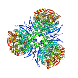
2H7C
DownloadVisualize
BU of 2h7c by Molmil
Crystal structure of human carboxylesterase in complex with Coenzyme A
Descriptor: 2-acetamido-2-deoxy-beta-D-glucopyranose, 2-acetamido-2-deoxy-beta-D-glucopyranose-(1-4)-2-acetamido-2-deoxy-beta-D-glucopyranose, COENZYME A, ...
Authors:Bencharit, S, Edwards, C.C, Morton, C.L, Howard-Williams, E.L, Potter, P.M, Redinbo, M.R.
Deposit date:2006-06-02
Release date:2006-08-29
Last modified:2023-10-25
Method:X-RAY DIFFRACTION (2 Å)
Cite:Multisite promiscuity in the processing of endogenous substrates by human carboxylesterase 1
J.Mol.Biol., 363, 2006
2H1N
DownloadVisualize
BU of 2h1n by Molmil
3.0 A X-ray structure of putative oligoendopeptidase F: crystals grown by vapor diffusion technique
Descriptor: Oligoendopeptidase F, UNKNOWN LIGAND, ZINC ION
Authors:Gerdts, C.J, Tereshko, V, Dementieva, I, Collart, F, Joachimiak, A, Kossiakoff, A, Ismagilov, R.F, Midwest Center for Structural Genomics (MCSG)
Deposit date:2006-05-16
Release date:2006-06-13
Last modified:2011-07-13
Method:X-RAY DIFFRACTION (3 Å)
Cite:Time-Controlled Microfluidic Seeding in nL-Volume Droplets To Separate Nucleation and Growth Stages of Protein Crystallization.
Angew.Chem.Int.Ed.Engl., 45, 2006
1LS1
DownloadVisualize
BU of 1ls1 by Molmil
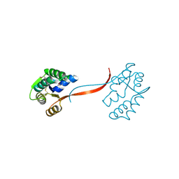
T. aquaticus Ffh NG Domain at 1.1A Resolution
Descriptor: MAGNESIUM ION, OXYGEN MOLECULE, SIGNAL RECOGNITION PARTICLE PROTEIN
Authors:Ramirez, U.D, Minasov, G, Freymann, D.M.
Deposit date:2002-05-16
Release date:2002-11-16
Last modified:2024-02-14
Method:X-RAY DIFFRACTION (1.1 Å)
Cite:Structural Basis for Mobility in the 1.1 A Crystal Structure of the NG Domain of Thermus aquaticus Ffh
J.Mol.Biol., 320, 2002
1O5U
DownloadVisualize
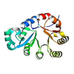
BU of 1o5u by Molmil
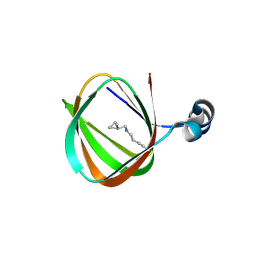
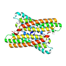
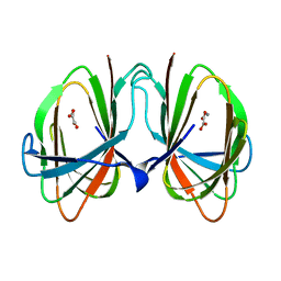
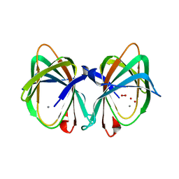
Crystal structure of a duf861 family protein (tm1112) from thermotoga maritima at 1.83 A resolution
Descriptor: UNKNOWN LIGAND, novel Thermotoga maritima enzyme TM1112
Authors:Joint Center for Structural Genomics (JCSG)
Deposit date:2003-10-06
Release date:2003-11-11
Last modified:2023-09-20
Method:X-RAY DIFFRACTION (1.83 Å)
Cite:Crystal structure of a novel Thermotoga maritima enzyme (TM1112) from the cupin family at 1.83 A resolution
Proteins, 56, 2004
1O4U
DownloadVisualize
BU of 1o4u by Molmil
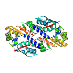
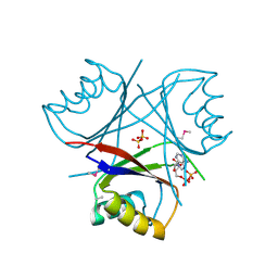
Crystal structure of a nicotinate nucleotide pyrophosphorylase (tm1645) from thermotoga maritima at 2.50 A resolution
Descriptor: Type II quinolic acid phosphoribosyltransferase
Authors:Joint Center for Structural Genomics (JCSG)
Deposit date:2003-07-09
Release date:2003-07-22
Last modified:2023-09-20
Method:X-RAY DIFFRACTION (2.5 Å)
Cite:Crystal structure of a type II quinolic acid phosphoribosyltransferase (TM1645) from Thermotoga maritima at 2.50 A resolution
Proteins, 55, 2004
1O4W
DownloadVisualize
BU of 1o4w by Molmil
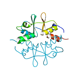
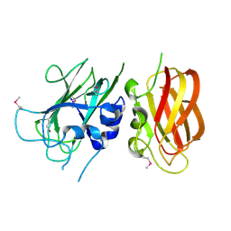
CRYSTAL STRUCTURE OF a PIN (PILT N-TERMINUS) DOMAIN CONTAINING PROTEIN (AF0591) FROM ARCHAEOGLOBUS FULGIDUS AT 1.90 A RESOLUTION
Descriptor: PIN (PilT N-terminus) domain
Authors:Joint Center for Structural Genomics (JCSG)
Deposit date:2003-07-16
Release date:2003-07-29
Last modified:2023-01-25
Method:X-RAY DIFFRACTION (1.9 Å)
Cite:Crystal structure of a PIN (PilT N-terminus) domain (AF0591) from Archaeoglobus fulgidus at 1.90 A resolution
Proteins, 56, 2004
1O50
DownloadVisualize
BU of 1o50 by Molmil
Crystal structure of a cbs domain-containing protein (tm0935) from thermotoga maritima at 1.87 A resolution
Descriptor: CBS domain-containing predicted protein TM0935
Authors:Joint Center for Structural Genomics (JCSG)
Deposit date:2003-07-31
Release date:2003-08-12
Last modified:2023-01-25
Method:X-RAY DIFFRACTION (1.87 Å)
Cite:Crystal structure of a tandem cystathionine-beta-synthase (CBS) domain protein (TM0935) from Thermotoga maritima at 1.87 A resolution
Proteins, 57, 2004
1O5H
DownloadVisualize
BU of 1o5h by Molmil
Crystal structure of formiminotetrahydrofolate cyclodeaminase (TM1560) from Thermotoga maritima at 2.80 A resolution
Descriptor: Formiminotetrahydrofolate cyclodeaminase
Authors:Joint Center for Structural Genomics (JCSG)
Deposit date:2003-09-17
Release date:2003-09-30
Last modified:2023-01-25
Method:X-RAY DIFFRACTION (2.8 Å)
Cite:Crystal structure of a formiminotetrahydrofolate cyclodeaminase (TM1560) from Thermotoga maritima at 2.80 A resolution reveals a new fold
Proteins, 58, 2005
1O4V
DownloadVisualize
BU of 1o4v by Molmil
Crystal structure of the catalytic subunit of a phosphoribosylaminoimidazole mutase (tm0446) from thermotoga maritima at 1.77 A resolution
Descriptor: SULFATE ION, phosphoribosylaminoimidazole mutase PurE
Authors:Joint Center for Structural Genomics (JCSG)
Deposit date:2003-07-11
Release date:2003-07-22
Last modified:2023-09-20
Method:X-RAY DIFFRACTION (1.77 Å)
Cite:Crystal structure of a phosphoribosylaminoimidazole mutase PurE (TM0446) from Thermotoga maritima at 1.77 A resolution
Proteins, 55, 2004
1O4S
DownloadVisualize
BU of 1o4s by Molmil
Crystal structure of Aspartate aminotransferase (TM1255) from Thermotoga maritima at 1.90 A resolution
Descriptor: Aspartate aminotransferase, PYRIDOXAL-5'-PHOSPHATE, SULFATE ION
Authors:Joint Center for Structural Genomics (JCSG)
Deposit date:2003-06-26
Release date:2003-07-08
Last modified:2023-09-20
Method:X-RAY DIFFRACTION (1.9 Å)
Cite:Crystal structure of an aspartate aminotransferase (TM1255) from Thermotoga maritima at 1.90 A resolution
Proteins, 55, 2004
1O4T
DownloadVisualize
BU of 1o4t by Molmil
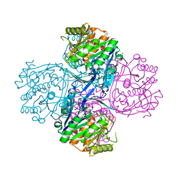
Crystal structure of a predicted oxalate decarboxylase (tm1287) from thermotoga maritima at 1.95 A resolution
Descriptor: MANGANESE (II) ION, OXALATE ION, putative oxalate decarboxylase
Authors:Joint Center for Structural Genomics (JCSG)
Deposit date:2003-07-03
Release date:2003-07-15
Last modified:2023-09-20
Method:X-RAY DIFFRACTION (1.95 Å)
Cite:Crystal structure of a putative oxalate decarboxylase (TM1287) from Thermotoga maritima at 1.95 A resolution
Proteins, 56, 2004
1O51
DownloadVisualize
BU of 1o51 by Molmil
Crystal structure of a putative PII-like signaling protein (TM0021) from Thermotoga maritima at 2.50 A resolution
Descriptor: ADENOSINE-5'-DIPHOSPHATE, Hypothetical protein TM0021, SULFATE ION
Authors:Joint Center for Structural Genomics (JCSG)
Deposit date:2003-08-01
Release date:2003-08-19
Last modified:2023-01-25
Method:X-RAY DIFFRACTION (2.5 Å)
Cite:Crystal structure of a putative PII-like signaling protein (TM0021) from Thermotoga maritima at 2.5 A resolution
Proteins, 54, 2004
1O59
DownloadVisualize
BU of 1o59 by Molmil
Crystal structure of Allantoicase (yir029w) from Saccharomyces cerevisiae at 2.40 A resolution
Descriptor: Allantoicase
Authors:Joint Center for Structural Genomics (JCSG)
Deposit date:2003-08-22
Release date:2003-09-02
Last modified:2023-01-25
Method:X-RAY DIFFRACTION (2.4 Å)
Cite:Crystal structure of an allantoicase (YIR029W) from Saccharomyces cerevisiae at 2.4 A resolution
Proteins, 56, 2004
1VJ2
DownloadVisualize
BU of 1vj2 by Molmil
Crystal structure of a novel family of manganese-containing cupin (tm1459) from thermotoga maritima at 1.65 A resolution
Descriptor: MANGANESE (II) ION, UNKNOWN LIGAND, novel manganese-containing cupin TM1459
Authors:Joint Center for Structural Genomics (JCSG)
Deposit date:2003-12-03
Release date:2003-12-09
Last modified:2023-09-20
Method:X-RAY DIFFRACTION (1.65 Å)
Cite:Crystal structure of a novel manganese-containing cupin (TM1459) from Thermotoga maritima at 1.65 A resolution.
Proteins, 56, 2004
2AFB
DownloadVisualize
BU of 2afb by Molmil
Crystal structure of 2-dehydro-3- deoxygluconokinase (EC 2.7.1.45) (tm0067) from THERMOTOGA MARITIMA at 2.05 A resolution
Descriptor: 2-keto-3-deoxygluconate kinase, CALCIUM ION, NICKEL (II) ION
Authors:Joint Center for Structural Genomics (JCSG)
Deposit date:2005-07-25
Release date:2006-02-14
Last modified:2023-01-25
Method:X-RAY DIFFRACTION (2.05 Å)
Cite:Crystal structure of 2-keto-3-deoxygluconate kinase (TM0067) from Thermotoga maritima at 2.05 A resolution.
Proteins, 70, 2007
2B8N
DownloadVisualize
BU of 2b8n by Molmil
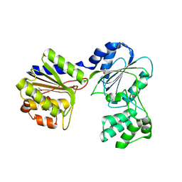
Crystal structure of Glycerate kinase (EC 2.7.1.31) (tm1585) from THERMOTOGA MARITIMA at 2.70 A resolution
Descriptor: glycerate kinase, putative
Authors:Joint Center for Structural Genomics (JCSG)
Deposit date:2005-10-07
Release date:2005-11-08
Last modified:2023-01-25
Method:X-RAY DIFFRACTION (2.53 Å)
Cite:Crystal structure of a glycerate kinase (TM1585) from Thermotoga maritima at 2.70 A resolution reveals a new fold
Proteins, 65, 2006
1KQ3
DownloadVisualize
BU of 1kq3 by Molmil
CRYSTAL STRUCTURE OF A GLYCEROL DEHYDROGENASE (TM0423) FROM THERMOTOGA MARITIMA AT 1.5 A RESOLUTION
Descriptor: 2-AMINO-2-HYDROXYMETHYL-PROPANE-1,3-DIOL, CHLORIDE ION, ZINC ION, ...
Authors:Wilson, I.A, Miller, M.D, Joint Center for Structural Genomics (JCSG)
Deposit date:2002-01-03
Release date:2002-02-27
Last modified:2024-02-14
Method:X-RAY DIFFRACTION (1.5 Å)
Cite:Structural genomics of the Thermotoga maritima proteome implemented in a high-throughput structure determination pipeline
Proc.Natl.Acad.Sci.USA, 99, 2002
1KQ4
DownloadVisualize
BU of 1kq4 by Molmil
CRYSTAL STRUCTURE OF A THY1-COMPLEMENTING PROTEIN (TM0449) FROM THERMOTOGA MARITIMA AT 2.25 A RESOLUTION
Descriptor: FLAVIN-ADENINE DINUCLEOTIDE, HYPOTHETICAL PROTEIN TM0449
Authors:Wilson, I.A, Miller, M.D, Joint Center for Structural Genomics (JCSG)
Deposit date:2002-01-03
Release date:2002-02-27
Last modified:2017-10-11
Method:X-RAY DIFFRACTION (2.25 Å)
Cite:Structural genomics of the Thermotoga maritima proteome implemented in a high-throughput structure determination pipeline
Proc.Natl.Acad.Sci.USA, 99, 2002
1O58
DownloadVisualize
BU of 1o58 by Molmil
Crystal structure of O-acetylserine sulfhydrylase (TM0665) from Thermotoga maritima at 1.80 A resolution
Descriptor: O-acetylserine sulfhydrylase, PHOSPHATE ION
Authors:Joint Center for Structural Genomics (JCSG)
Deposit date:2003-08-20
Release date:2003-09-02
Last modified:2023-01-25
Method:X-RAY DIFFRACTION (1.8 Å)
Cite:Crystal structure of O-acetylserine sulfhydrylase (TM0665) from Thermotoga maritima at 1.8 A resolution
Proteins, 56, 2004
1O1Z
DownloadVisualize
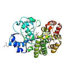
BU of 1o1z by Molmil
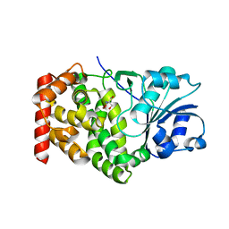
Crystal structure of glycerophosphodiester phosphodiesterase (GDPD) (TM1621) from Thermotoga maritima at 1.60 A resolution
Descriptor: SODIUM ION, glycerophosphodiester phosphodiesterase
Authors:Joint Center for Structural Genomics (JCSG)
Deposit date:2003-02-12
Release date:2003-04-01
Last modified:2023-01-25
Method:X-RAY DIFFRACTION (1.6 Å)
Cite:Crystal structure of a glycerophosphodiester phosphodiesterase (GDPD) from Thermotoga maritima (TM1621) at 1.60 A resolution.
Proteins, 56, 2004
1O2D
DownloadVisualize
BU of 1o2d by Molmil
Crystal structure of Alcohol dehydrogenase, iron-containing (TM0920) from Thermotoga maritima at 1.30 A resolution
Descriptor: 2-AMINO-2-HYDROXYMETHYL-PROPANE-1,3-DIOL, Alcohol dehydrogenase, iron-containing, ...
Authors:Joint Center for Structural Genomics (JCSG)
Deposit date:2003-02-27
Release date:2003-06-10
Last modified:2023-01-25
Method:X-RAY DIFFRACTION (1.3 Å)
Cite:Crystal structure of an iron-containing 1,3-propanediol dehydrogenase (TM0920) from Thermotoga maritima at 1.3 A resolution
Proteins, 54, 2004

219869

数据于2024-05-15公开中

PDB statisticsPDBj update infoContact PDBjnumon