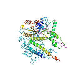
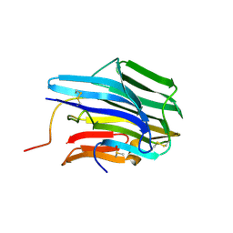
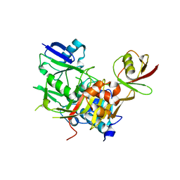
3AI2
 
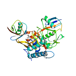
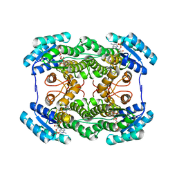 | | The crystal structure of L-sorbose reductase from Gluconobacter frateurii complexed with NADPH | | 分子名称: | NADPH DIHYDRO-NICOTINAMIDE-ADENINE-DINUCLEOTIDE PHOSPHATE, NADPH-sorbose reductase | | 著者 | Kubota, K, Nagata, K, Okai, M, Miyazono, K, Tanokura, M. | | 登録日 | 2010-05-07 | | 公開日 | 2011-02-09 | | 最終更新日 | 2023-11-01 | | 実験手法 | X-RAY DIFFRACTION (1.9 Å) | | 主引用文献 | The Crystal Structure of l-Sorbose Reductase from Gluconobacter frateurii Complexed with NADPH and l-Sorbose
J.Mol.Biol., 407, 2011
|
|
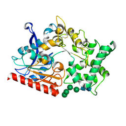
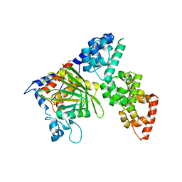
3AI3
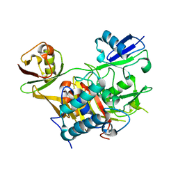
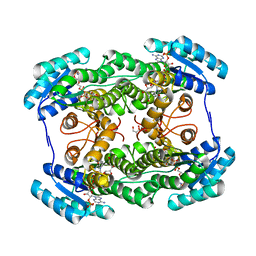 | | The crystal structure of L-Sorbose reductase from Gluconobacter frateurii complexed with NADPH and L-sorbose | | 分子名称: | L-sorbose, NADPH DIHYDRO-NICOTINAMIDE-ADENINE-DINUCLEOTIDE PHOSPHATE, NADPH-sorbose reductase, ... | | 著者 | Kubota, K, Nagata, K, Okai, M, Miyazono, K, Tanokura, M. | | 登録日 | 2010-05-07 | | 公開日 | 2011-02-09 | | 最終更新日 | 2023-11-01 | | 実験手法 | X-RAY DIFFRACTION (1.8 Å) | | 主引用文献 | The Crystal Structure of l-Sorbose Reductase from Gluconobacter frateurii Complexed with NADPH and l-Sorbose
J.Mol.Biol., 407, 2011
|
|
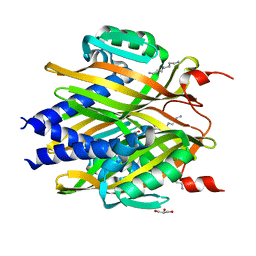
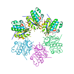
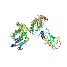
3AI1
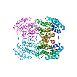 | | The crystal structure of L-sorbose reductase from Gluconobacter frateurii complexed with NADPH and L-sorbose reveals the structure bases of its catalytic mechanism and high substrate selectivity | | 分子名称: | NADPH-sorbose reductase | | 著者 | Kubota, K, Nagata, K, Okai, M, Miyazono, K, Tanokura, M. | | 登録日 | 2010-05-06 | | 公開日 | 2011-02-09 | | 最終更新日 | 2023-11-01 | | 実験手法 | X-RAY DIFFRACTION (2.38 Å) | | 主引用文献 | The Crystal Structure of l-Sorbose Reductase from Gluconobacter frateurii Complexed with NADPH and l-Sorbose
J.Mol.Biol., 407, 2011
|
|
3B2E
 | |
3VLC
 | |
3ORY
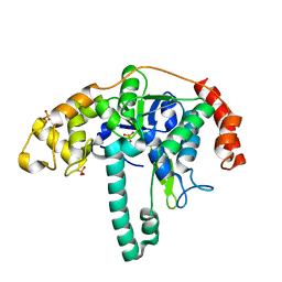 | | Crystal structure of Flap endonuclease 1 from hyperthermophilic archaeon Desulfurococcus amylolyticus | | 分子名称: | PHOSPHATE ION, flap endonuclease 1 | | 著者 | Mase, T, Kubota, K, Miyazono, K, Kawarabayashii, Y, Tanokura, M. | | 登録日 | 2010-09-08 | | 公開日 | 2011-02-09 | | 最終更新日 | 2024-03-20 | | 実験手法 | X-RAY DIFFRACTION (2 Å) | | 主引用文献 | Structure of flap endonuclease 1 from the hyperthermophilic archaeon Desulfurococcus amylolyticus
Acta Crystallogr.,Sect.F, 67, 2011
|
|
3JRS
 | | Crystal structure of (+)-ABA-bound PYL1 | | 分子名称: | (2Z,4E)-5-[(1S)-1-hydroxy-2,6,6-trimethyl-4-oxocyclohex-2-en-1-yl]-3-methylpenta-2,4-dienoic acid, Putative uncharacterized protein At5g46790 | | 著者 | Miyazono, K, Miyakawa, T, Sawano, Y, Kubota, K, Tanokura, M. | | 登録日 | 2009-09-08 | | 公開日 | 2009-11-03 | | 最終更新日 | 2024-03-20 | | 実験手法 | X-RAY DIFFRACTION (2.05 Å) | | 主引用文献 | Structural basis of abscisic acid signalling
Nature, 462, 2009
|
|
3JRQ
 | | Crystal structure of (+)-ABA-bound PYL1 in complex with ABI1 | | 分子名称: | (2Z,4E)-5-[(1S)-1-hydroxy-2,6,6-trimethyl-4-oxocyclohex-2-en-1-yl]-3-methylpenta-2,4-dienoic acid, Protein phosphatase 2C 56, Putative uncharacterized protein At5g46790 | | 著者 | Miyazono, K, Miyakawa, T, Sawano, Y, Kubota, K, Tanokura, M. | | 登録日 | 2009-09-08 | | 公開日 | 2009-11-03 | | 最終更新日 | 2023-11-01 | | 実験手法 | X-RAY DIFFRACTION (2.1 Å) | | 主引用文献 | Structural basis of abscisic acid signalling
Nature, 462, 2009
|
|
7WAB
 | | Crystal structure of the prolyl endoprotease, PEP, from Aspergillus niger | | 分子名称: | 2-acetamido-2-deoxy-beta-D-glucopyranose, 2-acetamido-2-deoxy-beta-D-glucopyranose-(1-4)-2-acetamido-2-deoxy-beta-D-glucopyranose, COMPASS (Complex proteins associated with Set1p) component shg1 family protein, ... | | 著者 | Miyazono, K, Kubota, K, Takahashi, K, Tanokura, M. | | 登録日 | 2021-12-14 | | 公開日 | 2022-01-12 | | 最終更新日 | 2022-02-16 | | 実験手法 | X-RAY DIFFRACTION (1.75 Å) | | 主引用文献 | Crystal structure and substrate recognition mechanism of the prolyl endoprotease PEP from Aspergillus niger.
Biochem.Biophys.Res.Commun., 591, 2022
|
|
1Y43
 | | crystal structure of aspergilloglutamic peptidase from Aspergillus niger | | 分子名称: | Aspergillopepsin II heavy chain, Aspergillopepsin II light chain, SULFATE ION | | 著者 | Sasaki, H, Nakagawa, A, Iwata, S, Muramatsu, T, Suganuma, M, Sawano, Y, Kojima, M, Kubota, K, Takahashi, K. | | 登録日 | 2004-11-30 | | 公開日 | 2005-12-13 | | 最終更新日 | 2013-02-27 | | 実験手法 | X-RAY DIFFRACTION (1.4 Å) | | 主引用文献 | The three-dimensional structure of aspergilloglutamic peptidase from Aspergillus niger
Proc.Jpn.Acad.,Ser.B, 80, 2004
|
|
3A76
 | | The crystal structure of LinA | | 分子名称: | GLYCEROL, Gamma-hexachlorocyclohexane dehydrochlorinase, SPERMIDINE | | 著者 | Okai, M, Kubota, K, Fukuda, M, Nagata, Y, Nagata, K, Tanokura, M. | | 登録日 | 2009-09-15 | | 公開日 | 2010-09-15 | | 最終更新日 | 2024-03-13 | | 実験手法 | X-RAY DIFFRACTION (2.25 Å) | | 主引用文献 | Crystal structure of g-hexachlorocyclohexane dehydrochlorinase LinA from Sphingobium japonicum UT26
J.Mol.Biol., 2010
|
|
3TRS
 | | The crystal structure of aspergilloglutamic peptidase from Aspergillus niger | | 分子名称: | Aspergillopepsin-2 heavy chain, Aspergillopepsin-2 light chain, DIMETHYL SULFOXIDE | | 著者 | Sasaki, H, Kubota, K, Lee, W.C, Ohtsuka, J, Kojima, M, Takahashi, K, Tanokura, M. | | 登録日 | 2011-09-10 | | 公開日 | 2012-08-22 | | 最終更新日 | 2023-11-01 | | 実験手法 | X-RAY DIFFRACTION (1.6 Å) | | 主引用文献 | The crystal structure of an intermediate dimer of aspergilloglutamic peptidase that mimics the enzyme-activation product complex produced upon autoproteolysis.
J.Biochem., 152, 2012
|
|
1ZOV
 | | Crystal Structure of Monomeric Sarcosine Oxidase from Bacillus sp. NS-129 | | 分子名称: | CHLORIDE ION, FLAVIN-ADENINE DINUCLEOTIDE, Monomeric sarcosine oxidase | | 著者 | Nagata, K, Sasaki, H, Ohtsuka, J, Hua, M, Okai, M, Kubota, K, Kamo, M, Ito, K, Ichikawa, T, Koyama, Y, Tanokura, M. | | 登録日 | 2005-05-14 | | 公開日 | 2006-05-23 | | 最終更新日 | 2023-10-25 | | 実験手法 | X-RAY DIFFRACTION (1.86 Å) | | 主引用文献 | Crystal structure of monomeric sarcosine oxidase from Bacillus sp. NS-129 reveals multiple conformations at the active-site loop
PROC.JPN.ACAD.,SER.B, 81, 2005
|
|
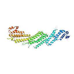
3LQB
 | | Crystal structure of the hatching enzyme ZHE1 from the zebrafish Danio rerio | | 分子名称: | 1,2-ETHANEDIOL, LOC792177 protein, SULFATE ION, ... | | 著者 | Tanokura, M, Okada, A, Nagata, K, Yasumasu, S, Ohtsuka, J, Iuchi, I. | | 登録日 | 2010-02-08 | | 公開日 | 2010-09-08 | | 最終更新日 | 2023-11-01 | | 実験手法 | X-RAY DIFFRACTION (1.1 Å) | | 主引用文献 | Crystal structure of zebrafish hatching enzyme 1 from the zebrafish Danio rerio
J.Mol.Biol., 402, 2010
|
|
2ZO4
 | | Crystal structure of metallo-beta-lactamase family protein TTHA1429 from Thermus thermophilus HB8 | | 分子名称: | Metallo-beta-lactamase family protein, ZINC ION | | 著者 | Yamamura, A, Nagata, K, Agari, Y, Ebihara, A, Nakagawa, N, Yokoyama, S, Kuramitsu, S, Tanokura, M. | | 登録日 | 2008-05-05 | | 公開日 | 2009-03-17 | | 最終更新日 | 2024-03-13 | | 実験手法 | X-RAY DIFFRACTION (2.1 Å) | | 主引用文献 | Crystal structure of TTHA1429, a novel metallo-beta-lactamase superfamily protein from Thermus thermophilus HB8.
Proteins, 73, 2008
|
|
2ZTS
 | |
5H11
 | |
3WXG
 | |
3WXE
 | |
3WXF
 | |
6KIP
 | |
2RTX
 | | Solution structure of the GGQ domain of YaeJ protein from Escherichia coli | | 分子名称: | Peptidyl-tRNA hydrolase YaeJ | | 著者 | Nameki, N, Enomoto, M, Kogure, H, Tochio, N, Guntert, P. | | 登録日 | 2013-09-21 | | 公開日 | 2013-12-25 | | 最終更新日 | 2023-06-14 | | 実験手法 | SOLUTION NMR | | 主引用文献 | Identification of residues required for stalled-ribosome rescue in the codon-independent release factor YaeJ
Nucleic Acids Res., 42, 2014
|
|
3VON
 | | Crystalstructure of the ubiquitin protease | | 分子名称: | Ubiquitin thioesterase OTUB1, Ubiquitin-conjugating enzyme E2 N, Ubiquitin-conjugating enzyme E2 variant 2 | | 著者 | Sato, Y, Fukai, S. | | 登録日 | 2012-01-30 | | 公開日 | 2012-05-30 | | 最終更新日 | 2023-11-08 | | 実験手法 | X-RAY DIFFRACTION (3.15 Å) | | 主引用文献 | Molecular basis of Lys-63-linked polyubiquitination inhibition by the interaction between human deubiquitinating enzyme OTUB1 and ubiquitin-conjugating enzyme UBC13.
J.Biol.Chem., 287, 2012
|
|


