Loading
PDBj
メニューPDBj@FacebookPDBj@TwitterPDBj@YouTubewwPDB FoundationwwPDB
RCSB PDBPDBeBMRBAdv. SearchSearch help
Search by PDB author
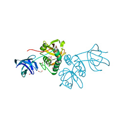
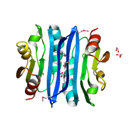
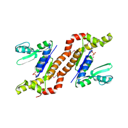
1S96
DownloadVisualize
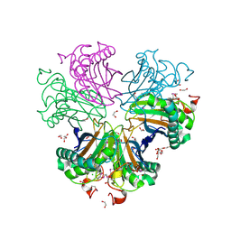
BU of 1s96 by Molmil
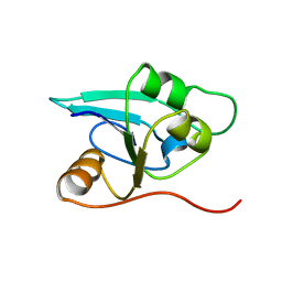
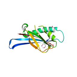
The 2.0 A X-ray structure of Guanylate Kinase from E.coli
分子名称: Guanylate kinase, PHOSPHATE ION, UNKNOWN ATOM OR ION
著者Kreusch, A, Spraggon, G, Klock, H.E, McMullan, D, Vincent, J, Rodrigues, K, Lesley, S.A.
登録日2004-02-03
公開日2004-02-10
最終更新日2011-07-13
実験手法X-RAY DIFFRACTION (2 Å)
主引用文献The Structure of Guanylate Kinase from Escherichia coli at 2.0 A resolution
To be Published
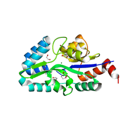
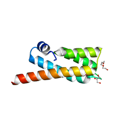
1R73
DownloadVisualize
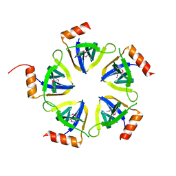
BU of 1r73 by Molmil
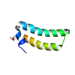
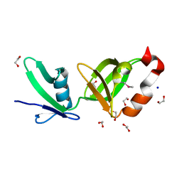
Solution Structure of TM1492, the L29 ribosomal protein from Thermotoga maritima
分子名称: 50S ribosomal protein L29
著者Peti, W, Etezady-Esfarjani, T, Herrmann, T, Klock, H.E, Lesley, S.A, Wuethrich, K, Joint Center for Structural Genomics (JCSG)
登録日2003-10-17
公開日2004-08-10
最終更新日2022-03-02
実験手法SOLUTION NMR
主引用文献NMR for structural proteomics of Thermotoga maritima: Screening and structure determination
J.STRUCT.FUNCT.GENOM., 5, 2004
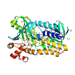
1RDU
DownloadVisualize
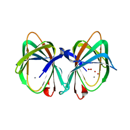
BU of 1rdu by Molmil
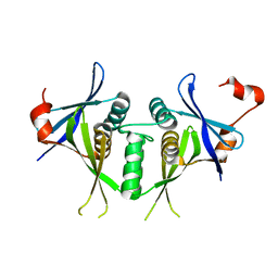
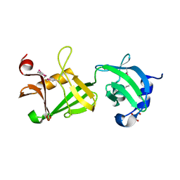
NMR STRUCTURE OF A PUTATIVE NIFB PROTEIN FROM THERMOTOGA (TM1290), WHICH BELONGS TO THE DUF35 FAMILY
分子名称: conserved hypothetical protein
著者Etezady-Esfarjani, T, Herrmann, T, Peti, W, Klock, H.E, Lesley, S.A, Wuthrich, K, Joint Center for Structural Genomics (JCSG)
登録日2003-11-06
公開日2004-07-06
最終更新日2022-03-02
実験手法SOLUTION NMR
主引用文献NMR Structure Determination of the Hypothetical Protein TM1290 from Thermotoga Maritima using Automated NOESY Analysis.
J.Biomol.NMR, 29, 2004
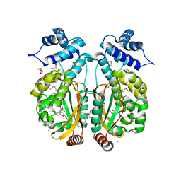
1SJ5
DownloadVisualize
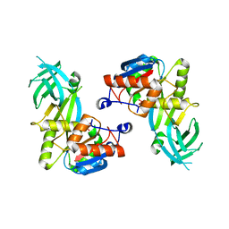
BU of 1sj5 by Molmil
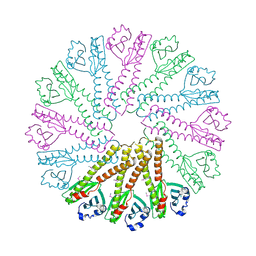
Crystal structure of a duf151 family protein (tm0160) from thermotoga maritima at 2.8 A resolution
分子名称: conserved hypothetical protein TM0160
著者Spraggon, G, Panatazatos, D, Klock, H.E, Wilson, I.A, Woods Jr, V.L, Lesley, S.A, Joint Center for Structural Genomics (JCSG)
登録日2004-03-02
公開日2005-03-01
最終更新日2023-08-23
実験手法X-RAY DIFFRACTION (2.8 Å)
主引用文献On the use of DXMS to produce more crystallizable proteins: structures of the T. maritima proteins TM0160 and TM1171.
Protein Sci., 13, 2004
1VKB
DownloadVisualize
BU of 1vkb by Molmil
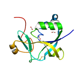
Crystal structure of an aig2-like protein (a2ld1, ggact, mgc7867) from mus musculus at 1.90 A resolution
分子名称: FORMIC ACID, hypothetical protein
著者Joint Center for Structural Genomics (JCSG)
登録日2004-05-07
公開日2004-05-18
最終更新日2023-01-25
実験手法X-RAY DIFFRACTION (1.9 Å)
主引用文献Crystal structure of a conserved hypothetical protein (gi: 13879369) from Mouse at 1.90 A resolution reveals a new fold.
Proteins, 61, 2005
3U21
DownloadVisualize
BU of 3u21 by Molmil
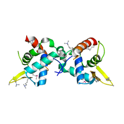
Crystal structure of a Fragment of Nuclear factor related to kappa-B-binding protein (residues 370-495) (NFRKB) from Homo sapiens at 2.18 A resolution
分子名称: Nuclear factor related to kappa-B-binding protein, SODIUM ION
著者Joint Center for Structural Genomics (JCSG), Partnership for Stem Cell Biology, Partnership for Stem Cell Biology (STEMCELL)
登録日2011-09-30
公開日2011-11-02
最終更新日2023-02-01
実験手法X-RAY DIFFRACTION (2.18 Å)
主引用文献Structure of a Novel Winged-Helix Like Domain from Human NFRKB Protein.
Plos One, 7, 2012
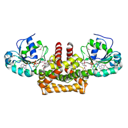
2Q8U
DownloadVisualize
BU of 2q8u by Molmil
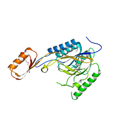
CRYSTAL STRUCTURE OF MRE11 FROM THERMOTOGA MARITIMA MSB8 (TM1635) AT 2.20 A RESOLUTION
分子名称: Exonuclease, putative
著者Joint Center for Structural Genomics (JCSG)
登録日2007-06-11
公開日2007-06-26
最終更新日2023-01-25
実験手法X-RAY DIFFRACTION (2.2 Å)
主引用文献Crystal structure of the first eubacterial Mre11 nuclease reveals novel features that may discriminate substrates during DNA repair.
J.Mol.Biol., 397, 2010
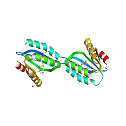
3BOS
DownloadVisualize
BU of 3bos by Molmil
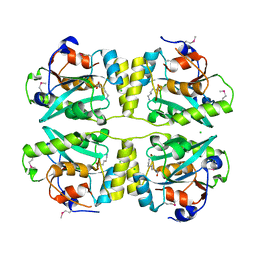
Crystal structure of a putative dna replication regulator HDA (SAMA_1916) from Shewanella amazonensis sb2b at 1.75 A resolution
分子名称: 1,2-ETHANEDIOL, CHLORIDE ION, CYTIDINE-5'-DIPHOSPHATE, ...
著者Joint Center for Structural Genomics (JCSG)
登録日2007-12-17
公開日2008-01-15
最終更新日2023-01-25
実験手法X-RAY DIFFRACTION (1.75 Å)
主引用文献A structural basis for the regulatory inactivation of DnaA.
J.Mol.Biol., 385, 2009
3KW0
DownloadVisualize
BU of 3kw0 by Molmil
Crystal structure of Cysteine peptidase (NP_982244.1) from BACILLUS CEREUS ATCC 10987 at 2.50 A resolution
分子名称: CHLORIDE ION, Cysteine peptidase, LYSINE
著者Joint Center for Structural Genomics (JCSG)
登録日2009-11-30
公開日2009-12-15
最終更新日2023-02-01
実験手法X-RAY DIFFRACTION (2.5 Å)
主引用文献Structural Analysis of Papain-Like NlpC/P60 Superfamily Enzymes with a Circularly Permuted Topology Reveals Potential Lipid Binding Sites.
Plos One, 6, 2011
2EVR
DownloadVisualize
BU of 2evr by Molmil
CRYSTAL STRUCTURE OF A PUTATIVE GAMMA-D-GLUTAMYL-L-DIAMINO ACID ENDOPEPTIDASE (NPUN_R0659) FROM NOSTOC PUNCTIFORME PCC 73102 AT 1.60 A RESOLUTION
分子名称: 1,2-ETHANEDIOL, CHLORIDE ION, COG0791: Cell wall-associated hydrolases (invasion-associated proteins), ...
著者Joint Center for Structural Genomics (JCSG)
登録日2005-10-31
公開日2005-11-22
最終更新日2023-01-25
実験手法X-RAY DIFFRACTION (1.6 Å)
主引用文献Structural Basis of Murein Peptide Specificity of a gamma-D-Glutamyl-L-Diamino Acid Endopeptidase.
Structure, 17, 2009
2FEA
DownloadVisualize
BU of 2fea by Molmil
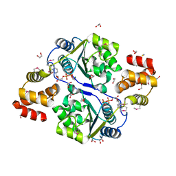
Crystal structure of MtnX phosphatase from Bacillus Subtilis at 2.00 A resolution
分子名称: 1,2-ETHANEDIOL, 2-hydroxy-3-keto-5-methylthiopentenyl-1-phosphate phosphatase, MAGNESIUM ION, ...
著者Joint Center for Structural Genomics (JCSG)
登録日2005-12-15
公開日2005-12-27
最終更新日2023-01-25
実験手法X-RAY DIFFRACTION (2 Å)
主引用文献Crystal structure of MtnX phosphatase from Bacillus subtilis at 2.0 A resolution provides a structural basis for bipartite phosphomonoester hydrolysis of 2-hydroxy-3-keto-5-methylthiopentenyl-1-phosphate.
Proteins, 69, 2007
3OZ2
DownloadVisualize
BU of 3oz2 by Molmil
Crystal structure of a geranylgeranyl bacteriochlorophyll reductase-like (Ta0516) from Thermoplasma acidophilum at 1.60 A resolution
分子名称: (2R)-3-{[(R)-{[(2S)-2,3-dihydroxypropyl]oxy}(hydroxy)phosphoryl]oxy}-2-[(6Z)-tridec-6-enoyloxy]propyl (9Z)-octadec-9-enoate, 1,2-ETHANEDIOL, Digeranylgeranylglycerophospholipid reductase, ...
著者Joint Center for Structural Genomics (JCSG)
登録日2010-09-24
公開日2010-10-27
最終更新日2023-02-01
実験手法X-RAY DIFFRACTION (1.6 Å)
主引用文献Insights into substrate specificity of geranylgeranyl reductases revealed by the structure of digeranylgeranylglycerophospholipid reductase, an essential enzyme in the biosynthesis of archaeal membrane lipids.
J.Mol.Biol., 404, 2010
3PXP
DownloadVisualize
BU of 3pxp by Molmil
Crystal structure of a PAS and DNA binding domain containing protein (Caur_2278) from CHLOROFLEXUS AURANTIACUS J-10-FL at 2.30 A resolution
分子名称: 1,2-ETHANEDIOL, CHLORIDE ION, Helix-turn-helix domain protein, ...
著者Joint Center for Structural Genomics (JCSG)
登録日2010-12-10
公開日2011-01-19
最終更新日2023-02-01
実験手法X-RAY DIFFRACTION (2.3 Å)
主引用文献Structure of an MmyB-Like Regulator from C. aurantiacus, Member of a New Transcription Factor Family Linked to Antibiotic Metabolism in Actinomycetes.
Plos One, 7, 2012
2OOK
DownloadVisualize
BU of 2ook by Molmil
Crystal structure of a protein with unknown function (YP_749275.1) from Shewanella Frigidimarina NCIMB 400 at 1.80 A resolution
分子名称: 1,2-ETHANEDIOL, Hypothetical protein
著者Joint Center for Structural Genomics (JCSG)
登録日2007-01-25
公開日2007-02-06
最終更新日2023-01-25
実験手法X-RAY DIFFRACTION (1.8 Å)
主引用文献Open and closed conformations of two SpoIIAA-like proteins (YP_749275.1 and YP_001095227.1) provide insights into membrane association and ligand binding.
Acta Crystallogr.,Sect.F, 66, 2010
2OOC
DownloadVisualize
BU of 2ooc by Molmil
Crystal structure of Histidine Phosphotransferase ShpA (NP_419930.1) from Caulobacter crescentus at 1.52 A resolution
分子名称: GLYCEROL, Histidine phosphotransferase, TETRAETHYLENE GLYCOL
著者Joint Center for Structural Genomics (JCSG)
登録日2007-01-25
公開日2007-02-06
最終更新日2023-01-25
実験手法X-RAY DIFFRACTION (1.52 Å)
主引用文献Crystal structure of histidine phosphotransfer protein ShpA, an essential regulator of stalk biogenesis in Caulobacter crescentus.
J.Mol.Biol., 390, 2009
2PV7
DownloadVisualize
BU of 2pv7 by Molmil
Crystal structure of chorismate mutase / prephenate dehydrogenase (tyrA) (1574749) from Haemophilus influenzae RD at 2.00 A resolution
分子名称: NICOTINAMIDE-ADENINE-DINUCLEOTIDE, T-protein [Includes: Chorismate mutase (EC 5.4.99.5) (CM) and Prephenate dehydrogenase (EC 1.3.1.12) (PDH)], TYROSINE
著者Joint Center for Structural Genomics (JCSG)
登録日2007-05-09
公開日2007-05-22
最終更新日2023-01-25
実験手法X-RAY DIFFRACTION (2 Å)
主引用文献The structure of Haemophilus influenzae prephenate dehydrogenase suggests unique features of bifunctional TyrA enzymes.
Acta Crystallogr.,Sect.F, 66, 2010
2Q3L
DownloadVisualize
BU of 2q3l by Molmil
CRYSTAL STRUCTURE OF AN UNCHARACTERIZED PROTEIN FROM DUF3478 FAMILY WITH A SPOIIAA-LIKE FOLD (SHEW_3102) FROM SHEWANELLA LOIHICA PV-4 AT 2.25 A RESOLUTION
分子名称: (4S)-2-METHYL-2,4-PENTANEDIOL, CHLORIDE ION, SODIUM ION, ...
著者Joint Center for Structural Genomics (JCSG)
登録日2007-05-30
公開日2007-06-12
最終更新日2023-01-25
実験手法X-RAY DIFFRACTION (2.25 Å)
主引用文献Open and closed conformations of two SpoIIAA-like proteins (YP_749275.1 and YP_001095227.1) provide insights into membrane association and ligand binding.
Acta Crystallogr.,Sect.F, 66, 2010
2QTP
DownloadVisualize
BU of 2qtp by Molmil
Crystal structure of a duf1185 family protein (spo0826) from silicibacter pomeroyi dss-3 at 2.10 A resolution
分子名称: Uncharacterized protein
著者Joint Center for Structural Genomics (JCSG)
登録日2007-08-02
公開日2007-08-21
最終更新日2023-01-25
実験手法X-RAY DIFFRACTION (2.1 Å)
主引用文献Structures of the first representatives of Pfam family PF06684 (DUF1185) reveal a novel variant of the Bacillus chorismate mutase fold and suggest a role in amino-acid metabolism.
Acta Crystallogr.,Sect.F, 66, 2010
2RA9
DownloadVisualize
BU of 2ra9 by Molmil
Crystal structure of a duf1285 family protein (sbal_2486) from shewanella baltica os155 at 1.40 A resolution
分子名称: 1,2-ETHANEDIOL, SODIUM ION, Uncharacterized protein DUF1285
著者Joint Center for Structural Genomics (JCSG)
登録日2007-09-14
公開日2007-10-16
最終更新日2023-01-25
実験手法X-RAY DIFFRACTION (1.4 Å)
主引用文献Structures of the first representatives of Pfam family PF06938 (DUF1285) reveal a new fold with repeated structural motifs and possible involvement in signal transduction.
Acta Crystallogr.,Sect.F, 66, 2010
2RE3
DownloadVisualize
BU of 2re3 by Molmil
CRYSTAL STRUCTURE OF a DUF1285 family protein (SPO_0140) FROM SILICIBACTER POMEROYI DSS-3 AT 2.50 A RESOLUTION
分子名称: GLYCEROL, Uncharacterized protein
著者Joint Center for Structural Genomics (JCSG)
登録日2007-09-25
公開日2007-10-09
最終更新日2023-01-25
実験手法X-RAY DIFFRACTION (2.5 Å)
主引用文献Structures of the first representatives of Pfam family PF06938 (DUF1285) reveal a new fold with repeated structural motifs and possible involvement in signal transduction.
Acta Crystallogr.,Sect.F, 66, 2010
3B77
DownloadVisualize
BU of 3b77 by Molmil
Crystal structure of a ph domain containing bacterial protein (exig_2160) from exiguobacterium sibiricum 255-15 at 2.42 A resolution
分子名称: Uncharacterized protein
著者Joint Center for Structural Genomics (JCSG)
登録日2007-10-30
公開日2007-11-13
最終更新日2023-01-25
実験手法X-RAY DIFFRACTION (2.42 Å)
主引用文献Bacterial pleckstrin homology domains: a prokaryotic origin for the PH domain.
J.Mol.Biol., 396, 2010
3BYQ
DownloadVisualize
BU of 3byq by Molmil
Crystal structure of a duf1185 family protein (bb2672) from bordetella bronchiseptica rb50 at 1.70 A resolution
分子名称: 1,2-ETHANEDIOL, CHLORIDE ION, TETRAETHYLENE GLYCOL, ...
著者Joint Center for Structural Genomics (JCSG)
登録日2008-01-16
公開日2008-01-29
最終更新日2023-02-01
実験手法X-RAY DIFFRACTION (1.7 Å)
主引用文献Structures of the first representatives of Pfam family PF06684 (DUF1185) reveal a novel variant of the Bacillus chorismate mutase fold and suggest a role in amino-acid metabolism.
Acta Crystallogr.,Sect.F, 66, 2010
3BY7
DownloadVisualize
BU of 3by7 by Molmil
CRYSTAL STRUCTURE OF A PROTEIN STRUCTURALLY SIMILAR TO SM/LSM-LIKE RNA-BINDING PROTEINS (JCVI_PEP_1096686650277) FROM UNCULTURED MARINE ORGANISM AT 2.60 A RESOLUTION
分子名称: uncharacterized protein
著者Joint Center for Structural Genomics (JCSG)
登録日2008-01-15
公開日2008-01-29
最終更新日2023-02-01
実験手法X-RAY DIFFRACTION (2.6 Å)
主引用文献Crystal structure of a novel Sm-like protein of putative cyanophage origin at 2.60 A resolution.
Proteins, 75, 2009
1VJ2
DownloadVisualize
BU of 1vj2 by Molmil
Crystal structure of a novel family of manganese-containing cupin (tm1459) from thermotoga maritima at 1.65 A resolution
分子名称: MANGANESE (II) ION, UNKNOWN LIGAND, novel manganese-containing cupin TM1459
著者Joint Center for Structural Genomics (JCSG)
登録日2003-12-03
公開日2003-12-09
最終更新日2023-09-20
実験手法X-RAY DIFFRACTION (1.65 Å)
主引用文献Crystal structure of a novel manganese-containing cupin (TM1459) from Thermotoga maritima at 1.65 A resolution.
Proteins, 56, 2004
1VKY
DownloadVisualize
BU of 1vky by Molmil
Crystal structure of S-adenosylmethionine tRNA ribosyltransferase (TM0574) from Thermotoga maritima at 2.00 A resolution
分子名称: S-adenosylmethionine:tRNA ribosyltransferase-isomerase, UNKNOWN LIGAND
著者Joint Center for Structural Genomics (JCSG)
登録日2004-06-28
公開日2004-08-17
最終更新日2023-01-25
実験手法X-RAY DIFFRACTION (2 Å)
主引用文献Crystal structure of S-adenosylmethionine:tRNA ribosyltransferase-isomerase (QueA) from Thermotoga maritima at 2.0 A resolution reveals a new fold.
Proteins, 59, 2005

219140

件を2024-05-01に公開中

PDB statisticsPDBj update infoContact PDBjnumon