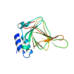
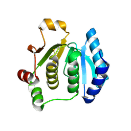
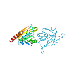
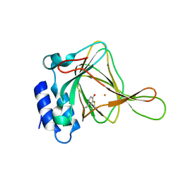
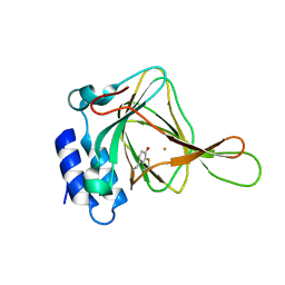
5I0T
 
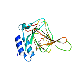 | | Thiosulfate bound Cysteine Dioxygenase at pH 6.8 | | Descriptor: | Cysteine dioxygenase type 1, FE (III) ION, THIOSULFATE | | Authors: | Kean, K.M, Driggers, C.M, Karplus, P.A. | | Deposit date: | 2016-02-04 | | Release date: | 2016-12-14 | | Last modified: | 2023-11-15 | | Method: | X-RAY DIFFRACTION (1.37 Å) | | Cite: | Structure-Based Insights into the Role of the Cys-Tyr Crosslink and Inhibitor Recognition by Mammalian Cysteine Dioxygenase.
J. Mol. Biol., 428, 2016
|
|
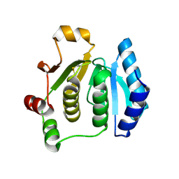
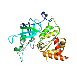
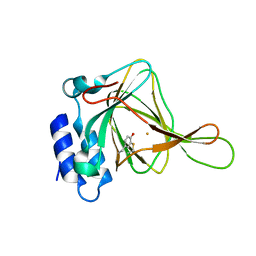
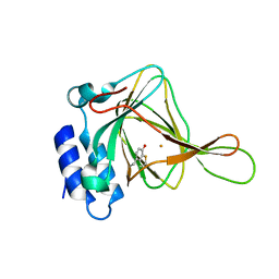
5I0S
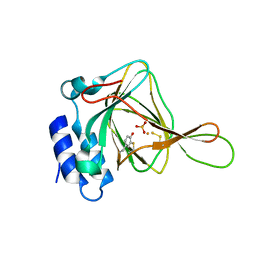 | | Thiosulfate bound Cysteine Dioxygenase at pH 6.2 | | Descriptor: | Cysteine dioxygenase type 1, FE (III) ION, THIOSULFATE | | Authors: | Kean, K.M, Driggers, C.M, Karplus, P.A. | | Deposit date: | 2016-02-04 | | Release date: | 2016-12-14 | | Last modified: | 2023-11-15 | | Method: | X-RAY DIFFRACTION (1.3 Å) | | Cite: | Structure-Based Insights into the Role of the Cys-Tyr Crosslink and Inhibitor Recognition by Mammalian Cysteine Dioxygenase.
J. Mol. Biol., 428, 2016
|
|
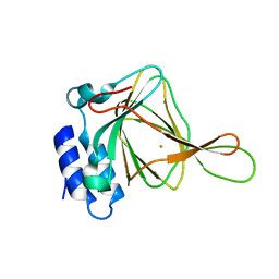
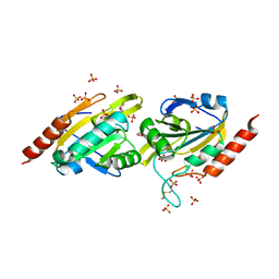
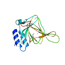
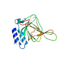
5I0R
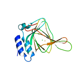 | | D-cysteine bound C93A mutant of Cysteine Dioxygenase at pH 8 | | Descriptor: | CHLORIDE ION, Cysteine dioxygenase type 1, D-CYSTEINE, ... | | Authors: | Kean, K.M, Driggers, C.M, Karplus, P.A. | | Deposit date: | 2016-02-04 | | Release date: | 2016-12-14 | | Last modified: | 2023-09-27 | | Method: | X-RAY DIFFRACTION (1.35 Å) | | Cite: | Structure-Based Insights into the Role of the Cys-Tyr Crosslink and Inhibitor Recognition by Mammalian Cysteine Dioxygenase.
J. Mol. Biol., 428, 2016
|
|
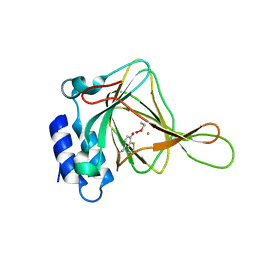
5I0U
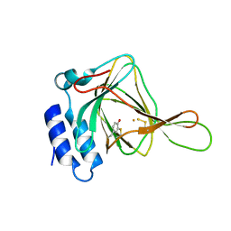 | |
2GH2
 | |
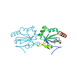
2KLO
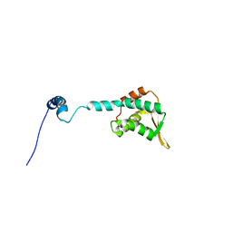 | | Structure of the Cdt1 C-terminal domain | | Descriptor: | DNA replication factor Cdt1 | | Authors: | Khayrutdinov, B.I, Bae, W.J, Yun, Y.M, Tsuyama, T, Kim, J.J, Hwang, E, Ryu, K.-S, Cheong, H.-K, Cheong, C, Karplus, P.A, Guntert, P, Tada, S, Jeon, Y.H, Cho, Y. | | Deposit date: | 2009-07-06 | | Release date: | 2009-10-13 | | Last modified: | 2022-03-16 | | Method: | SOLUTION NMR | | Cite: | Structure of the Cdt1 C-terminal domain: Conservation of the winged helix fold in replication licensing factors
Protein Sci., 18, 2009
|
|
1ZLE
 | | Crystal structure of a RGD-containing host-selective toxin: Pyrenophora tritici-repentis Ptr ToxA | | Descriptor: | NICKEL (II) ION, Ptr necrosis toxin | | Authors: | Sarma, G.N, Manning, V.A, Ciuffetti, L.M, Karplus, P.A. | | Deposit date: | 2005-05-06 | | Release date: | 2005-08-16 | | Last modified: | 2019-12-25 | | Method: | X-RAY DIFFRACTION (1.9 Å) | | Cite: | Structure of Ptr ToxA: An RGD-Containing Host-Selective Toxin from Pyrenophora tritici-repentis
Plant Cell, 17, 2005
|
|
1ZLD
 | | Crystal structure of a RGD-containing host-selective toxin: Pyrenophora tritici-repentis Ptr ToxA | | Descriptor: | Ptr necrosis toxin, SULFATE ION | | Authors: | Sarma, G.N, Manning, V.A, Ciuffetti, L.M, Karplus, P.A. | | Deposit date: | 2005-05-06 | | Release date: | 2005-08-16 | | Last modified: | 2019-12-25 | | Method: | X-RAY DIFFRACTION (1.65 Å) | | Cite: | Structure of Ptr ToxA: An RGD-Containing Host-Selective Toxin from Pyrenophora tritici-repentis
Plant Cell, 17, 2005
|
|
1ZYN
 | | Oxidized structure of the N-terminal domain of Salmonella typhimurium AhpF | | Descriptor: | Alkyl hydroperoxide reductase subunit F | | Authors: | Roberts, B.R, Wood, Z.A, Jonsson, T.J, Poole, L.B, Karplus, P.A. | | Deposit date: | 2005-06-10 | | Release date: | 2005-06-21 | | Last modified: | 2018-04-18 | | Method: | X-RAY DIFFRACTION (2.3 Å) | | Cite: | Oxidized and synchrotron cleaved structures of the disulfide redox center in the N-terminal domain of Salmonella typhimurium AhpF
Protein Sci., 14, 2005
|
|
1ZYP
 | | Synchrotron reduced form of the N-terminal domain of Salmonella typhimurium AhpF | | Descriptor: | Alkyl hydroperoxide reductase subunit F | | Authors: | Roberts, B.R, Wood, Z.A, Jonsson, T.J, Poole, L.B, Karplus, P.A. | | Deposit date: | 2005-06-10 | | Release date: | 2005-06-21 | | Last modified: | 2023-12-20 | | Method: | X-RAY DIFFRACTION (2.4 Å) | | Cite: | Oxidized and synchrotron cleaved structures of the disulfide redox center in the N-terminal domain of Salmonella typhimurium AhpF
Protein Sci., 14, 2005
|
|
2B5H
 | | 1.5 A Resolution Crystal Structure of Recombinant R. Norvegicus Cysteine Dioxygenase | | Descriptor: | Cysteine dioxygenase type I, FE (III) ION | | Authors: | Simmons, C.R, Liu, Q, Huang, Q, Hao, Q, Begley, T.P, Karplus, P.A, Stipanuk, M.H. | | Deposit date: | 2005-09-28 | | Release date: | 2006-04-11 | | Last modified: | 2024-02-14 | | Method: | X-RAY DIFFRACTION (1.5 Å) | | Cite: | Crystal Structure of Mammalian Cysteine Dioxygenase: A NOVEL MONONUCLEAR IRON CENTER FOR CYSTEINE THIOL OXIDATION.
J.Biol.Chem., 281, 2006
|
|
4G2E
 | |
4GQF
 | | Aeropyrum pernix Peroxiredoxin Q Enzyme in the Locally Unfolded Conformation | | Descriptor: | GLYCEROL, SULFATE ION, Thiol peroxidase | | Authors: | Perkins, A, Karplus, P.A, Gretes, M.C, Nelson, K.J, Poole, L.B. | | Deposit date: | 2012-08-22 | | Release date: | 2012-10-24 | | Last modified: | 2023-12-06 | | Method: | X-RAY DIFFRACTION (2.3 Å) | | Cite: | Mapping the Active Site Helix-to-Strand Conversion of CxxxxC Peroxiredoxin Q Enzymes.
Biochemistry, 51, 2012
|
|
1JB9
 | | Crystal Structure of The Ferredoxin:NADP+ Reductase From Maize Root AT 1.7 Angstroms | | Descriptor: | FLAVIN-ADENINE DINUCLEOTIDE, ferredoxin-NADP reductase | | Authors: | Faber, H.R, Karplus, P.A, Aliverti, A, Ferioli, C, Spinola, M. | | Deposit date: | 2001-06-03 | | Release date: | 2001-07-04 | | Last modified: | 2023-08-16 | | Method: | X-RAY DIFFRACTION (1.7 Å) | | Cite: | Biochemical and crystallographic characterization of ferredoxin-NADP(+) reductase from nonphotosynthetic tissues.
Biochemistry, 40, 2001
|
|
4GQC
 | | Crystal Structure of Aeropyrum pernix Peroxiredoxin Q Enzyme in Fully-Folded and Locally-Unfolded Conformations | | Descriptor: | DITHIANE DIOL, GLYCEROL, SULFATE ION, ... | | Authors: | Perkins, A, Karplus, P.A, Gretes, M.C, Nelson, K.J, Poole, L.B. | | Deposit date: | 2012-08-22 | | Release date: | 2012-10-24 | | Last modified: | 2023-09-13 | | Method: | X-RAY DIFFRACTION (2 Å) | | Cite: | Mapping the Active Site Helix-to-Strand Conversion of CxxxxC Peroxiredoxin Q Enzymes.
Biochemistry, 51, 2012
|
|
4IES
 | | Cys-persulfenate bound Cysteine Dioxygenase at pH 6.2 in the presence of Cys | | Descriptor: | Cysteine dioxygenase type 1, FE (III) ION, S-HYDROPEROXYCYSTEINE | | Authors: | Driggers, C.M, Cooley, R.B, Sankaran, B, Karplus, P.A. | | Deposit date: | 2012-12-13 | | Release date: | 2013-06-26 | | Last modified: | 2013-09-11 | | Method: | X-RAY DIFFRACTION (1.4 Å) | | Cite: | Cysteine Dioxygenase Structures from pH4 to 9: Consistent Cys-Persulfenate Formation at Intermediate pH and a Cys-Bound Enzyme at Higher pH.
J.Mol.Biol., 425, 2013
|
|
4IEW
 | |
4IEX
 | |
4IEO
 | |
4IEV
 | |
4IER
 | |
4IEZ
 | | unliganded Cysteine Dioxygenase at pH 8.0 | | Descriptor: | Cysteine dioxygenase type 1, FE (II) ION | | Authors: | Driggers, C.M, Cooley, R.B, Karplus, P.A. | | Deposit date: | 2012-12-13 | | Release date: | 2013-06-26 | | Last modified: | 2013-09-11 | | Method: | X-RAY DIFFRACTION (1.39 Å) | | Cite: | Cysteine Dioxygenase Structures from pH4 to 9: Consistent Cys-Persulfenate Formation at Intermediate pH and a Cys-Bound Enzyme at Higher pH.
J.Mol.Biol., 425, 2013
|
|
4IEQ
 | |
4IEP
 | |
4IEU
 | |


