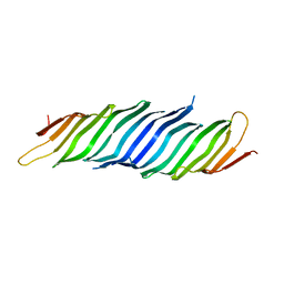
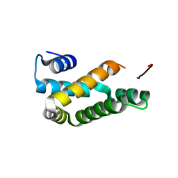
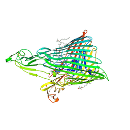
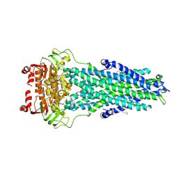
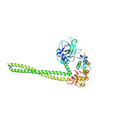
5EDL
 
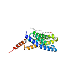 | | Crystal structure of an S-component of ECF transporter | | 分子名称: | 3-(4-AMINO-2-METHYL-PYRIMIDIN-5-YLMETHYL)-5-(2-HYDROXY-ETHYL)-4-METHYL-THIAZOL-3-IUM, Putative HMP/thiamine permease protein YkoE, [(Z)-octadec-9-enyl] (2R)-2,3-bis(oxidanyl)propanoate | | 著者 | Josts, I, Tidow, H. | | 登録日 | 2015-10-21 | | 公開日 | 2016-08-17 | | 実験手法 | X-RAY DIFFRACTION (1.95 Å) | | 主引用文献 | Crystal Structure of a Group I Energy Coupling Factor Vitamin Transporter S Component in Complex with Its Cognate Substrate.
Cell Chem Biol, 23, 2016
|
|
7ABW
 | | Crystal structure of siderophore reductase FoxB | | 分子名称: | 3,6,9,12,15,18,21,24-OCTAOXAHEXACOSAN-1-OL, DECYL-BETA-D-MALTOPYRANOSIDE, PROTOPORPHYRIN IX CONTAINING FE, ... | | 著者 | Josts, I, Tidow, H. | | 登録日 | 2020-09-09 | | 公開日 | 2021-09-01 | | 実験手法 | X-RAY DIFFRACTION (3.35 Å) | | 主引用文献 | Structural insights into a novel family of integral membrane siderophore reductases.
Proc.Natl.Acad.Sci.USA, 118, 2021
|
|
6I96
 | |
6I97
 | |
6I98
 | |
5VTG
 | |
8B43
 | | Crystal structure of ferrioxamine transporter | | 分子名称: | 1,12-bis(oxidanyl)-1,6,12,17-tetrazacyclodocosane-2,5,13,16-tetrone, FE (III) ION, Ferrichrome-iron receptor, ... | | 著者 | Josts, I, Tidow, H. | | 登録日 | 2022-09-19 | | 公開日 | 2023-04-26 | | 最終更新日 | 2024-02-07 | | 実験手法 | X-RAY DIFFRACTION (2.49 Å) | | 主引用文献 | Interactions of TonB-dependent transporter FoxA with siderophores and antibiotics that affect binding, uptake, and signal transduction.
Proc.Natl.Acad.Sci.USA, 120, 2023
|
|
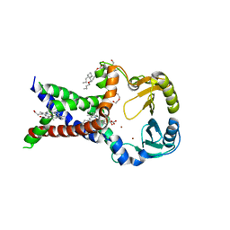
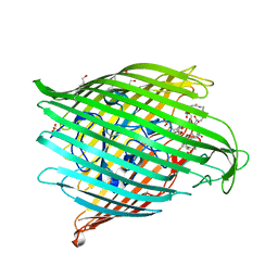
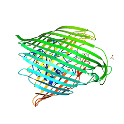
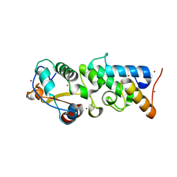
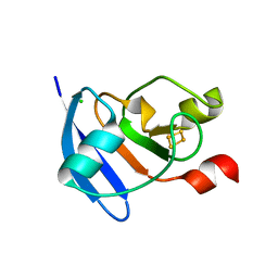
5NOH
 | | HRSV M2-1 core domain | | 分子名称: | Transcription elongation factor M2-1 | | 著者 | Josts, I, Almeida Hernandez, Y, Molina, I.G, de Prat-Gay, G, Tidow, H. | | 登録日 | 2017-04-12 | | 公開日 | 2018-01-03 | | 最終更新日 | 2019-10-16 | | 実験手法 | X-RAY DIFFRACTION (1.8 Å) | | 主引用文献 | Structure and stability of the Human respiratory syncytial virus M2-1RNA-binding core domain reveals a compact and cooperative folding unit.
Acta Crystallogr F Struct Biol Commun, 74, 2018
|
|
6Z8A
 | | Outer membrane FoxA in complex with nocardamine | | 分子名称: | (8E)-6,17,28-trihydroxy-1,6,12,17,23,28-hexaazacyclotritriacont-8-ene-2,5,13,16,24,27-hexone, DIMETHYL SULFOXIDE, FE (III) ION, ... | | 著者 | Josts, I, Tidow, H. | | 登録日 | 2020-06-02 | | 公開日 | 2020-11-25 | | 最終更新日 | 2024-01-24 | | 実験手法 | X-RAY DIFFRACTION (2.95 Å) | | 主引用文献 | Nocardamine-Dependent Iron Uptake in Pseudomonas aeruginosa : Exclusive Involvement of the FoxA Outer Membrane Transporter.
Acs Chem.Biol., 15, 2020
|
|
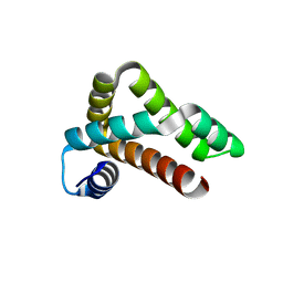
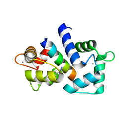
5NKX
 | | HRSV M2-1 core domain, P3221 crystal form | | 分子名称: | M2-1 | | 著者 | Almeida Hernandez, Y, Josts, I, Molina, I.G, de Pray-Gay, G, Tidow, H. | | 登録日 | 2017-04-03 | | 公開日 | 2018-01-03 | | 最終更新日 | 2024-01-17 | | 実験手法 | X-RAY DIFFRACTION (2.00008678 Å) | | 主引用文献 | Structure and stability of the Human respiratory syncytial virus M2-1RNA-binding core domain reveals a compact and cooperative folding unit.
Acta Crystallogr F Struct Biol Commun, 74, 2018
|
|
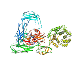
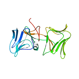
4QKO
 | | The Crystal Structure of the Pyocin S2 Nuclease Domain, Immunity Protein Complex at 1.8 Angstroms | | 分子名称: | BROMIDE ION, MAGNESIUM ION, Pyocin-S2, ... | | 著者 | Grinter, R, Josts, I, Roszak, A.W, Cogdell, C.J, Walker, D. | | 登録日 | 2014-06-07 | | 公開日 | 2015-06-10 | | 最終更新日 | 2023-11-08 | | 実験手法 | X-RAY DIFFRACTION (1.8 Å) | | 主引用文献 | Structural Insights into pyocin S2
To be Published
|
|
4RTD
 | | Escherichia coli alpha-2-macroglobulin activated by porcine elastase | | 分子名称: | Uncharacterized lipoprotein YfhM | | 著者 | Fyfe, C.D, Grinter, R, Roszak, A.W, Josts, I, Cogdell, R.J, Walker, D. | | 登録日 | 2014-11-14 | | 公開日 | 2015-07-15 | | 最終更新日 | 2015-07-29 | | 実験手法 | X-RAY DIFFRACTION (3.65 Å) | | 主引用文献 | Structure of protease-cleaved Escherichia coli alpha-2-macroglobulin reveals a putative mechanism of conformational activation for protease entrapment.
Acta Crystallogr.,Sect.D, 71, 2015
|
|
4ZHP
 | | The crystal structure of Potato ferredoxin I with 2Fe-2S cluster | | 分子名称: | FE2/S2 (INORGANIC) CLUSTER, Potato Ferredoxin I | | 著者 | Grinter, R, Josts, I, Roszak, A.W, Cogdell, R.J, Walker, D. | | 登録日 | 2015-04-26 | | 公開日 | 2016-08-31 | | 最終更新日 | 2024-01-10 | | 実験手法 | X-RAY DIFFRACTION (2.46 Å) | | 主引用文献 | Structure of the bacterial plant-ferredoxin receptor FusA.
Nat Commun, 7, 2016
|
|
4ZGV
 | | The Crystal Structure of the Ferredoxin Receptor FusA from Pectobacterium atrosepticum SCRI1043 | | 分子名称: | Ferredoxin receptor, LAURYL DIMETHYLAMINE-N-OXIDE, octyl beta-D-glucopyranoside | | 著者 | Grinter, R, Josts, I, Roszak, A.W, Cogdell, R.J, Walker, D. | | 登録日 | 2015-04-24 | | 公開日 | 2016-08-31 | | 最終更新日 | 2020-07-29 | | 実験手法 | X-RAY DIFFRACTION (3.2 Å) | | 主引用文献 | Structure of the bacterial plant-ferredoxin receptor FusA.
Nat Commun, 7, 2016
|
|
4ZHO
 | | The crystal structure of Arabidopsis ferredoxin 2 with 2Fe-2S cluster | | 分子名称: | CHLORIDE ION, FE2/S2 (INORGANIC) CLUSTER, Ferredoxin-2, ... | | 著者 | Grinter, R, Josts, I, Roszak, A.W, Cogdell, R.J, Walker, D. | | 登録日 | 2015-04-26 | | 公開日 | 2016-08-31 | | 最終更新日 | 2017-08-30 | | 実験手法 | X-RAY DIFFRACTION (2.34 Å) | | 主引用文献 | Structure of the bacterial plant-ferredoxin receptor FusA.
Nat Commun, 7, 2016
|
|
6HCS
 | |
4QAY
 | |
7BCW
 | | Structure of MsbA in Salipro with ADP vanadate | | 分子名称: | (2S)-3-(hexadecanoyloxy)-2-[(9Z)-octadec-9-enoyloxy]propyl 2-(trimethylammonio)ethyl phosphate, ADENOSINE-5'-DIPHOSPHATE, ATP-dependent lipid A-core flippase, ... | | 著者 | Traore, D.A.K, Tidow, H. | | 登録日 | 2020-12-21 | | 公開日 | 2022-01-12 | | 最終更新日 | 2022-05-25 | | 実験手法 | ELECTRON MICROSCOPY (3.5 Å) | | 主引用文献 | Cryo-EM structure of MsbA in saposin-lipid nanoparticles (Salipro) provides insights into nucleotide coordination.
Febs J., 289, 2022
|
|
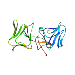
4UHP
 | | Crystal structure of the pyocin AP41 DNase-Immunity complex | | 分子名称: | BACTERIOCIN IMMUNITY PROTEIN, LARGE COMPONENT OF PYOCIN AP41 | | 著者 | Joshi, A, Chen, S, Wojdyla, J.A, Kaminska, R, Kleanthous, C. | | 登録日 | 2015-03-25 | | 公開日 | 2015-08-05 | | 最終更新日 | 2024-01-10 | | 実験手法 | X-RAY DIFFRACTION (2 Å) | | 主引用文献 | Structures of the Ultra-High Affinity Protein-Protein Complexes of Pyocins S2 and Ap41 and Their Cognate Immunity Proteins from Pseudomonas Aeruginosa
J.Mol.Biol., 427, 2015
|
|
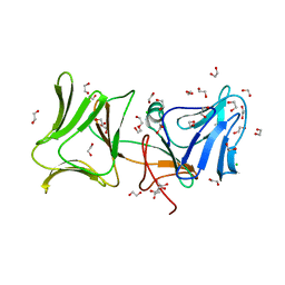
4UHQ
 | | Crystal structure of the pyocin AP41 DNase | | 分子名称: | CITRIC ACID, LARGE COMPONENT OF PYOCIN AP41, NICKEL (II) ION | | 著者 | Joshi, A, Chen, S, Wojdyla, J.A, Kaminska, R, Kleanthous, C. | | 登録日 | 2015-03-25 | | 公開日 | 2015-08-05 | | 最終更新日 | 2024-01-10 | | 実験手法 | X-RAY DIFFRACTION (1.5 Å) | | 主引用文献 | Structures of the Ultra-High Affinity Protein-Protein Complexes of Pyocins S2 and Ap41 and Their Cognate Immunity Proteins from Pseudomonas Aeruginosa
J.Mol.Biol., 427, 2015
|
|
7MHU
 | | Sialidase24 apo | | 分子名称: | Exo-alpha-sialidase | | 著者 | Rees, S.D, Chang, G.A. | | 登録日 | 2021-04-15 | | 公開日 | 2022-02-23 | | 最終更新日 | 2024-04-03 | | 実験手法 | X-RAY DIFFRACTION (2 Å) | | 主引用文献 | Computational models in the service of X-ray and cryo-electron microscopy structure determination.
Proteins, 89, 2021
|
|
5EW5
 | |
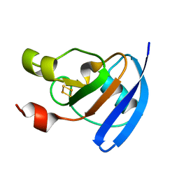
4LED
 | | The Crystal Structure of Pyocin L1 bound to D-rhamnose at 2.37 Angstroms | | 分子名称: | Pyocin L1, alpha-D-rhamnopyranose | | 著者 | Grinter, R, Roszak, A.W, Mccaughey, L, Cogdell, C.J, Walker, D. | | 登録日 | 2013-06-25 | | 公開日 | 2014-02-19 | | 最終更新日 | 2023-09-20 | | 実験手法 | X-RAY DIFFRACTION (2.37 Å) | | 主引用文献 | Lectin-Like Bacteriocins from Pseudomonas spp. Utilise D-Rhamnose Containing Lipopolysaccharide as a Cellular Receptor.
Plos Pathog., 10, 2014
|
|
4LEA
 | | The Crystal Structure of Pyocin L1 bound to D-mannose at 2.55 Angstroms | | 分子名称: | Pyocin L1, beta-D-mannopyranose | | 著者 | Grinter, R, Roszak, A.W, Mccaughey, L, Cogdell, C.J, Walker, D. | | 登録日 | 2013-06-25 | | 公開日 | 2014-02-19 | | 最終更新日 | 2023-09-20 | | 実験手法 | X-RAY DIFFRACTION (2.55 Å) | | 主引用文献 | Lectin-Like Bacteriocins from Pseudomonas spp. Utilise D-Rhamnose Containing Lipopolysaccharide as a Cellular Receptor.
Plos Pathog., 10, 2014
|
|
4LE7
 | | The Crystal Structure of Pyocin L1 at 2.09 Angstroms | | 分子名称: | 1,2-ETHANEDIOL, CHLORIDE ION, Pyocin L1 | | 著者 | Grinter, R, Roszak, A.W, Mccaughey, L, Cogdell, R.J, Walker, D. | | 登録日 | 2013-06-25 | | 公開日 | 2014-02-19 | | 最終更新日 | 2023-09-20 | | 実験手法 | X-RAY DIFFRACTION (2.09 Å) | | 主引用文献 | Lectin-Like Bacteriocins from Pseudomonas spp. Utilise D-Rhamnose Containing Lipopolysaccharide as a Cellular Receptor.
Plos Pathog., 10, 2014
|
|


