3POS
 
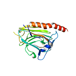 | | Crystal structure of the globular domain of human calreticulin | | Descriptor: | CALCIUM ION, Calreticulin | | Authors: | Chouquet, A, Paidassi, H, Ling, W.-L, Frachet, P, Houen, G, Arlaud, G.J, Gaboriaud, C. | | Deposit date: | 2010-11-23 | | Release date: | 2011-03-09 | | Last modified: | 2017-08-09 | | Method: | X-RAY DIFFRACTION (1.65 Å) | | Cite: | X-ray structure of the human calreticulin globular domain reveals a Peptide-binding area and suggests a multi-molecular mechanism
Plos One, 6, 2011
|
|
5LK5
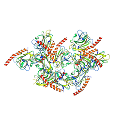 | |
3POW
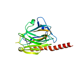 | |


