Loading
PDBj
MenuPDBj@FacebookPDBj@TwitterPDBj@YouTubewwPDB FoundationwwPDB
RCSB PDBPDBeBMRBAdv. SearchSearch help
Search by PDB author
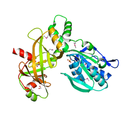
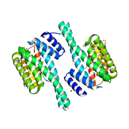
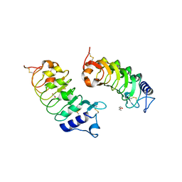
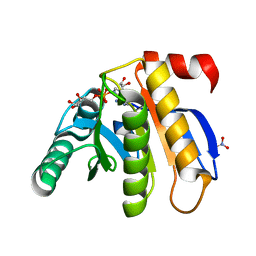
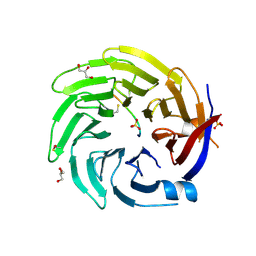
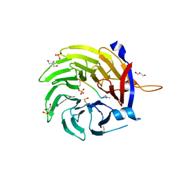
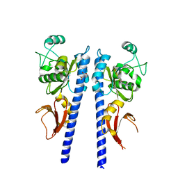
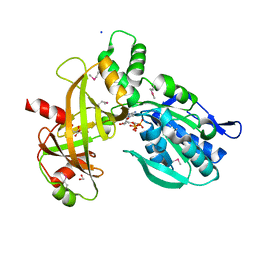
3T4S
DownloadVisualize
BU of 3t4s by Molmil
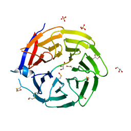
Arabidopsis histidine kinase 4 sensor domain in complex with kinetin
Descriptor: Histidine kinase 4, MALONATE ION, N-(FURAN-2-YLMETHYL)-7H-PURIN-6-AMINE
Authors:Hothorn, M.
Deposit date:2011-07-26
Release date:2011-10-05
Last modified:2023-09-13
Method:X-RAY DIFFRACTION (1.6 Å)
Cite:Structural basis for cytokinin recognition by Arabidopsis thaliana histidine kinase 4.
Nat.Chem.Biol., 7, 2011
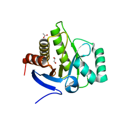
7Z67
DownloadVisualize
BU of 7z67 by Molmil
Crystal structure of the tandem kinase & triphosphate tunnel metalloenzyme domain module of the TTM1 protein from Arabidoposis thaliana in complex with a adenosine nucleotide analog.
Descriptor: 1,2-ETHANEDIOL, CITRIC ACID, Inorganic pyrophosphatase TTM1, ...
Authors:Hothorn, M, Martinez, J.
Deposit date:2022-03-11
Release date:2022-03-23
Method:X-RAY DIFFRACTION (2.65 Å)
Cite:Structural and biological insight into the plant unique multimodular triphosphosphate tunnel metalloenzymes of Arabidopsis thaliana
To Be Published
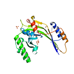
7Z66
DownloadVisualize
BU of 7z66 by Molmil
Crystal structure of the tandem kinase & triphosphate tunnel metalloenzyme domain module of the TTM1 protein from Arabidoposis thaliana in complex with inorganic phosphate and citric acid.
Descriptor: 1,2-ETHANEDIOL, CITRIC ACID, Inorganic pyrophosphatase TTM1, ...
Authors:Hothorn, M, Martinez, J.
Deposit date:2022-03-11
Release date:2022-04-06
Method:X-RAY DIFFRACTION (2.7 Å)
Cite:Structural and biological insight into the plant unique multimodular triphosphosphate tunnel metalloenzymes of Arabidopsis thaliana
To Be Published
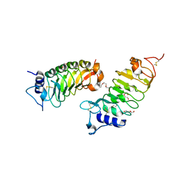
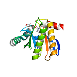
7A0K
DownloadVisualize
BU of 7a0k by Molmil
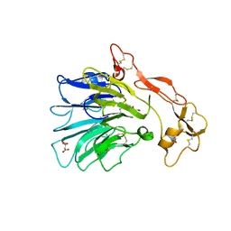
Crystal structure of the entire ectodomain from the Physcomitrella patens receptor kinase CR4
Descriptor: 1,2-ETHANEDIOL, 2-acetamido-2-deoxy-beta-D-glucopyranose, 2-acetamido-2-deoxy-beta-D-glucopyranose-(1-4)-2-acetamido-2-deoxy-beta-D-glucopyranose, ...
Authors:Hothorn, M, Okuda, S.
Deposit date:2020-08-09
Release date:2021-08-18
Last modified:2024-01-31
Method:X-RAY DIFFRACTION (2.7 Å)
Cite:Crystal structures of Arabidopsis and Physcomitrella CR4 reveal the molecular architecture of CRINKLY4 receptor kinases.
To Be Published
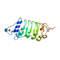
7A0J
DownloadVisualize
BU of 7a0j by Molmil
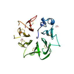
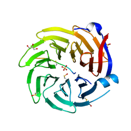
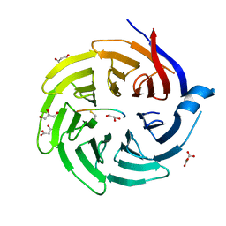
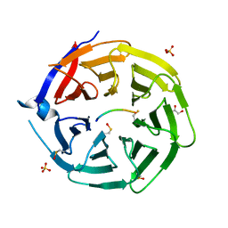
Crystal structure of the CRINKLY WD40 ectodomain from the Arabidopsis thaliana receptor kinase ACR4
Descriptor: 1,2-ETHANEDIOL, 2-acetamido-2-deoxy-beta-D-glucopyranose, Serine/threonine-protein kinase-like protein ACR4, ...
Authors:Hothorn, M, Okuda, S.
Deposit date:2020-08-09
Release date:2021-08-18
Method:X-RAY DIFFRACTION (1.95 Å)
Cite:Crystal structures of Arabidopsis and Physcomitrella CR4 reveal the molecular architecture of CRINKLY4 receptor kinases.
To Be Published
8QTT
DownloadVisualize
BU of 8qtt by Molmil
Crystal structure of a C-terminally truncated version of Arabidopsis thaliana 14-3-3 omega in complex with a phosphopeptide from the inhibitor protein BKI1.
Descriptor: 1,2-ETHANEDIOL, 14-3-3-like protein GF14 omega, BRI1 kinase inhibitor 1, ...
Authors:Hothorn, M, Obergfell, E.
Deposit date:2023-10-13
Release date:2023-11-15
Method:X-RAY DIFFRACTION (2.35 Å)
Cite:Mechanistic insights into the function of 14-3-3 proteins as negative regulators of brassinosteroid signaling.
To Be Published
8QTF
DownloadVisualize
BU of 8qtf by Molmil
Crystal structure of a C-terminally truncated version of Arabidopsis thaliana 14-3-3 omega in complex with a phosphopeptide from the transcription factor BZR1.
Descriptor: (4S)-2-METHYL-2,4-PENTANEDIOL, 1,2-ETHANEDIOL, 14-3-3-like protein GF14 omega, ...
Authors:Hothorn, M, Obergfell, E.
Deposit date:2023-10-12
Release date:2023-11-22
Method:X-RAY DIFFRACTION (1.9 Å)
Cite:Mechanistic insights into the function of 14-3-3 proteins as negative regulators of brassinosteroid signaling.
To Be Published
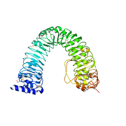
6FIF
DownloadVisualize
BU of 6fif by Molmil
Crystal structure of the BRI1 Gly644-Asp (bri1-6) mutant from Arabidopsis thaliana.
Descriptor: 1,2-ETHANEDIOL, 2-acetamido-2-deoxy-beta-D-glucopyranose, 2-acetamido-2-deoxy-beta-D-glucopyranose-(1-4)-2-acetamido-2-deoxy-beta-D-glucopyranose, ...
Authors:Hothorn, M, Santiago, J, Hohmann, U.
Deposit date:2018-01-18
Release date:2018-01-31
Last modified:2024-01-17
Method:X-RAY DIFFRACTION (2.54 Å)
Cite:Mechanistic basis for the activation of plant membrane receptor kinases by SERK-family coreceptors.
Proc. Natl. Acad. Sci. U.S.A., 115, 2018
6FG8
DownloadVisualize
BU of 6fg8 by Molmil
Crystal structure of the BIR3 - SERK1 complex from Arabidopsis thaliana.
Descriptor: 2-acetamido-2-deoxy-beta-D-glucopyranose-(1-4)-2-acetamido-2-deoxy-beta-D-glucopyranose, DI(HYDROXYETHYL)ETHER, PENTAETHYLENE GLYCOL, ...
Authors:Hothorn, M, Hohmann, U.
Deposit date:2018-01-10
Release date:2018-01-24
Last modified:2024-01-17
Method:X-RAY DIFFRACTION (1.25 Å)
Cite:The SERK3 elongated allele defines a role for BIR ectodomains in brassinosteroid signalling.
Nat Plants, 4, 2018
6FG7
DownloadVisualize
BU of 6fg7 by Molmil
Crystal structure of the BIR2 ectodomain from Arabidopsis thaliana.
Descriptor: 2-acetamido-2-deoxy-beta-D-glucopyranose, 2-acetamido-2-deoxy-beta-D-glucopyranose-(1-4)-[alpha-L-fucopyranose-(1-6)]2-acetamido-2-deoxy-beta-D-glucopyranose, Inactive LRR receptor-like serine/threonine-protein kinase BIR2, ...
Authors:Hothorn, M, Hohmann, U.
Deposit date:2018-01-10
Release date:2018-01-24
Last modified:2020-07-29
Method:X-RAY DIFFRACTION (1.9 Å)
Cite:The SERK3 elongated allele defines a role for BIR ectodomains in brassinosteroid signalling.
Nat Plants, 4, 2018
6G3W
DownloadVisualize
BU of 6g3w by Molmil
Crystal structure of the BIR3 - SERK2 complex from Arabidopsis thaliana.
Descriptor: 1,2-ETHANEDIOL, 2-acetamido-2-deoxy-beta-D-glucopyranose, DI(HYDROXYETHYL)ETHER, ...
Authors:Hothorn, M, Hohmann, U.
Deposit date:2018-03-26
Release date:2018-04-04
Last modified:2024-01-17
Method:X-RAY DIFFRACTION (2.2 Å)
Cite:The SERK3 elongated allele defines a role for BIR ectodomains in brassinosteroid signalling.
Nat Plants, 4, 2018
6FY5
DownloadVisualize
BU of 6fy5 by Molmil
Crystal structure of the macro domain of human macroh2a2
Descriptor: 1,2-ETHANEDIOL, ACETATE ION, Core histone macro-H2A.2
Authors:Hothorn, M.
Deposit date:2018-03-10
Release date:2018-04-04
Last modified:2024-01-17
Method:X-RAY DIFFRACTION (1.65 Å)
Cite:MacroH2A histone variants limit chromatin plasticity through two distinct mechanisms.
EMBO Rep., 19, 2018
3G3T
DownloadVisualize
BU of 3g3t by Molmil
Crystal structure of a eukaryotic polyphosphate polymerase in complex with orthophosphate
Descriptor: 1,2-ETHANEDIOL, PHOSPHATE ION, Vacuolar transporter chaperone 4
Authors:Lenherr, E.D, Hothorn, M, Scheffzek, K.
Deposit date:2009-02-02
Release date:2009-05-05
Last modified:2023-09-06
Method:X-RAY DIFFRACTION (1.85 Å)
Cite:Catalytic core of a membrane-associated eukaryotic polyphosphate polymerase.
Science, 324, 2009
3IIF
DownloadVisualize
BU of 3iif by Molmil
Crystal structure of the macro domain of human histone macroH2A1.1 in complex with ADP-ribose (form B)
Descriptor: ADENOSINE-5-DIPHOSPHORIBOSE, Core histone macro-H2A.1, Isoform 1
Authors:Hothorn, M, Bortfeld, M, Ladurner, A.G, Scheffzek, K.
Deposit date:2009-07-31
Release date:2009-08-18
Last modified:2023-09-06
Method:X-RAY DIFFRACTION (2.1 Å)
Cite:A macrodomain-containing histone rearranges chromatin upon sensing PARP1 activation.
Nat.Struct.Mol.Biol., 16, 2009
3IID
DownloadVisualize
BU of 3iid by Molmil
Crystal structure of the macro domain of human histone macroH2A1.1 in complex with ADP-ribose (form A)
Descriptor: ADENOSINE-5-DIPHOSPHORIBOSE, Core histone macro-H2A.1, Isoform 1, ...
Authors:Hothorn, M, Bortfeld, M, Ladurner, A.G, Scheffzek, K.
Deposit date:2009-07-31
Release date:2009-08-18
Last modified:2023-09-06
Method:X-RAY DIFFRACTION (1.9 Å)
Cite:A macrodomain-containing histone rearranges chromatin upon sensing PARP1 activation.
Nat.Struct.Mol.Biol., 16, 2009
6TO9
DownloadVisualize
BU of 6to9 by Molmil
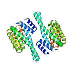
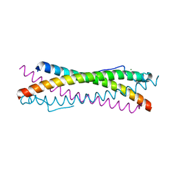
Crystal structure of the oligomerisation domain of the transcription factor PHOSPHATE STARVATION RESPONSE 1 from Arabidopsis (crystal form 2)
Descriptor: MAGNESIUM ION, Protein PHOSPHATE STARVATION RESPONSE 1
Authors:Hothorn, M.
Deposit date:2019-12-11
Release date:2021-01-13
Last modified:2024-01-24
Method:X-RAY DIFFRACTION (2.45 Å)
Cite:Inositol pyrophosphates promote the interaction of SPX domains with the coiled-coil motif of PHR transcription factors to regulate plant phosphate homeostasis.
Nat Commun, 12, 2021
6TO5
DownloadVisualize
BU of 6to5 by Molmil
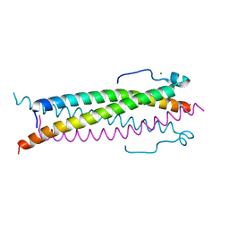
Crystal structure of the oligomerisation domain of the transcription factor PHOSPHATE STARVATION RESPONSE 1 from Arabidopsis.
Descriptor: MAGNESIUM ION, Protein PHOSPHATE STARVATION RESPONSE 1
Authors:Hothorn, M.
Deposit date:2019-12-11
Release date:2021-01-13
Last modified:2024-01-24
Method:X-RAY DIFFRACTION (2.38 Å)
Cite:Inositol pyrophosphates promote the interaction of SPX domains with the coiled-coil motif of PHR transcription factors to regulate plant phosphate homeostasis.
Nat Commun, 12, 2021
6TOC
DownloadVisualize
BU of 6toc by Molmil
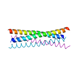
Crystal structure of the oligomerisation domain of the transcription factor PHOSPHATE STARVATION RESPONSE 1 from Arabidopsis (crystal form 3).
Descriptor: Protein PHOSPHATE STARVATION RESPONSE 1
Authors:Hothorn, M.
Deposit date:2019-12-11
Release date:2021-01-13
Last modified:2024-01-24
Method:X-RAY DIFFRACTION (1.853 Å)
Cite:Inositol pyrophosphates promote the interaction of SPX domains with the coiled-coil motif of PHR transcription factors to regulate plant phosphate homeostasis.
Nat Commun, 12, 2021
6QTS
DownloadVisualize
BU of 6qts by Molmil
Crystal structure of a mutant Arabidopsis WD40 domain in complex with a photoreceptor
Descriptor: E3 ubiquitin-protein ligase COP1, GLYCEROL, SULFATE ION, ...
Authors:Hothorn, M, Lau, K.
Deposit date:2019-02-25
Release date:2019-07-10
Last modified:2024-01-24
Method:X-RAY DIFFRACTION (1.11 Å)
Cite:Plant photoreceptors and their signaling components compete for COP1 binding via VP peptide motifs.
Embo J., 38, 2019
6QTR
DownloadVisualize
BU of 6qtr by Molmil
Crystal structure of a mutant Arabidopsis WD40 domain in complex with a transcription factor
Descriptor: E3 ubiquitin-protein ligase COP1, GLYCEROL, SULFATE ION, ...
Authors:Hothorn, M, Lau, K.
Deposit date:2019-02-25
Release date:2019-07-10
Last modified:2024-01-24
Method:X-RAY DIFFRACTION (1.37 Å)
Cite:Plant photoreceptors and their signaling components compete for COP1 binding via VP peptide motifs.
Embo J., 38, 2019
6QTT
DownloadVisualize
BU of 6qtt by Molmil
Crystal structure of an Arabidopsis WD40 domain in complex with a transcription factor homolog
Descriptor: E3 ubiquitin-protein ligase COP1, GLYCEROL, MALONATE ION, ...
Authors:Hothorn, M, Lau, K.
Deposit date:2019-02-25
Release date:2019-07-10
Last modified:2024-01-24
Method:X-RAY DIFFRACTION (1.51 Å)
Cite:Plant photoreceptors and their signaling components compete for COP1 binding via VP peptide motifs.
Embo J., 38, 2019
6QTV
DownloadVisualize
BU of 6qtv by Molmil
Crystal structure of an Arabidopsis WD40 domain in complex with an atypical bHLH transcription factor
Descriptor: E3 ubiquitin-protein ligase COP1, GLYCEROL, MALONATE ION, ...
Authors:Hothorn, M, Lau, K.
Deposit date:2019-02-25
Release date:2019-07-10
Last modified:2024-01-24
Method:X-RAY DIFFRACTION (1.31 Å)
Cite:Plant photoreceptors and their signaling components compete for COP1 binding via VP peptide motifs.
Embo J., 38, 2019
6QTQ
DownloadVisualize
BU of 6qtq by Molmil
Crystal structure of an Arabidopsis WD40 domain in complex with photoreceptor
Descriptor: E3 ubiquitin-protein ligase COP1, GLYCEROL, SULFATE ION, ...
Authors:Hothorn, M, Lau, K.
Deposit date:2019-02-25
Release date:2019-07-10
Last modified:2024-01-24
Method:X-RAY DIFFRACTION (1.3 Å)
Cite:Plant photoreceptors and their signaling components compete for COP1 binding via VP peptide motifs.
Embo J., 38, 2019
6QTX
DownloadVisualize
BU of 6qtx by Molmil
Crystal structure of an Arabidopsis WD40 domain in complex with a flowering transcription factor homolog
Descriptor: E3 ubiquitin-protein ligase COP1, GLYCEROL, SULFATE ION, ...
Authors:Hothorn, M, Lau, K.
Deposit date:2019-02-25
Release date:2019-07-10
Last modified:2019-10-02
Method:X-RAY DIFFRACTION (1.95 Å)
Cite:Plant photoreceptors and their signaling components compete for COP1 binding via VP peptide motifs.
Embo J., 38, 2019
6QTO
DownloadVisualize
BU of 6qto by Molmil
Crystal structure of an Arabidopsis WD40 domain in complex with a transcription factor
Descriptor: E3 ubiquitin-protein ligase COP1, GLYCEROL, SULFATE ION, ...
Authors:Hothorn, M, Lau, K.
Deposit date:2019-02-25
Release date:2019-07-10
Last modified:2024-01-24
Method:X-RAY DIFFRACTION (1.27 Å)
Cite:Plant photoreceptors and their signaling components compete for COP1 binding via VP peptide motifs.
Embo J., 38, 2019

218853

PDB entries from 2024-04-24

PDB statisticsPDBj update infoContact PDBjnumon