Loading
PDBj
メニューPDBj@FacebookPDBj@TwitterPDBj@YouTubewwPDB FoundationwwPDB
RCSB PDBPDBeBMRBAdv. SearchSearch help
Search by PDB author
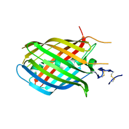
1H4U
DownloadVisualize
BU of 1h4u by Molmil
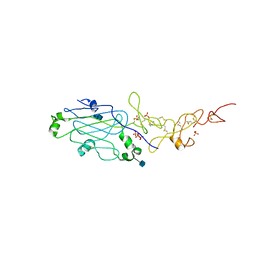
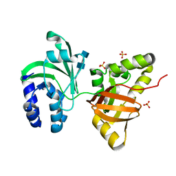
Domain G2 of mouse nidogen-1
分子名称: NIDOGEN-1
著者Hopf, M, Gohring, W, Ries, A, Timpl, R, Hohenester, E.
登録日2001-05-14
公開日2001-06-28
最終更新日2011-07-13
実験手法X-RAY DIFFRACTION (2.2 Å)
主引用文献Crystal Structure and Mutational Analysis of a Perlecan-Binding Fragment of Nidogen-1
Nat.Struct.Biol., 8, 2001
1H30
DownloadVisualize
BU of 1h30 by Molmil
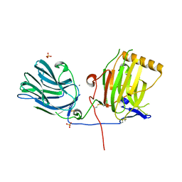
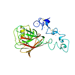
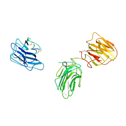
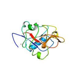
C-terminal LG domain pair of human Gas6
分子名称: CALCIUM ION, GROWTH-ARREST-SPECIFIC PROTEIN, SULFATE ION
著者Sasaki, T, Knyazev, P.G, Cheburkin, Y, Gohring, W, Tisi, D, Ullrich, A, Timpl, R, Hohenester, E.
登録日2002-08-21
公開日2003-01-30
最終更新日2011-07-13
実験手法X-RAY DIFFRACTION (2.2 Å)
主引用文献Crystal Structure of a Carboxy-Terminal Fragment of Growth-Arrest-Specific Protein Gas6: Receptor Tyrosine Kinase Activation by Laminin G-Like Domains
J.Biol.Chem., 277, 2002
6EJC
DownloadVisualize
BU of 6ejc by Molmil
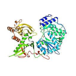
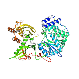
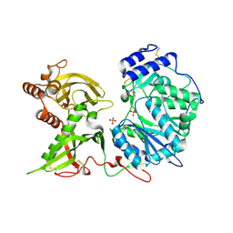
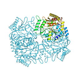
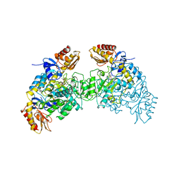
Human Xylosyltransferase 1 in complex with peptide QEEEGSGVGQGG
分子名称: PHOSPHATE ION, Protein AMBP, SODIUM ION, ...
著者Briggs, D.C, Hohenester, E.
登録日2017-09-20
公開日2018-05-02
最終更新日2024-01-17
実験手法X-RAY DIFFRACTION (2.057 Å)
主引用文献Structural Basis for the Initiation of Glycosaminoglycan Biosynthesis by Human Xylosyltransferase 1.
Structure, 26, 2018
6EJE
DownloadVisualize
BU of 6eje by Molmil
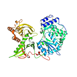
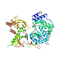
Human Xylosyltransferase 1 in complex with peptide PAAEGSGEQDFT
分子名称: PHOSPHATE ION, SODIUM ION, Syndecan-1, ...
著者Briggs, D.C, Hohenester, E.
登録日2017-09-20
公開日2018-05-02
最終更新日2024-01-17
実験手法X-RAY DIFFRACTION (2.43 Å)
主引用文献Structural Basis for the Initiation of Glycosaminoglycan Biosynthesis by Human Xylosyltransferase 1.
Structure, 26, 2018
6EJ8
DownloadVisualize
BU of 6ej8 by Molmil
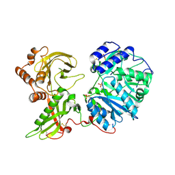
Human Xylosyltransferase 1 in complex with peptide QEEEGSGGGQGG
分子名称: 2-acetamido-2-deoxy-beta-D-glucopyranose, PHOSPHATE ION, Protein AMBP, ...
著者Briggs, D.C, Hohenester, E.
登録日2017-09-20
公開日2018-05-02
最終更新日2024-01-17
実験手法X-RAY DIFFRACTION (2.09 Å)
主引用文献Structural Basis for the Initiation of Glycosaminoglycan Biosynthesis by Human Xylosyltransferase 1.
Structure, 26, 2018
6EJD
DownloadVisualize
BU of 6ejd by Molmil
Human Xylosyltransferase 1 in complex with peptide QEEEGSGGPQGG
分子名称: PHOSPHATE ION, Protein AMBP, Xylosyltransferase 1
著者Briggs, D.C, Hohenester, E.
登録日2017-09-20
公開日2018-05-02
最終更新日2024-01-17
実験手法X-RAY DIFFRACTION (2.684 Å)
主引用文献Structural Basis for the Initiation of Glycosaminoglycan Biosynthesis by Human Xylosyltransferase 1.
Structure, 26, 2018
6EJB
DownloadVisualize
BU of 6ejb by Molmil
Human Xylosyltransferase 1 in complex with peptide QEEEGSAGGQGG
分子名称: PHOSPHATE ION, Protein AMBP, Xylosyltransferase 1
著者Briggs, D.C, Hohenester, E.
登録日2017-09-20
公開日2018-05-02
最終更新日2024-01-17
実験手法X-RAY DIFFRACTION (2.557 Å)
主引用文献Structural Basis for the Initiation of Glycosaminoglycan Biosynthesis by Human Xylosyltransferase 1.
Structure, 26, 2018
6EJ7
DownloadVisualize
BU of 6ej7 by Molmil
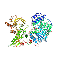
Human Xylosyltransferase 1 in complex with UDP-xylose and peptide QEEEGAGGGQGG
分子名称: 1,2-ETHANEDIOL, 2-acetamido-2-deoxy-beta-D-glucopyranose, DI(HYDROXYETHYL)ETHER, ...
著者Briggs, D.C, Hohenester, E.
登録日2017-09-20
公開日2018-05-02
最終更新日2024-01-17
実験手法X-RAY DIFFRACTION (2 Å)
主引用文献Structural Basis for the Initiation of Glycosaminoglycan Biosynthesis by Human Xylosyltransferase 1.
Structure, 26, 2018
6EJA
DownloadVisualize
BU of 6eja by Molmil
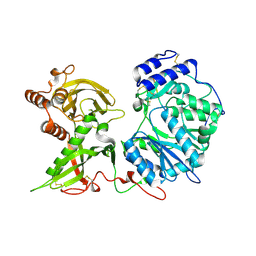
Human Xylosyltransferase 1 in complex with peptide QEEEYSGGGQGG
分子名称: Protein AMBP, SODIUM ION, Xylosyltransferase 1
著者Briggs, D.C, Hohenester, E.
登録日2017-09-20
公開日2018-05-02
最終更新日2024-01-17
実験手法X-RAY DIFFRACTION (1.94 Å)
主引用文献Structural Basis for the Initiation of Glycosaminoglycan Biosynthesis by Human Xylosyltransferase 1.
Structure, 26, 2018
6EJ9
DownloadVisualize
BU of 6ej9 by Molmil
Human Xylosyltransferase 1 in complex with peptide QEPEGSGGGQGG
分子名称: PHOSPHATE ION, Protein AMBP, SODIUM ION, ...
著者Briggs, D.C, Hohenester, E.
登録日2017-09-20
公開日2018-05-02
最終更新日2024-01-17
実験手法X-RAY DIFFRACTION (2.02 Å)
主引用文献Structural Basis for the Initiation of Glycosaminoglycan Biosynthesis by Human Xylosyltransferase 1.
Structure, 26, 2018
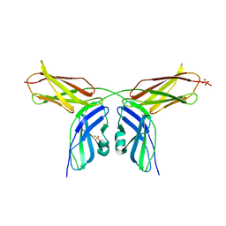
1UX6
DownloadVisualize
BU of 1ux6 by Molmil
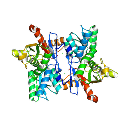
Structure of a thrombospondin C-terminal fragment reveals a novel calcium core in the type 3 repeats
分子名称: CALCIUM ION, THROMBOSPONDIN-1
著者Kvansakul, M, Adams, J.C, Hohenester, E.
登録日2004-02-19
公開日2004-03-18
最終更新日2017-07-12
実験手法X-RAY DIFFRACTION (1.9 Å)
主引用文献Structure of a Thrombospondin C-Terminal Fragment Reveals a Novel Calcium Core in the Type 3 Repeats
Embo J., 23, 2004
1W8A
DownloadVisualize
BU of 1w8a by Molmil
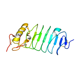
Third LRR domain of Drosophila Slit
分子名称: SLIT PROTEIN
著者Howitt, J.A, Clout, N.J, Hohenester, E.
登録日2004-09-17
公開日2004-10-25
最終更新日2023-12-13
実験手法X-RAY DIFFRACTION (2.8 Å)
主引用文献Binding Site for Robo Receptors Revealed by Dissection of the Leucine-Rich Repeat Region of Slit.
Embo J., 23, 2004
2VKW
DownloadVisualize
BU of 2vkw by Molmil
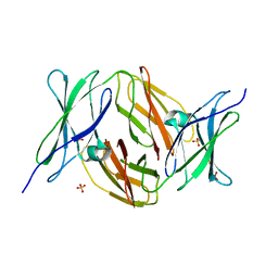
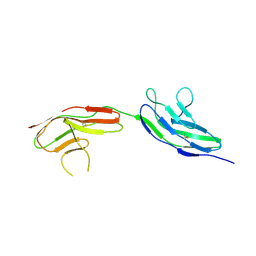
Human NCAM, FN3 domains 1 and 2
分子名称: NEURAL CELL ADHESION MOLECULE 1,140 KDA ISOFORM, SULFATE ION
著者Carafoli, F, Saffell, J.L, Hohenester, E.
登録日2008-01-04
公開日2008-02-26
最終更新日2024-05-08
実験手法X-RAY DIFFRACTION (2.3 Å)
主引用文献Structure of the Tandem Fibronectin Type 3 Domains of Neural Cell Adhesion Molecule
J.Mol.Biol., 377, 2008
2VKX
DownloadVisualize
BU of 2vkx by Molmil
Human NCAM, FN3 domains 1 and 2, M610R mutant
分子名称: NEURAL CELL ADHESION MOLECULE, SULFATE ION
著者Carafoli, F, Saffell, J.L, Hohenester, E.
登録日2008-01-04
公開日2008-02-26
最終更新日2024-05-01
実験手法X-RAY DIFFRACTION (2.7 Å)
主引用文献Structure of the Tandem Fibronectin Type 3 Domains of Neural Cell Adhesion Molecule
J.Mol.Biol., 377, 2008
2VR9
DownloadVisualize
BU of 2vr9 by Molmil
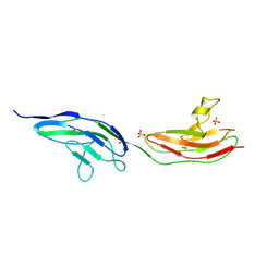
Drosophila Robo IG1-2 (tetragonal form)
分子名称: ROUNDABOUT 1
著者Fukuhara, N, Howitt, J.A, Hussain, S, Hohenester, E.
登録日2008-03-28
公開日2008-04-08
最終更新日2023-12-13
実験手法X-RAY DIFFRACTION (3.2 Å)
主引用文献Structural and Functional Analysis of Slit and Heparin Binding to Immunoglobulin-Like Domains 1 and 2 of Drosophila Robo
J.Biol.Chem., 283, 2008
2VRA
DownloadVisualize
BU of 2vra by Molmil
Drosophila Robo IG1-2 (monoclinic form)
分子名称: 2-O-sulfo-alpha-L-idopyranuronic acid-(1-4)-2-deoxy-6-O-sulfo-2-(sulfoamino)-alpha-D-glucopyranose-(1-4)-2-O-sulfo-alpha-L-idopyranuronic acid-(1-4)-2-deoxy-6-O-sulfo-2-(sulfoamino)-alpha-D-glucopyranose, ROUNDABOUT 1, SULFATE ION
著者Fukuhara, N, Howitt, J.A, Hussain, S, Hohenester, E.
登録日2008-03-28
公開日2008-04-08
最終更新日2023-12-13
実験手法X-RAY DIFFRACTION (3.2 Å)
主引用文献Structural and Functional Analysis of Slit and Heparin Binding to Immunoglobulin-Like Domains 1 and 2 of Drosophila Robo
J.Biol.Chem., 283, 2008
2WJS
DownloadVisualize
BU of 2wjs by Molmil
Crystal structure of the LG1-3 region of the laminin alpha2 chain
分子名称: 2-acetamido-2-deoxy-beta-D-glucopyranose, CALCIUM ION, LAMININ SUBUNIT ALPHA-2
著者Carafoli, F, Clout, N.J, Hohenester, E.
登録日2009-05-28
公開日2009-06-23
最終更新日2023-12-13
実験手法X-RAY DIFFRACTION (2.8 Å)
主引用文献Crystal Structure of the Lg1-3 Region of the Laminin {Alpha}2 Chain.
J.Biol.Chem., 284, 2009
2Y38
DownloadVisualize
BU of 2y38 by Molmil
LAMININ ALPHA5 CHAIN N-TERMINAL FRAGMENT
分子名称: 2-acetamido-2-deoxy-beta-D-glucopyranose, 2-acetamido-2-deoxy-beta-D-glucopyranose-(1-4)-2-acetamido-2-deoxy-beta-D-glucopyranose, LAMININ SUBUNIT ALPHA-5, ...
著者Hussain, S.A, Carafoli, F, Hohenester, E.
登録日2010-12-19
公開日2011-02-23
最終更新日2020-07-29
実験手法X-RAY DIFFRACTION (2.9 Å)
主引用文献Determinants of Laminin Polymerisation Revealed by the Crystal Structure of the Alpha5 Chain Amino-Terminal Region
Embo Rep., 12, 2011
1BNL
DownloadVisualize
BU of 1bnl by Molmil
ZINC DEPENDENT DIMERS OBSERVED IN CRYSTALS OF HUMAN ENDOSTATIN
分子名称: COLLAGEN XVIII, ZINC ION
著者Ding, Y.-H, Javaherian, K, Lo, K.-M, Chopra, R, Boehm, T, Lanciotti, J, Harris, B.A, Li, Y, Shapiro, R, Hohenester, E, Timpl, R, Folkman, J, Wiley, D.C.
登録日1998-07-30
公開日1998-10-14
最終更新日2011-07-13
実験手法X-RAY DIFFRACTION (2.9 Å)
主引用文献Zinc-dependent dimers observed in crystals of human endostatin.
Proc.Natl.Acad.Sci.USA, 95, 1998
1DKA
DownloadVisualize
BU of 1dka by Molmil
DIALKYLGLYCINE DECARBOXYLASE STRUCTURE: BIFUNCTIONAL ACTIVE SITE AND ALKALI METAL BINDING SITES
分子名称: 2,2-DIALKYLGLYCINE DECARBOXYLASE (PYRUVATE), 2-(N-MORPHOLINO)-ETHANESULFONIC ACID, POTASSIUM ION, ...
著者Toney, M.D, Hohenester, E, Jansonius, J.N.
登録日1993-06-18
公開日1994-10-15
最終更新日2017-11-29
実験手法X-RAY DIFFRACTION (2.6 Å)
主引用文献Dialkylglycine decarboxylase structure: bifunctional active site and alkali metal sites.
Science, 261, 1993
1OAS
DownloadVisualize
BU of 1oas by Molmil
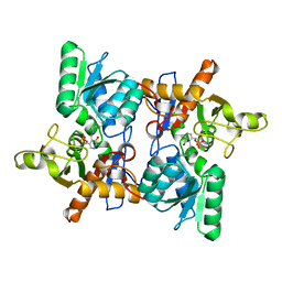
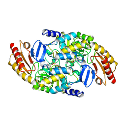
O-ACETYLSERINE SULFHYDRYLASE FROM SALMONELLA TYPHIMURIUM
分子名称: O-ACETYLSERINE SULFHYDRYLASE, PYRIDOXAL-5'-PHOSPHATE
著者Burkhard, P, Rao, G.S.J, Hohenester, E, Schnackerz, K.D, Cook, P.F, Jansonius, J.N.
登録日1999-01-29
公開日2000-01-28
最終更新日2023-12-27
実験手法X-RAY DIFFRACTION (2.2 Å)
主引用文献Three-dimensional structure of O-acetylserine sulfhydrylase from Salmonella typhimurium.
J.Mol.Biol., 283, 1998
1O70
DownloadVisualize
BU of 1o70 by Molmil
Novel Fold Revealed by the Structure of a FAS1 Domain Pair from the Insect Cell Adhesion Molecule Fasciclin I
分子名称: 2-acetamido-2-deoxy-beta-D-glucopyranose-(1-4)-2-acetamido-2-deoxy-beta-D-glucopyranose, FASCICLIN I, SULFATE ION
著者Clout, N.J, Tisi, D, Hohenester, E.
登録日2002-10-23
公開日2003-02-13
最終更新日2020-07-29
実験手法X-RAY DIFFRACTION (2.6 Å)
主引用文献Novel Fold Revealed by the Structure of a Fas1 Domain Pair from the Insect Cell Adhesion Molecule Fasciclin I
Structure, 11, 2003
2OAT
DownloadVisualize
BU of 2oat by Molmil
ORNITHINE AMINOTRANSFERASE COMPLEXED WITH 5-FLUOROMETHYLORNITHINE
分子名称: 1-AMINO-7-(2-METHYL-3-OXIDO-5-((PHOSPHONOXY)METHYL)-4-PYRIDOXAL-5-OXO-6-HEPTENATE, ORNITHINE AMINOTRANSFERASE
著者Storici, P, Schirmer, T.
登録日1998-05-07
公開日1998-12-09
最終更新日2024-05-22
実験手法X-RAY DIFFRACTION (1.95 Å)
主引用文献Crystal structure of human ornithine aminotransferase complexed with the highly specific and potent inhibitor 5-fluoromethylornithine.
J.Mol.Biol., 285, 1999
1OAT
DownloadVisualize
BU of 1oat by Molmil
ORNITHINE AMINOTRANSFERASE
分子名称: ORNITHINE AMINOTRANSFERASE, PYRIDOXAL-5'-PHOSPHATE
著者Shen, B.W, Schirmer, T, Jansonius, J.N.
登録日1997-03-26
公開日1998-04-01
最終更新日2023-08-09
実験手法X-RAY DIFFRACTION (2.5 Å)
主引用文献Crystal structure of human recombinant ornithine aminotransferase.
J.Mol.Biol., 277, 1998
1FCJ
DownloadVisualize
BU of 1fcj by Molmil
CRYSTAL STRUCTURE OF OASS COMPLEXED WITH CHLORIDE AND SULFATE
分子名称: CHLORIDE ION, O-ACETYLSERINE SULFHYDRYLASE, PYRIDOXAL-5'-PHOSPHATE, ...
著者Burkhard, P, Tai, C, Jansonius, J.N, Cook, P.F.
登録日2000-07-18
公開日2000-10-18
最終更新日2018-01-31
実験手法X-RAY DIFFRACTION (2 Å)
主引用文献Identification of an allosteric anion-binding site on O-acetylserine sulfhydrylase: structure of the enzyme with chloride bound.
J.Mol.Biol., 303, 2000

220113

件を2024-05-22に公開中

PDB statisticsPDBj update infoContact PDBjnumon