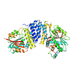
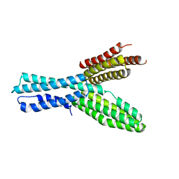
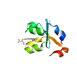
1VD2
 
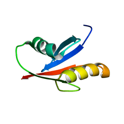 | | Solution Structure of the PB1 domain of PKCiota | | 分子名称: | Protein kinase C, iota type | | 著者 | Hirano, Y, Yoshinaga, S, Yokochi, M, Ogura, K, Noda, Y, Sumimoto, H, Inagaki, F. | | 登録日 | 2004-03-18 | | 公開日 | 2004-09-14 | | 最終更新日 | 2023-12-27 | | 実験手法 | SOLUTION NMR | | 主引用文献 | Solution structure of atypical protein kinase C PB1 domain and its mode of interaction with ZIP/p62 and MEK5
J.Biol.Chem., 279, 2004
|
|
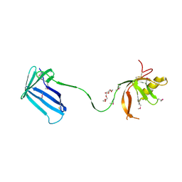
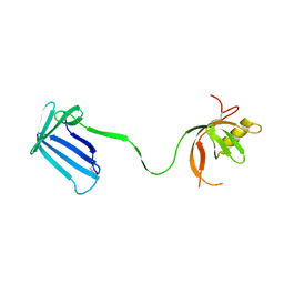
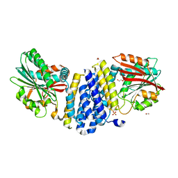
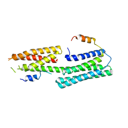
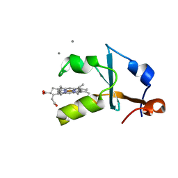
1WMH
 | | Crystal structure of a PB1 domain complex of Protein kinase c iota and Par6 alpha | | 分子名称: | Partitioning defective-6 homolog alpha, Protein kinase C, iota type | | 著者 | Hirano, Y, Yoshinaga, S, Suzuki, N.N, Horiuchi, M, Kohjima, M, Takeya, R, Sumimoto, H, Inagaki, F. | | 登録日 | 2004-07-09 | | 公開日 | 2004-12-07 | | 最終更新日 | 2024-03-13 | | 実験手法 | X-RAY DIFFRACTION (1.5 Å) | | 主引用文献 | Structure of a Cell Polarity Regulator, a Complex between Atypical PKC and Par6 PB1 Domains
J.Biol.Chem., 280, 2005
|
|
2Z4I
 | | Crystal structure of the Cpx pathway activator NlpE from Escherichia coli | | 分子名称: | Copper homeostasis protein cutF, HEXAETHYLENE GLYCOL, SULFATE ION | | 著者 | Hirano, Y, Hossain, M.M, Takeda, K, Tokuda, H, Miki, K. | | 登録日 | 2007-06-18 | | 公開日 | 2007-09-04 | | 最終更新日 | 2021-11-10 | | 実験手法 | X-RAY DIFFRACTION (2.6 Å) | | 主引用文献 | Structural Studies of the Cpx Pathway Activator NlpE on the Outer Membrane of Escherichia coli
Structure, 15, 2007
|
|
2Z4H
 | | Crystal structure of the Cpx pathway activator NlpE from Escherichia coli | | 分子名称: | Copper homeostasis protein cutF, SULFATE ION | | 著者 | Hirano, Y, Hossain, M.M, Takeda, K, Tokuda, H, Miki, K. | | 登録日 | 2007-06-18 | | 公開日 | 2007-09-04 | | 最終更新日 | 2021-11-10 | | 実験手法 | X-RAY DIFFRACTION (2.8 Å) | | 主引用文献 | Structural Studies of the Cpx Pathway Activator NlpE on the Outer Membrane of Escherichia coli
Structure, 15, 2007
|
|
3A9F
 | | Crystal structure of the C-terminal domain of cytochrome cz from Chlorobium tepidum | | 分子名称: | 3,6,9,12,15,18-HEXAOXAICOSANE-1,20-DIOL, Cytochrome c, HEME C, ... | | 著者 | Hirano, Y, Higuchi, M, Azai, C, Oh-oka, H, Miki, K, Wang, Z.-Y. | | 登録日 | 2009-10-25 | | 公開日 | 2010-03-02 | | 最終更新日 | 2024-03-13 | | 実験手法 | X-RAY DIFFRACTION (1.3 Å) | | 主引用文献 | Crystal structure of the electron carrier domain of the reaction center cytochrome c(z) subunit from green photosynthetic bacterium Chlorobium tepidum
J.Mol.Biol., 397, 2010
|
|
5B3G
 | | The crystal structure of the heterodimer of SHORT-ROOT and SCARECROW GRAS domains | | 分子名称: | 1,2-ETHANEDIOL, DI(HYDROXYETHYL)ETHER, PHOSPHATE ION, ... | | 著者 | Hirano, Y, Nakagawa, M, Hakoshima, T. | | 登録日 | 2016-02-29 | | 公開日 | 2017-03-01 | | 最終更新日 | 2024-03-20 | | 実験手法 | X-RAY DIFFRACTION (2 Å) | | 主引用文献 | Structure of the SHR-SCR heterodimer bound to the BIRD/IDD transcriptional factor JKD
Nat Plants, 3, 2017
|
|
5B3H
 | | The crystal structure of the JACKDAW/IDD10 bound to the heterodimeric SHR-SCR complex | | 分子名称: | Protein SCARECROW, Protein SHORT-ROOT, ZINC ION, ... | | 著者 | Hirano, Y, Suyama, T, Nakagawa, M, Hakoshima, T. | | 登録日 | 2016-02-29 | | 公開日 | 2017-03-01 | | 最終更新日 | 2023-11-08 | | 実験手法 | X-RAY DIFFRACTION (2.7 Å) | | 主引用文献 | Structure of the SHR-SCR heterodimer bound to the BIRD/IDD transcriptional factor JKD
Nat Plants, 3, 2017
|
|
5D8V
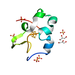 | | Ultra-high resolution structure of high-potential iron-sulfur protein | | 分子名称: | GLYCEROL, High-potential iron-sulfur protein, IRON/SULFUR CLUSTER, ... | | 著者 | Hirano, Y, Takeda, K, Miki, K. | | 登録日 | 2015-08-18 | | 公開日 | 2016-05-25 | | 最終更新日 | 2023-11-08 | | 実験手法 | X-RAY DIFFRACTION (0.48 Å) | | 主引用文献 | Charge-density analysis of an iron-sulfur protein at an ultra-high resolution of 0.48 angstrom
Nature, 534, 2016
|
|
3AU5
 | |
3AU4
 | |
5XFL
 | |
5Y04
 | |
3VRC
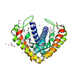 | | Crystal structure of cytochrome c' from Thermochromatium tepidum | | 分子名称: | CADMIUM ION, CHLORIDE ION, Cytochrome c', ... | | 著者 | Hirano, Y, Kimura, Y, Suzuki, H, Miki, K, Wang, Z.-Y. | | 登録日 | 2012-04-09 | | 公開日 | 2012-09-12 | | 最終更新日 | 2023-11-08 | | 実験手法 | X-RAY DIFFRACTION (1 Å) | | 主引用文献 | Structure Analysis and Comparative Characterization of the Cytochrome c' and Flavocytochrome c from Thermophilic Purple Photosynthetic Bacterium Thermochromatium tepidum
Biochemistry, 51, 2012
|
|
3VRD
 | | Crystal structure of flavocytochrome c from Thermochromatium tepidum | | 分子名称: | FLAVIN-ADENINE DINUCLEOTIDE, Flavocytochrome c flavin subunit, Flavocytochrome c heme subunit, ... | | 著者 | Hirano, Y, Kimura, Y, Suzuki, H, Miki, K, Wang, Z.-Y. | | 登録日 | 2012-04-09 | | 公開日 | 2012-09-12 | | 最終更新日 | 2023-12-06 | | 実験手法 | X-RAY DIFFRACTION (1.5 Å) | | 主引用文献 | Structure Analysis and Comparative Characterization of the Cytochrome c' and Flavocytochrome c from Thermophilic Purple Photosynthetic Bacterium Thermochromatium tepidum
Biochemistry, 51, 2012
|
|
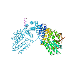
6M4K
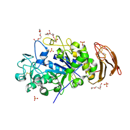 | | X-ray crystal structure of wild type alpha-amylase I from Eisenia fetida | | 分子名称: | ACETATE ION, Alpha-amylase, CALCIUM ION, ... | | 著者 | Hirano, Y, Tsukamoto, K, Ariki, S, Naka, Y, Ueda, M, Tamada, T. | | 登録日 | 2020-03-07 | | 公開日 | 2020-09-16 | | 最終更新日 | 2023-11-29 | | 実験手法 | X-RAY DIFFRACTION (1.3 Å) | | 主引用文献 | X-ray crystallographic structural studies of alpha-amylase I from Eisenia fetida.
Acta Crystallogr D Struct Biol, 76, 2020
|
|
6M4L
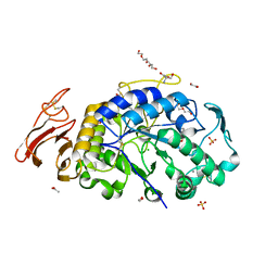 | | X-ray crystal structure of the E249Q mutant of alpha-amylase I from Eisenia fetida | | 分子名称: | 1,2-ETHANEDIOL, 2-AMINO-2-HYDROXYMETHYL-PROPANE-1,3-DIOL, ACETATE ION, ... | | 著者 | Hirano, Y, Tsukamoto, K, Ariki, S, Naka, Y, Ueda, M, Tamada, T. | | 登録日 | 2020-03-07 | | 公開日 | 2020-09-16 | | 最終更新日 | 2023-11-29 | | 実験手法 | X-RAY DIFFRACTION (1.6 Å) | | 主引用文献 | X-ray crystallographic structural studies of alpha-amylase I from Eisenia fetida.
Acta Crystallogr D Struct Biol, 76, 2020
|
|
6M4M
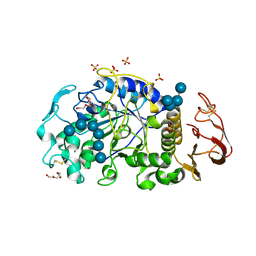 | | X-ray crystal structure of the E249Q mutan of alpha-amylase I and maltohexaose complex from Eisenia fetida | | 分子名称: | Alpha-amylase, CALCIUM ION, CHLORIDE ION, ... | | 著者 | Hirano, Y, Tsukamoto, K, Ariki, S, Naka, Y, Ueda, M, Tamada, T. | | 登録日 | 2020-03-07 | | 公開日 | 2020-09-16 | | 最終更新日 | 2023-11-29 | | 実験手法 | X-RAY DIFFRACTION (1.7 Å) | | 主引用文献 | X-ray crystallographic structural studies of alpha-amylase I from Eisenia fetida.
Acta Crystallogr D Struct Biol, 76, 2020
|
|
3X35
 | | Crystal structure of the reduced form of the solubilized domain of porcine cytochrome b5 in form 2 crystal | | 分子名称: | ACETATE ION, Cytochrome b5, PROTOPORPHYRIN IX CONTAINING FE | | 著者 | Hirano, Y, Kimura, S, Tamada, T. | | 登録日 | 2015-01-14 | | 公開日 | 2015-07-15 | | 最終更新日 | 2023-11-08 | | 実験手法 | X-RAY DIFFRACTION (0.95 Å) | | 主引用文献 | High-resolution crystal structures of the solubilized domain of porcine cytochrome b5.
Acta Crystallogr.,Sect.D, 71, 2015
|
|
3X33
 | | Crystal structure of the oxidized form of the solubilized domain of porcine cytochrome b5 in form 2 crystal | | 分子名称: | ACETATE ION, Cytochrome b5, PROTOPORPHYRIN IX CONTAINING FE | | 著者 | Hirano, Y, Kimura, S, Tamada, T. | | 登録日 | 2015-01-14 | | 公開日 | 2015-07-15 | | 最終更新日 | 2023-11-08 | | 実験手法 | X-RAY DIFFRACTION (0.93 Å) | | 主引用文献 | High-resolution crystal structures of the solubilized domain of porcine cytochrome b5.
Acta Crystallogr.,Sect.D, 71, 2015
|
|
3X34
 | | Crystal structure of the reduced form of the solubilized domain of porcine cytochrome b5 in form 1 crystal | | 分子名称: | CALCIUM ION, Cytochrome b5, PROTOPORPHYRIN IX CONTAINING FE | | 著者 | Hirano, Y, Kimura, S, Tamada, T. | | 登録日 | 2015-01-14 | | 公開日 | 2015-07-15 | | 最終更新日 | 2023-11-08 | | 実験手法 | X-RAY DIFFRACTION (0.76 Å) | | 主引用文献 | High-resolution crystal structures of the solubilized domain of porcine cytochrome b5.
Acta Crystallogr.,Sect.D, 71, 2015
|
|
3X32
 | | Crystal structure of the oxidized form of the solubilized domain of porcine cytochrome b5 in form 1 crystal | | 分子名称: | 4-(2-HYDROXYETHYL)-1-PIPERAZINE ETHANESULFONIC ACID, CALCIUM ION, Cytochrome b5, ... | | 著者 | Hirano, Y, Kimura, S, Tamada, T. | | 登録日 | 2015-01-14 | | 公開日 | 2015-07-15 | | 最終更新日 | 2023-11-08 | | 実験手法 | X-RAY DIFFRACTION (0.83 Å) | | 主引用文献 | High-resolution crystal structures of the solubilized domain of porcine cytochrome b5.
Acta Crystallogr.,Sect.D, 71, 2015
|
|
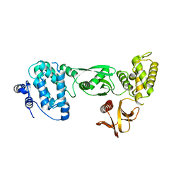
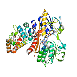
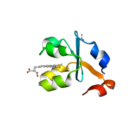
6IEJ
 | | The C2 domain of cytosolic phospholipase A2 alpha bound to phosphatidylcholine | | 分子名称: | 1,2-dihexanoyl-sn-glycero-3-phosphocholine, CALCIUM ION, Cytosolic phospholipase A2, ... | | 著者 | Hirano, Y, Gao, Y.G, Stephenson, D.J, Vu, N.T, Malinina, L, Chalfant, C.E, Patel, D.J, Brown, R.E. | | 登録日 | 2018-09-14 | | 公開日 | 2019-05-22 | | 最終更新日 | 2023-11-22 | | 実験手法 | X-RAY DIFFRACTION (2.206 Å) | | 主引用文献 | Structural basis of phosphatidylcholine recognition by the C2-domain of cytosolic phospholipase A2alpha.
Elife, 8, 2019
|
|
6KPD
 | |
6KPB
 | |
5Y6T
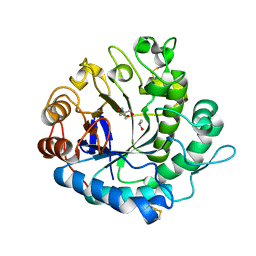 | | Crystal structure of endo-1,4-beta-mannanase from Eisenia fetida | | 分子名称: | 2-AMINO-2-HYDROXYMETHYL-PROPANE-1,3-DIOL, ISOPROPYL ALCOHOL, endo-1,4-beta-mannanase | | 著者 | Hirano, Y, Ueda, M, Tamada, T. | | 登録日 | 2017-08-15 | | 公開日 | 2018-06-27 | | 最終更新日 | 2023-11-22 | | 実験手法 | X-RAY DIFFRACTION (1.7 Å) | | 主引用文献 | Gene cloning, expression, and X-ray crystallographic analysis of a beta-mannanase from Eisenia fetida.
Enzyme.Microb.Technol., 117, 2018
|
|


