9AYK
 
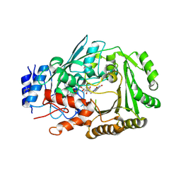
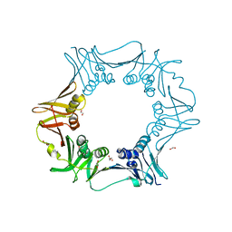
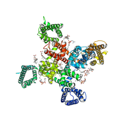 | | Cryo-EM structure of human Cav3.2 with ML218 | | 分子名称: | 1-O-OCTADECYL-SN-GLYCERO-3-PHOSPHOCHOLINE, 2-acetamido-2-deoxy-beta-D-glucopyranose, 3,5-dichloro-N-{[(1R,5S,6r)-3-(3,3-dimethylbutyl)-3-azabicyclo[3.1.0]hexan-6-yl]methyl}benzamide, ... | | 著者 | Fan, X, Huang, J, Yan, N. | | 登録日 | 2024-03-08 | | 公開日 | 2024-04-24 | | 実験手法 | ELECTRON MICROSCOPY (3 Å) | | 主引用文献 | Structural basis for human Ca v 3.2 inhibition by selective antagonists.
Cell Res., 2024
|
|
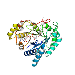
2FXE
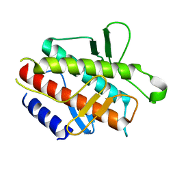
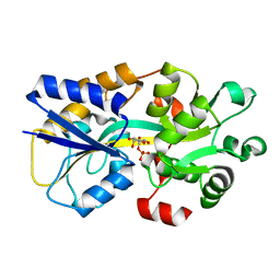
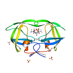 | | X-ray crystal structure of HIV-1 protease CRM mutant complexed with atazanavir (BMS-232632) | | 分子名称: | (3S,8S,9S,12S)-3,12-BIS(1,1-DIMETHYLETHYL)-8-HYDROXY-4,11-DIOXO-9-(PHENYLMETHYL)-6-[[4-(2-PYRIDINYL)PHENYL]METHYL]-2,5, 6,10,13-PENTAAZATETRADECANEDIOIC ACID DIMETHYL ESTER, ACETATE ION, ... | | 著者 | Sheriff, S, Klei, H.E. | | 登録日 | 2006-02-05 | | 公開日 | 2007-02-20 | | 最終更新日 | 2023-08-30 | | 実験手法 | X-RAY DIFFRACTION (1.8 Å) | | 主引用文献 | X-ray crystal structures of human immunodeficiency virus type 1 protease mutants complexed with atazanavir.
J.Virol., 81, 2007
|
|
2FXD
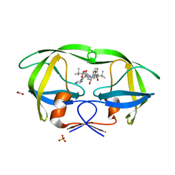 | | X-ray crystal structure of HIV-1 protease IRM mutant complexed with atazanavir (BMS-232632) | | 分子名称: | (3S,8S,9S,12S)-3,12-BIS(1,1-DIMETHYLETHYL)-8-HYDROXY-4,11-DIOXO-9-(PHENYLMETHYL)-6-[[4-(2-PYRIDINYL)PHENYL]METHYL]-2,5, 6,10,13-PENTAAZATETRADECANEDIOIC ACID DIMETHYL ESTER, ACETATE ION, ... | | 著者 | Klei, H.E, Sheriff, S. | | 登録日 | 2006-02-04 | | 公開日 | 2007-02-20 | | 最終更新日 | 2023-08-30 | | 実験手法 | X-RAY DIFFRACTION (1.6 Å) | | 主引用文献 | X-ray crystal structures of human immunodeficiency virus type 1 protease mutants complexed with atazanavir.
J.Virol., 81, 2007
|
|
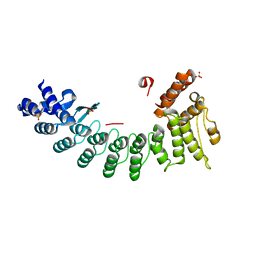
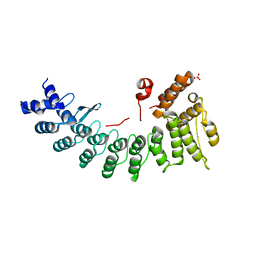
7CMZ
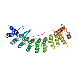
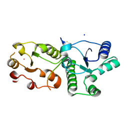 | | Crystal Structure of BRCT7/8 in Complex with the APS Motif of PHF8 | | 分子名称: | DNA topoisomerase 2-binding protein 1, Histone lysine demethylase PHF8, POTASSIUM ION, ... | | 著者 | Che, S.Y, Ma, S, Cao, C, Yao, Z, Shi, L, Yang, N. | | 登録日 | 2020-07-29 | | 公開日 | 2021-03-17 | | 最終更新日 | 2023-11-29 | | 実験手法 | X-RAY DIFFRACTION (1.695 Å) | | 主引用文献 | PHF8-promoted TOPBP1 demethylation drives ATR activation and preserves genome stability.
Sci Adv, 7, 2021
|
|
6JQH
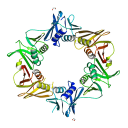
 | | Crystal structure of MaDA | | 分子名称: | FLAVIN-ADENINE DINUCLEOTIDE, MaDA | | 著者 | Du, X.X, Lei, X.G. | | 登録日 | 2019-03-31 | | 公開日 | 2020-04-08 | | 最終更新日 | 2023-11-22 | | 実験手法 | X-RAY DIFFRACTION (2.303 Å) | | 主引用文献 | FAD-dependent enzyme-catalysed intermolecular [4+2] cycloaddition in natural product biosynthesis.
Nat.Chem., 12, 2020
|
|
4DDP
 | |
7CNG
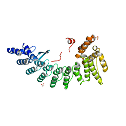
 | | Structure of CDK5R1 bound FEM1B | | 分子名称: | Protein fem-1 homolog B,Peptide from Cyclin-dependent kinase 5 activator 1, SULFATE ION | | 著者 | Chen, X, Liao, S, Xu, C. | | 登録日 | 2020-07-31 | | 公開日 | 2020-10-21 | | 最終更新日 | 2023-11-29 | | 実験手法 | X-RAY DIFFRACTION (3.49 Å) | | 主引用文献 | Molecular basis for arginine C-terminal degron recognition by Cul2 FEM1 E3 ligase.
Nat.Chem.Biol., 17, 2021
|
|
6IZO
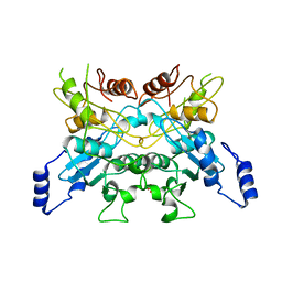
 | | Crystal structure of DNA polymerase sliding clamp from Caulobacter crescentus | | 分子名称: | 1,2-ETHANEDIOL, Beta sliding clamp, DI(HYDROXYETHYL)ETHER | | 著者 | Jiang, X, Zhang, L, Teng, M, Li, X. | | 登録日 | 2018-12-20 | | 公開日 | 2019-11-27 | | 最終更新日 | 2023-11-22 | | 実験手法 | X-RAY DIFFRACTION (1.94 Å) | | 主引用文献 | Caulobacter crescentus beta sliding clamp employs a noncanonical regulatory model of DNA replication.
Febs J., 287, 2020
|
|
6JIR
 | | Crystal structure of C. crescentus beta sliding clamp with PEG bound to putative beta-motif tethering region | | 分子名称: | 1,2-ETHANEDIOL, Beta sliding clamp, DI(HYDROXYETHYL)ETHER, ... | | 著者 | Jiang, X, Teng, M, Li, X. | | 登録日 | 2019-02-23 | | 公開日 | 2019-11-27 | | 最終更新日 | 2023-11-22 | | 実験手法 | X-RAY DIFFRACTION (1.95 Å) | | 主引用文献 | Caulobacter crescentus beta sliding clamp employs a noncanonical regulatory model of DNA replication.
Febs J., 287, 2020
|
|
8I0C
 | | Crystal structure of Aldo-keto reductase 1C3 complexed with compound S0703 | | 分子名称: | 1-[4-[3,5-bis(chloranyl)phenyl]-3-fluoranyl-phenyl]cyclopropane-1-carboxylic acid, Aldo-keto reductase family 1 member C3, NADP NICOTINAMIDE-ADENINE-DINUCLEOTIDE PHOSPHATE | | 著者 | Jiang, J, He, S, Liu, Y, Fang, P, Sun, H. | | 登録日 | 2023-01-10 | | 公開日 | 2023-09-20 | | 実験手法 | X-RAY DIFFRACTION (2.33 Å) | | 主引用文献 | Development of Biaryl-Containing Aldo-Keto Reductase 1C3 (AKR1C3) Inhibitors for Reversing AKR1C3-Mediated Drug Resistance in Cancer Treatment.
J.Med.Chem., 66, 2023
|
|
6LBG
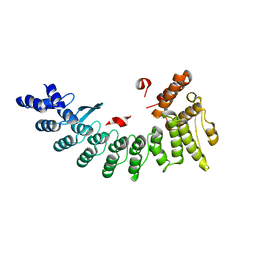
 | | Structure of OR51B2 bound FEM1C | | 分子名称: | Protein fem-1 homolog C,Peptide from Olfactory receptor 51B2, SULFATE ION | | 著者 | Chen, X, Liao, S, Xu, C. | | 登録日 | 2019-11-14 | | 公開日 | 2020-10-21 | | 最終更新日 | 2023-11-22 | | 実験手法 | X-RAY DIFFRACTION (2.51 Å) | | 主引用文献 | Molecular basis for arginine C-terminal degron recognition by Cul2 FEM1 E3 ligase.
Nat.Chem.Biol., 17, 2021
|
|
6LE6
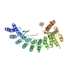
 | | Structure of LNLPTQGRAR bound FEM1C | | 分子名称: | Protein fem-1 homolog C,10-mer peptide, SULFATE ION | | 著者 | Chen, X, Liao, S, Xu, C. | | 登録日 | 2019-11-24 | | 公開日 | 2020-10-21 | | 最終更新日 | 2023-11-22 | | 実験手法 | X-RAY DIFFRACTION (2.33 Å) | | 主引用文献 | Molecular basis for arginine C-terminal degron recognition by Cul2 FEM1 E3 ligase.
Nat.Chem.Biol., 17, 2021
|
|
6LBF
 | | Crystal structure of FEM1B | | 分子名称: | Protein fem-1 homolog B, SULFATE ION | | 著者 | Chen, X, Liao, S, Xu, C. | | 登録日 | 2019-11-14 | | 公開日 | 2020-10-21 | | 最終更新日 | 2024-03-27 | | 実験手法 | X-RAY DIFFRACTION (3.252 Å) | | 主引用文献 | Molecular basis for arginine C-terminal degron recognition by Cul2 FEM1 E3 ligase.
Nat.Chem.Biol., 17, 2021
|
|
6LDP
 | | Structure of CDK5R1-bound FEM1C | | 分子名称: | Protein fem-1 homolog C,Peptide from Cyclin-dependent kinase 5 activator 1, SULFATE ION | | 著者 | Chen, X, Liao, S, Xu, C. | | 登録日 | 2019-11-22 | | 公開日 | 2020-10-21 | | 最終更新日 | 2024-03-20 | | 実験手法 | X-RAY DIFFRACTION (2.35 Å) | | 主引用文献 | Molecular basis for arginine C-terminal degron recognition by Cul2 FEM1 E3 ligase.
Nat.Chem.Biol., 17, 2021
|
|
6L08
 | |
6LEN
 | | Structure of NS11 bound FEM1C | | 分子名称: | Protein fem-1 homolog C,NS11 peptide | | 著者 | Chen, x, Liao, S, Xu, C. | | 登録日 | 2019-11-25 | | 公開日 | 2020-10-21 | | 最終更新日 | 2023-11-22 | | 実験手法 | X-RAY DIFFRACTION (2.383 Å) | | 主引用文献 | Molecular basis for arginine C-terminal degron recognition by Cul2 FEM1 E3 ligase.
Nat.Chem.Biol., 17, 2021
|
|
6LF0
 | | Structure of FEM1C | | 分子名称: | Protein fem-1 homolog C, SULFATE ION | | 著者 | Chen, X, Liao, S, Xu, C. | | 登録日 | 2019-11-27 | | 公開日 | 2020-10-21 | | 最終更新日 | 2023-11-22 | | 実験手法 | X-RAY DIFFRACTION (2.11 Å) | | 主引用文献 | Molecular basis for arginine C-terminal degron recognition by Cul2 FEM1 E3 ligase.
Nat.Chem.Biol., 17, 2021
|
|
6LBN
 | | Structure of SIL1-bound FEM1C | | 分子名称: | Protein fem-1 homolog C,Peptide from Nucleotide exchange factor SIL1 | | 著者 | Chen, X, Liao, S, Xu, C. | | 登録日 | 2019-11-14 | | 公開日 | 2020-10-21 | | 最終更新日 | 2023-11-22 | | 実験手法 | X-RAY DIFFRACTION (2.899 Å) | | 主引用文献 | Molecular basis for arginine C-terminal degron recognition by Cul2 FEM1 E3 ligase.
Nat.Chem.Biol., 17, 2021
|
|
6LEY
 | | Structure of Sil1G bound FEM1C | | 分子名称: | Protein fem-1 homolog C,Peptide from Nucleotide exchange factor SIL1 | | 著者 | Chen, X, Liao, S, Xu, C. | | 登録日 | 2019-11-27 | | 公開日 | 2020-10-21 | | 最終更新日 | 2023-11-22 | | 実験手法 | X-RAY DIFFRACTION (2.39 Å) | | 主引用文献 | Molecular basis for arginine C-terminal degron recognition by Cul2 FEM1 E3 ligase.
Nat.Chem.Biol., 17, 2021
|
|
6LKG
 | |
6LKK
 | |
6LKL
 | |
6LKI
 | |
6M0A
 | |
6LKJ
 | |


