3DQL
 
 | |
3DQ3
 | |
3DQD
 | |
3DQN
 | |
2HMW
 | |
2HMT
 | |
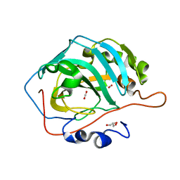
3D92
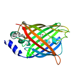
 | | Human carbonic anhydrase II bound with substrate carbon dioxide | | Descriptor: | CARBON DIOXIDE, GLYCEROL, ZINC ION, ... | | Authors: | Domsic, J.F, Avvaru, B.S, McKenna, R. | | Deposit date: | 2008-05-26 | | Release date: | 2008-09-02 | | Last modified: | 2023-08-30 | | Method: | X-RAY DIFFRACTION (1.1 Å) | | Cite: | Entrapment of carbon dioxide in the active site of carbonic anhydrase II
J.Biol.Chem., 283, 2008
|
|
3D93
 | |
5T8B
 | |
5T8C
 | |


