Loading
PDBj
MenuPDBj@FacebookPDBj@TwitterPDBj@YouTubewwPDB FoundationwwPDB
RCSB PDBPDBeBMRBAdv. SearchSearch help
Search by PDB author
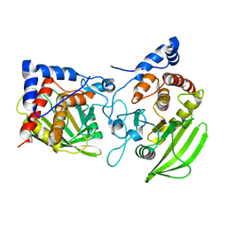
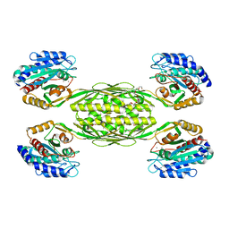
4EWT
DownloadVisualize
BU of 4ewt by Molmil
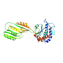
The crystal structure of a putative aminohydrolase from methicillin resistant Staphylococcus aureus
Descriptor: 1-DEOXY-1-THIO-HEPTAETHYLENE GLYCOL, DI(HYDROXYETHYL)ETHER, MANGANESE (II) ION, ...
Authors:Girish, T.S, Vivek, B, Colaco, M, Misquith, S, Gopal, B.
Deposit date:2012-04-27
Release date:2013-02-20
Last modified:2023-11-08
Method:X-RAY DIFFRACTION (2.1 Å)
Cite:Structure of an amidohydrolase, SACOL0085, from methicillin-resistant Staphylococcus aureus COL
Acta Crystallogr.,Sect.F, 69, 2013
2PI7
DownloadVisualize
BU of 2pi7 by Molmil
Structure of the catalytic domain of the chick retinal neurite inhibitor-Receptor Protein Tyrosine Phosphatase CRYP-2/cPTPRO
Descriptor: NITRATE ION, Protein tyrosine phosphatase CRYP-2
Authors:Girish, T.S, Gopal, B.
Deposit date:2007-04-13
Release date:2007-05-01
Last modified:2023-10-25
Method:X-RAY DIFFRACTION (2.59 Å)
Cite:The crystal structure of the catalytic domain of the chick retinal neurite inhibitor-receptor protein tyrosine phosphatase CRYP-2/cPTPRO
Proteins, 68, 2007
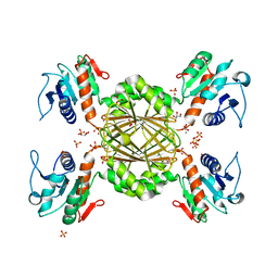
3QY9
DownloadVisualize
BU of 3qy9 by Molmil
The Crystal Structure of Dihydrodipicolinate reductase from Staphylococcus aureus
Descriptor: ACETATE ION, Dihydrodipicolinate reductase, GLYCEROL, ...
Authors:Girish, T.S, Gopal, B.
Deposit date:2011-03-03
Release date:2011-08-03
Last modified:2023-11-01
Method:X-RAY DIFFRACTION (1.8 Å)
Cite:Structure and nucleotide specificity of Staphylococcus aureus dihydrodipicolinate reductase (DapB)
Febs Lett., 585, 2011
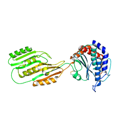
3KHZ
DownloadVisualize
BU of 3khz by Molmil
Crystal Structure of R350A mutant of Staphylococcus aureus metallopeptidase (Sapep/DapE) in the apo-form
Descriptor: Putative dipeptidase SACOL1801
Authors:Girish, T.S, Gopal, B.
Deposit date:2009-10-31
Release date:2010-07-07
Last modified:2023-11-01
Method:X-RAY DIFFRACTION (2.5 Å)
Cite:Crystal structure of Staphylococcus aureus metallopeptidase (Sapep) reveals large domain motions between the manganese-bound and apo-states
J.Biol.Chem., 285, 2010
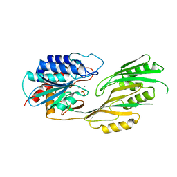
3KI9
DownloadVisualize
BU of 3ki9 by Molmil
Crystal structure of Staphylococcus aureus metallopeptidase (Sapep/DapE) in the Mn2+ bound form
Descriptor: MANGANESE (II) ION, PHOSPHATE ION, Putative dipeptidase SACOL1801
Authors:Girish, T.S, Gopal, B.
Deposit date:2009-11-01
Release date:2010-07-07
Last modified:2023-11-01
Method:X-RAY DIFFRACTION (2.9 Å)
Cite:Crystal structure of Staphylococcus aureus metallopeptidase (Sapep) reveals large domain motions between the manganese-bound and apo-states
J.Biol.Chem., 285, 2010
3KHX
DownloadVisualize
BU of 3khx by Molmil
Crystal structure of Staphylococcus aureus metallopeptidase (Sapep/DapE) in the apo-form
Descriptor: Putative dipeptidase SACOL1801
Authors:Girish, T.S, Gopal, B.
Deposit date:2009-10-31
Release date:2010-07-07
Last modified:2023-11-01
Method:X-RAY DIFFRACTION (2.3 Å)
Cite:Crystal structure of Staphylococcus aureus metallopeptidase (Sapep) reveals large domain motions between the manganese-bound and apo-states
J.Biol.Chem., 285, 2010
5HQ2
DownloadVisualize
BU of 5hq2 by Molmil
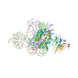
Structural model of Set8 histone H4 Lys20 methyltransferase bound to nucleosome core particle
Descriptor: DNA (149-MER), Guanine nucleotide exchange factor SRM1, Histone H2A, ...
Authors:Tavarekere, G, McGinty, R.K, Tan, S.
Deposit date:2016-01-21
Release date:2016-03-23
Last modified:2024-03-06
Method:X-RAY DIFFRACTION (4.5 Å)
Cite:Multivalent Interactions by the Set8 Histone Methyltransferase With Its Nucleosome Substrate.
J.Mol.Biol., 428, 2016
3DI0
DownloadVisualize
BU of 3di0 by Molmil
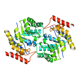
Crystal Structure of Dihydrodipicolinate synthase from Staphylococcus aureus
Descriptor: Dihydrodipicolinate synthase, GLYCEROL
Authors:Tavarekere, G.S.
Deposit date:2008-06-19
Release date:2008-08-12
Last modified:2023-11-01
Method:X-RAY DIFFRACTION (2.38 Å)
Cite:Structural and functional characterization of Staphylococcus aureus dihydrodipicolinate synthase
Febs Lett., 582, 2008
3DI1
DownloadVisualize
BU of 3di1 by Molmil
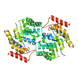
Crystal structure of the Staphylococcus aureus Dihydrodipicolinate synthase-pyruvate complex
Descriptor: Dihydrodipicolinate synthase, PYRUVIC ACID
Authors:Tavarekere, G.S.
Deposit date:2008-06-19
Release date:2008-08-12
Last modified:2023-11-15
Method:X-RAY DIFFRACTION (2.2 Å)
Cite:Structural and functional characterization of Staphylococcus aureus dihydrodipicolinate synthase
Febs Lett., 582, 2008

222926

PDB entries from 2024-07-24

PDB statisticsPDBj update infoContact PDBjnumon