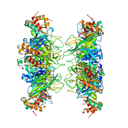
6TUW
 
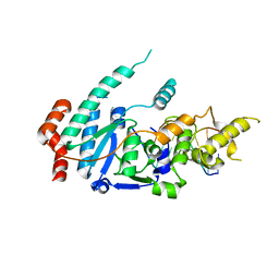 | | human XPG-DNA, Complex 1 | | 分子名称: | DNA (5'-D(P*GP*AP*AP*CP*TP*CP*TP*G)-3'), DNA (5'-D(P*TP*GP*CP*AP*GP*AP*GP*TP*TP*C)-3'), DNA repair protein complementing XP-G cells,DNA repair protein complementing XP-G cells | | 著者 | Ruiz, F.M, Fernandez-Tornero, C. | | 登録日 | 2020-01-08 | | 公開日 | 2020-09-16 | | 最終更新日 | 2024-01-24 | | 実験手法 | X-RAY DIFFRACTION (3.5 Å) | | 主引用文献 | The crystal structure of human XPG, the xeroderma pigmentosum group G endonuclease, provides insight into nucleotide excision DNA repair.
Nucleic Acids Res., 48, 2020
|
|
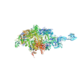
6TUS
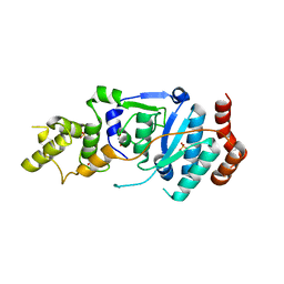 | | human XPG, Apo2 form | | 分子名称: | DNA repair protein complementing XP-G cells,DNA repair protein complementing XP-G cells, GLYCEROL, SULFATE ION | | 著者 | Ruiz, F.M, Fernandez-Tornero, C. | | 登録日 | 2020-01-08 | | 公開日 | 2020-09-16 | | 最終更新日 | 2024-01-24 | | 実験手法 | X-RAY DIFFRACTION (2.5 Å) | | 主引用文献 | The crystal structure of human XPG, the xeroderma pigmentosum group G endonuclease, provides insight into nucleotide excision DNA repair.
Nucleic Acids Res., 48, 2020
|
|
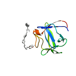
1H8P
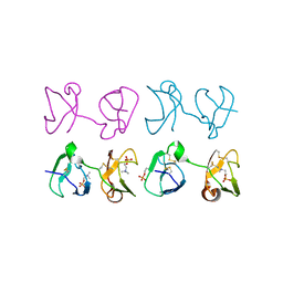 | | Bull seminal plasma PDC-109 fibronectin type II module | | 分子名称: | PHOSPHOCHOLINE, SEMINAL PLASMA PROTEIN PDC-109 | | 著者 | Wah, D.A, Fernandez-Tornero, C, Calvete, J.J, Romero, A. | | 登録日 | 2001-02-14 | | 公開日 | 2002-04-12 | | 最終更新日 | 2019-07-24 | | 実験手法 | X-RAY DIFFRACTION (1.82 Å) | | 主引用文献 | Sperm Coating Mechanism from the 1.8 A Crystal Structure of Pdc-109-Phosphorylcholine Complex
Structure, 10, 2002
|
|
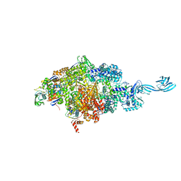
2PYO
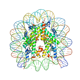 | | Drosophila nucleosome core | | 分子名称: | CHLORIDE ION, DNA (147-MER), Histone H2A, ... | | 著者 | Clapier, C.R, Petosa, C, Mueller, C.W. | | 登録日 | 2007-05-16 | | 公開日 | 2007-11-06 | | 最終更新日 | 2024-02-21 | | 実験手法 | X-RAY DIFFRACTION (2.43 Å) | | 主引用文献 | Structure of the Drosophila nucleosome core particle highlights evolutionary constraints on the H2A-H2B histone dimer.
Proteins, 71, 2007
|
|
4IS4
 | |
4Q5S
 | |
4Q4Z
 | |
2ERM
 | | Solution structure of a biologically active human FGF-1 monomer, complexed to a hexasaccharide heparin-analogue | | 分子名称: | 2-deoxy-2-(sulfoamino)-alpha-D-glucopyranose-(1-4)-2-O-sulfo-alpha-L-idopyranuronic acid-(1-4)-2-acetamido-2-deoxy-6-O-sulfo-alpha-D-glucopyranose-(1-4)-alpha-L-idopyranuronic acid-(1-4)-2-deoxy-2-(sulfoamino)-alpha-D-glucopyranose-(1-4)-2-O-sulfo-alpha-L-idopyranuronic acid, Heparin-binding growth factor 1, ISOPROPYL ALCOHOL | | 著者 | Canales, A, Lozano, R, Nieto, P.M, Martin-Lomas, M, Gimenez-Gallego, G, Jimenez-Barbero, J. | | 登録日 | 2005-10-25 | | 公開日 | 2006-10-03 | | 最終更新日 | 2024-05-01 | | 実験手法 | SOLUTION NMR | | 主引用文献 | Solution NMR structure of a human FGF-1 monomer, activated by a hexasaccharide heparin-analogue.
Febs J., 273, 2006
|
|
1WRA
 | |


