Loading
PDBj
MenuPDBj@FacebookPDBj@TwitterPDBj@YouTubewwPDB FoundationwwPDB
RCSB PDBPDBeBMRBAdv. SearchSearch help
Search by PDB author
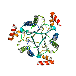
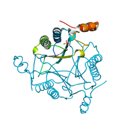
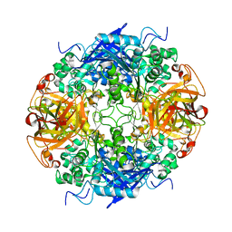
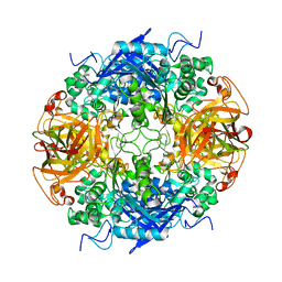
3N4G
DownloadVisualize
BU of 3n4g by Molmil
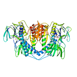
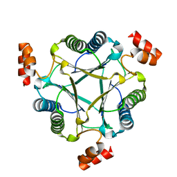
Crystal structure of native Cg10062
Descriptor: Putative tautomerase
Authors:Guo, Y, Robertson, B.A, Hackert, M.L, Whitman, C.P.
Deposit date:2010-05-21
Release date:2011-06-01
Last modified:2023-09-06
Method:X-RAY DIFFRACTION (2.44 Å)
Cite:Crystal Structures of the Native and Inactivated Cg10062, a cis-3-Chloroacrylic Acid Dehalogenase from Corynebacterium glutamicum: Implications for the Evolution of cis-3-Chloroacrylic Acid Dehalogenase Activity in the Tautomerase Superfamily
To be Published
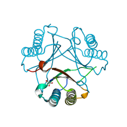
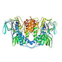
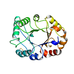
3N4D
DownloadVisualize
BU of 3n4d by Molmil
Crystal structure of Cg10062 inactivated by(R)-oxirane-2-carboxylate
Descriptor: Putative tautomerase
Authors:Guo, Y, Robertson, B.A, Hackert, M.L, Whitman, C.P.
Deposit date:2010-05-21
Release date:2011-06-01
Last modified:2023-09-06
Method:X-RAY DIFFRACTION (2.54 Å)
Cite:Crystal Structures of the Native and Inactivated Cg10062, a cis-3-Chloroacrylic Acid Dehalogenase from Corynebacterium glutamicum: Implications for the Evolution of cis-3-Chloroacrylic Acid Dehalogenase Activity in the Tautomerase Superfamily
To be Published
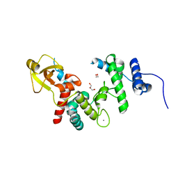
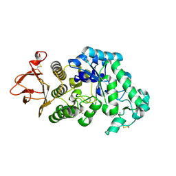
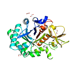
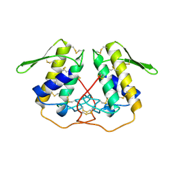
1QDT
DownloadVisualize
BU of 1qdt by Molmil
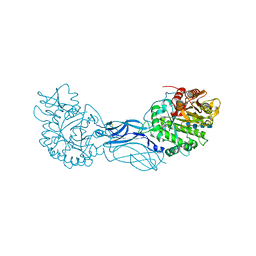
2.1 A RESOLUTION STRUCTURE OF ESCHERICHIA COLI LYTIC TRANSGLYCOYSLASE SLT35 IN COMPLEX WITH CALCIUM
Descriptor: 1,2-ETHANEDIOL, CALCIUM ION, GLYCEROL, ...
Authors:van Asselt, E.J, Dijkstra, A.J.
Deposit date:1999-07-10
Release date:2000-01-24
Last modified:2024-02-14
Method:X-RAY DIFFRACTION (2.1 Å)
Cite:Binding of calcium in the EF-hand of Escherichia coli lytic transglycosylase Slt35 is important for stability.
FEBS Lett., 458, 1999
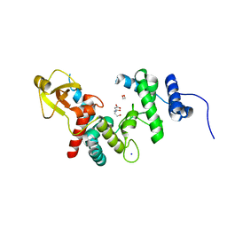
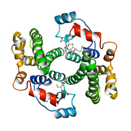
1QDR
DownloadVisualize
BU of 1qdr by Molmil
2.1 A RESOLUTION STRUCTURE OF ESCHERICHIA COLI LYTIC TRANSGLYCOSYLASE SLT35
Descriptor: 1,2-ETHANEDIOL, BICINE, LYTIC MUREIN TRANSGLYCOSYLASE B, ...
Authors:van Asselt, E.J, Dijkstra, A.J.
Deposit date:1999-07-10
Release date:2000-01-24
Last modified:2024-02-14
Method:X-RAY DIFFRACTION (2.1 Å)
Cite:Binding of calcium in the EF-hand of Escherichia coli lytic transglycosylase Slt35 is important for stability.
FEBS Lett., 458, 1999
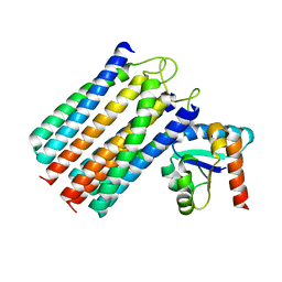
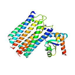
2WWV
DownloadVisualize
BU of 2wwv by Molmil
NMR structure of the IIAchitobiose-IIBchitobiose complex of the N,N'- diacetylchitoboise brance of the E. coli phosphotransferase system.
Descriptor: N,N'-DIACETYLCHITOBIOSE-SPECIFIC PHOSPHOTRANSFERASE ENZYME IIA COMPONENT, N,N'-DIACETYLCHITOBIOSE-SPECIFIC PHOSPHOTRANSFERASE ENZYME IIB COMPONENT
Authors:Sang, Y.S, Cai, M, Clore, G.M.
Deposit date:2009-10-29
Release date:2009-12-08
Last modified:2024-05-15
Method:SOLUTION NMR
Cite:Solution Structure of the Iiachitobose-Iibchitobiose Complex of the N,N'-Diacetylchitobiose Branch of the Escherichia Coli Phosphotransfer System
J.Biol.Chem., 285, 2010
2WY2
DownloadVisualize
BU of 2wy2 by Molmil
NMR structure of the IIAchitobiose-IIBchitobiose phosphoryl transition state complex of the N,N'-diacetylchitoboise brance of the E. coli phosphotransferase system.
Descriptor: N,N'-DIACETYLCHITOBIOSE-SPECIFIC PHOSPHOTRANSFERASE ENZYME IIA COMPONENT, N,N'-DIACETYLCHITOBIOSE-SPECIFIC PHOSPHOTRANSFERASE ENZYME IIB COMPONENT, PHOSPHITE ION
Authors:Sang, Y.S, Cai, M, Clore, G.M.
Deposit date:2009-11-11
Release date:2009-12-08
Last modified:2024-05-15
Method:SOLUTION NMR
Cite:Solution Structure of the Iiachitobose-Iibchitobiose Complex of the N,N'-Diacetylchitobiose Branch of the Escherichia Coli Phosphotransfer System
J.Biol.Chem., 285, 2010
2FLZ
DownloadVisualize
BU of 2flz by Molmil
The X-ray structure of cis-3-chloroacrylic acid dehalogenase (cis-CaaD) with a sulfate ion bound in the active site
Descriptor: SULFATE ION, cis-3-chloroacrylic acid dehalogenase
Authors:de Jong, R.M.
Deposit date:2006-01-06
Release date:2006-11-21
Last modified:2023-08-30
Method:X-RAY DIFFRACTION (2.75 Å)
Cite:Crystal Structures of Native and Inactivated cis-3-Chloroacrylic Acid Dehalogenase: STRUCTURAL BASIS FOR SUBSTRATE SPECIFICITY AND INACTIVATION BY (R)-OXIRANE-2-CARBOXYLATE.
J.Biol.Chem., 282, 2007
2FLT
DownloadVisualize
BU of 2flt by Molmil
The X-ray structure of the cis-3-chloroacrylic acid dehalogenase cis-CaaD inactivated with (R)-Oxirane-2-carboxylate
Descriptor: LACTIC ACID, cis-3-chloroacrylic acid dehalogenase
Authors:de Jong, R.M.
Deposit date:2006-01-06
Release date:2006-11-21
Last modified:2023-11-15
Method:X-RAY DIFFRACTION (2.1 Å)
Cite:Crystal Structures of Native and Inactivated cis-3-Chloroacrylic Acid Dehalogenase: STRUCTURAL BASIS FOR SUBSTRATE SPECIFICITY AND INACTIVATION BY (R)-OXIRANE-2-CARBOXYLATE.
J.Biol.Chem., 282, 2007
2GUY
DownloadVisualize
BU of 2guy by Molmil
Orthorhombic crystal structure (space group P21212) of Aspergillus niger alpha-amylase at 1.6 A resolution
Descriptor: Alpha-amylase A, CALCIUM ION, beta-D-mannopyranose-(1-4)-2-acetamido-2-deoxy-beta-D-glucopyranose-(1-4)-2-acetamido-2-deoxy-beta-D-glucopyranose
Authors:Vujicic Zagar, A.
Deposit date:2006-05-02
Release date:2006-08-15
Last modified:2023-08-30
Method:X-RAY DIFFRACTION (1.59 Å)
Cite:Monoclinic crystal form of Aspergillus niger alpha-amylase in complex with maltose at 1.8 angstroms resolution.
Acta Crystallogr.,Sect.F, 62, 2006
2B9V
DownloadVisualize
BU of 2b9v by Molmil
Acetobacter turbidans alpha-amino acid ester hydrolase
Descriptor: Alpha-amino acid ester hydrolase
Authors:Barends, T.R.M.
Deposit date:2005-10-13
Release date:2005-12-27
Last modified:2023-09-20
Method:X-RAY DIFFRACTION (2 Å)
Cite:Acetobacter turbidans alpha-amino acid ester hydrolase: how a single mutation improves an antibiotic-producing enzyme.
J.Biol.Chem., 281, 2006
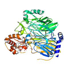
3LAD
DownloadVisualize
BU of 3lad by Molmil
REFINED CRYSTAL STRUCTURE OF LIPOAMIDE DEHYDROGENASE FROM AZOTOBACTER VINELANDII AT 2.2 ANGSTROMS RESOLUTION. A COMPARISON WITH THE STRUCTURE OF GLUTATHIONE REDUCTASE
Descriptor: DIHYDROLIPOAMIDE DEHYDROGENASE, FLAVIN-ADENINE DINUCLEOTIDE
Authors:Mattevi, A, Schierbeek, A.J, Hol, W.G.J.
Deposit date:1991-12-11
Release date:1994-01-31
Last modified:2011-07-13
Method:X-RAY DIFFRACTION (2.2 Å)
Cite:Refined crystal structure of lipoamide dehydrogenase from Azotobacter vinelandii at 2.2 A resolution. A comparison with the structure of glutathione reductase.
J.Mol.Biol., 220, 1991
1GUV
DownloadVisualize
BU of 1guv by Molmil
Structure of human chitotriosidase
Descriptor: 1,2-ETHANEDIOL, CHITOTRIOSIDASE
Authors:Von Moeller, H, Houston, D, Boot, R.G, Aerts, J.M.F.G, Van Aalten, D.M.F.
Deposit date:2002-01-31
Release date:2003-03-30
Last modified:2023-12-13
Method:X-RAY DIFFRACTION (2.35 Å)
Cite:Structure of Human Chitotriosidase - Implications for Specific Inhibitor Design and Function of Mammalian Chitinase-Like Lectins
J.Biol.Chem., 277, 2002
1KB0
DownloadVisualize
BU of 1kb0 by Molmil
Crystal Structure of Quinohemoprotein Alcohol Dehydrogenase from Comamonas testosteroni
Descriptor: CALCIUM ION, GLYCEROL, HEME C, ...
Authors:Rozeboom, H.J, Oubrie, A.
Deposit date:2001-11-05
Release date:2001-12-28
Last modified:2023-08-16
Method:X-RAY DIFFRACTION (1.44 Å)
Cite:Crystal structure of quinohemoprotein alcohol dehydrogenase from Comamonas testosteroni: structural basis for substrate oxidation and electron transfer.
J.Biol.Chem., 277, 2002
1RYY
DownloadVisualize
BU of 1ryy by Molmil
Acetobacter turbidans alpha-amino acid ester hydrolase Y206A mutant
Descriptor: alpha-amino acid ester hydrolase
Authors:Barends, T.R.M.
Deposit date:2003-12-23
Release date:2005-02-22
Last modified:2024-02-14
Method:X-RAY DIFFRACTION (2.8 Å)
Cite:Acetobacter turbidans {alpha}-Amino Acid Ester Hydrolase: HOW A SINGLE MUTATION IMPROVES AN ANTIBIOTIC-PRODUCING ENZYME.
J.Biol.Chem., 281, 2006
1CNV
DownloadVisualize
BU of 1cnv by Molmil
CRYSTAL STRUCTURE OF CONCANAVALIN B AT 1.65 A RESOLUTION
Descriptor: CONCANAVALIN B
Authors:Hennig, M.
Deposit date:1995-02-20
Release date:1996-03-08
Last modified:2011-07-13
Method:X-RAY DIFFRACTION (1.65 Å)
Cite:Crystal structure of concanavalin B at 1.65 A resolution. An "inactivated" chitinase from seeds of Canavalia ensiformis.
J.Mol.Biol., 254, 1995
1PP2
DownloadVisualize
BU of 1pp2 by Molmil
THE REFINED CRYSTAL STRUCTURE OF DIMERIC PHOSPHOLIPASE A2 AT 2.5 ANGSTROMS. ACCESS TO A SHIELDED CATALYTIC CENTER
Descriptor: CALCIUM-FREE PHOSPHOLIPASE A2
Authors:Brunie, S, Sigler, P.B.
Deposit date:1986-03-10
Release date:1986-05-07
Last modified:2011-07-13
Method:X-RAY DIFFRACTION (2.5 Å)
Cite:The refined crystal structure of dimeric phospholipase A2 at 2.5 A. Access to a shielded catalytic center.
J.Biol.Chem., 260, 1985
1B48
DownloadVisualize
BU of 1b48 by Molmil
CRYSTAL STRUCTURE OF MGSTA4-4 IN COMPLEX WITH GSH CONJUGATE OF 4-HYDROXYNONENAL IN ONE SUBUNIT AND GSH IN THE OTHER: EVIDENCE OF SIGNALING ACROSS DIMER INTERFACE IN MGSTA4-4
Descriptor: 4-S-GLUTATHIONYL-5-PENTYL-TETRAHYDRO-FURAN-2-OL, GLUTATHIONE, PROTEIN (GLUTATHIONE S-TRANSFERASE)
Authors:Xiao, B, Zimniak, P, Ji, X.
Deposit date:1999-01-06
Release date:1999-09-29
Last modified:2023-08-30
Method:X-RAY DIFFRACTION (2.6 Å)
Cite:Crystal structure of a murine glutathione S-transferase in complex with a glutathione conjugate of 4-hydroxynon-2-enal in one subunit and glutathione in the other: evidence of signaling across the dimer interface.
Biochemistry, 38, 1999
1K9T
DownloadVisualize
BU of 1k9t by Molmil
Chitinase a complexed with tetra-N-acetylchitotriose
Descriptor: 2-acetamido-2-deoxy-beta-D-glucopyranose-(1-4)-2-acetamido-2-deoxy-beta-D-glucopyranose-(1-4)-2-acetamido-2-deoxy-beta-D-glucopyranose-(1-4)-2-acetamido-2-deoxy-beta-D-glucopyranose, CHITINASE A
Authors:Prag, G, Tucker, P.A, Oppenheim, A.B.
Deposit date:2001-10-30
Release date:2002-11-06
Last modified:2023-10-25
Method:X-RAY DIFFRACTION (1.8 Å)
Cite:Complex Structures of Chitinase A Mutant with Oligonag Provide Insight Into the Enzymatic Mechanism
To be Published
1LPF
DownloadVisualize
BU of 1lpf by Molmil
THREE-DIMENSIONAL STRUCTURE OF LIPOAMIDE DEHYDROGENASE FROM PSEUDOMONAS FLUORESCENS AT 2.8 ANGSTROMS RESOLUTION. ANALYSIS OF REDOX AND THERMOSTABILITY PROPERTIES
Descriptor: DIHYDROLIPOAMIDE DEHYDROGENASE, FLAVIN-ADENINE DINUCLEOTIDE
Authors:Mattevi, A, Hol, W.
Deposit date:1992-10-26
Release date:1994-01-31
Last modified:2011-07-13
Method:X-RAY DIFFRACTION (2.8 Å)
Cite:Three-dimensional structure of lipoamide dehydrogenase from Pseudomonas fluorescens at 2.8 A resolution. Analysis of redox and thermostability properties.
J.Mol.Biol., 230, 1993

219869

數據於2024-05-15公開中

PDB statisticsPDBj update infoContact PDBjnumon