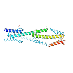
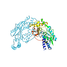
Loading
PDBj
MenuPDBj@FacebookPDBj@TwitterPDBj@YouTubewwPDB FoundationwwPDB
RCSB PDBPDBeBMRBAdv. SearchSearch help
Search by PDB author
3RTY
DownloadVisualize
BU of 3rty by Molmil
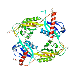
Structure of an Enclosed Dimer Formed by The Drosophila Period Protein
Descriptor: 2,3-DIHYDROXY-1,4-DITHIOBUTANE, Period circadian protein
Authors:King, H.A, Hoelz, A, Crane, B.R, Young, M.W.
Deposit date:2011-05-04
Release date:2011-12-21
Method:X-RAY DIFFRACTION (2.85 Å)
Cite:Structure of an enclosed dimer formed by the Drosophila period protein.
J.Mol.Biol., 413, 2011
3SFT
DownloadVisualize
BU of 3sft by Molmil
Crystal structure of Thermotoga maritima CheB methylesterase catalytic domain
Descriptor: Chemotaxis response regulator protein-glutamate methylesterase
Authors:Park, S.Y, Crane, B.R.
Deposit date:2011-06-14
Release date:2011-07-20
Last modified:2023-11-01
Method:X-RAY DIFFRACTION (2.15 Å)
Cite:An insight into the interaction mode between CheB and chemoreceptor from two crystal structures of CheB methylesterase catalytic domain
Biochem.Biophys.Res.Commun., 411, 2011
4HI4
DownloadVisualize
BU of 4hi4 by Molmil
Crystal structure of the 5-coordinate ferric heme-binding PAS domain of Aer2 from P. aeruginosa
Descriptor: Aerotaxis transducer Aer2, CHLORIDE ION, GLYCEROL, ...
Authors:Airola, M.V, Crane, B.R.
Deposit date:2012-10-11
Release date:2013-01-09
Last modified:2024-02-28
Method:X-RAY DIFFRACTION (2.304 Å)
Cite:Architecture of the soluble receptor Aer2 indicates an in-line mechanism for PAS and HAMP domain signaling.
J.Mol.Biol., 425, 2013
4I44
DownloadVisualize
BU of 4i44 by Molmil
Aer2 poly-HAMP domains: V33G HAMP1 inverted signaling mutant
Descriptor: Aerotaxis transducer Aer2, CHLORIDE ION
Authors:Airola, M.V, Sukomon, N, Crane, B.R.
Deposit date:2012-11-27
Release date:2013-02-27
Last modified:2024-02-28
Method:X-RAY DIFFRACTION (2.88 Å)
Cite:HAMP Domain Conformers That Propagate Opposite Signals in Bacterial Chemoreceptors.
Plos Biol., 11, 2013
4I3M
DownloadVisualize
BU of 4i3m by Molmil
Aer2 poly-HAMP domains: L44H HAMP1 CW-lock mutant
Descriptor: Aerotaxis transducer Aer2, CHLORIDE ION, MAGNESIUM ION, ...
Authors:Airola, M.V, Sukomon, N, Crane, B.R.
Deposit date:2012-11-26
Release date:2013-02-27
Last modified:2024-02-28
Method:X-RAY DIFFRACTION (1.95 Å)
Cite:HAMP Domain Conformers That Propagate Opposite Signals in Bacterial Chemoreceptors.
Plos Biol., 11, 2013
4HZ1
DownloadVisualize
BU of 4hz1 by Molmil
Crystal Structure of Pseudomonas aeruginosa azurin with iron(II) at the copper-binding site.
Descriptor: ACETATE ION, Azurin, FE (II) ION
Authors:McLaughlin, M.P, Retegan, M, Bill, E, Payne, T.M, Shafaat, H.S, Pea, S, Sudhamsu, J, Ensign, A.A, Crane, B.R, Neese, F, Holland, P.L.
Deposit date:2012-11-14
Release date:2012-12-12
Last modified:2023-09-20
Method:X-RAY DIFFRACTION (2.2 Å)
Cite:Azurin as a Protein Scaffold for a Low-coordinate Nonheme Iron Site with a Small-molecule Binding Pocket.
J.Am.Chem.Soc., 134, 2012
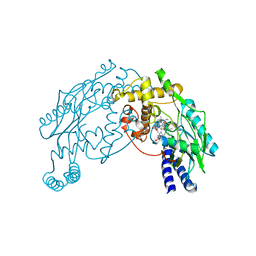
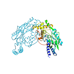
4JPB
DownloadVisualize
BU of 4jpb by Molmil
The structure of a ternary complex between CheA domains P4 and P5 with CheW and with an unzipped fragment of TM14, a chemoreceptor analog from Thermotoga maritima.
Descriptor: Chemotaxis protein CheA, Chemotaxis protein CheW, Methyl-accepting chemotaxis protein
Authors:Li, X, Bayas, C, Bilwes, A.M, Crane, B.R.
Deposit date:2013-03-19
Release date:2013-08-28
Last modified:2024-02-28
Method:X-RAY DIFFRACTION (3.186 Å)
Cite:The 3.2 angstrom resolution structure of a receptor: CheA:CheW signaling complex defines overlapping binding sites and key residue interactions within bacterial chemosensory arrays.
Biochemistry, 52, 2013
1I5B
DownloadVisualize
BU of 1i5b by Molmil
STRUCTURE OF CHEA DOMAIN P4 IN COMPLEX WITH ADPNP AND MANGANESE
Descriptor: ACETATE ION, CHEMOTAXIS PROTEIN CHEA, MANGANESE (II) ION, ...
Authors:Bilwes, A.M, Quezada, C.M, Croal, L.R, Crane, B.R, Simon, M.I.
Deposit date:2001-02-26
Release date:2001-08-26
Last modified:2023-08-09
Method:X-RAY DIFFRACTION (1.94 Å)
Cite:Nucleotide binding by the histidine kinase CheA.
Nat.Struct.Biol., 8, 2001
1I53
DownloadVisualize
BU of 1i53 by Molmil
RE(I)-TRICARBONYL DIIMINE (Q107H)) AZURIN
Descriptor: 4,7-DIMETHYL-[1,10]PHENANTHROLINE, AZURIN, COPPER (II) ION, ...
Authors:Di Bilio, A.J, Crane, B.R, Wehbi, W.A, Kiser, C.N, Abu-Omar, M.M, Carlos, R.M, Richards, J.H, Winkler, J.R, Gray, H.B.
Deposit date:2001-02-24
Release date:2001-10-17
Last modified:2023-08-09
Method:X-RAY DIFFRACTION (1.8 Å)
Cite:Properties of photogenerated tryptophan and tyrosyl radicals in structurally characterized proteins containing rhenium(I) tricarbonyl diimines.
J.Am.Chem.Soc., 123, 2001
1I5C
DownloadVisualize
BU of 1i5c by Molmil
STRUCTURE OF CHEA DOMAIN P4 IN COMPLEX WITH ADP
Descriptor: ADENOSINE-5'-DIPHOSPHATE, CHEMOTAXIS PROTEIN CHEA
Authors:Bilwes, A.M, Quezada, C.M, Croal, L.R, Crane, B.R, Simon, M.I.
Deposit date:2001-02-26
Release date:2001-08-26
Last modified:2023-08-09
Method:X-RAY DIFFRACTION (1.9 Å)
Cite:Nucleotide binding by the histidine kinase CheA.
Nat.Struct.Biol., 8, 2001
1I59
DownloadVisualize
BU of 1i59 by Molmil
STRUCTURE OF THE HISTIDINE KINASE CHEA ATP-BINDING DOMAIN IN COMPLEX WITH ADPNP AND MAGENSIUM
Descriptor: ACETATE ION, ADENOSINE-5'-DIPHOSPHATE, CHEMOTAXIS PROTEIN CHEA, ...
Authors:Bilwes, A.M, Quezada, C.M, Croal, L.R, Crane, B.R, Simon, M.I.
Deposit date:2001-02-26
Release date:2001-08-26
Last modified:2023-08-09
Method:X-RAY DIFFRACTION (1.8 Å)
Cite:Nucleotide binding by the histidine kinase CheA.
Nat.Struct.Biol., 8, 2001
2NOX
DownloadVisualize
BU of 2nox by Molmil
Crystal structure of tryptophan 2,3-dioxygenase from Ralstonia metallidurans
Descriptor: PROTOPORPHYRIN IX CONTAINING FE, Tryptophan 2,3-dioxygenase
Authors:Zhang, Y, Kang, S.A, Mukherjee, T, Bale, S, Crane, B.R, Begley, T.P, Ealick, S.E.
Deposit date:2006-10-26
Release date:2006-12-19
Last modified:2023-08-30
Method:X-RAY DIFFRACTION (2.4 Å)
Cite:Crystal structure and mechanism of tryptophan 2,3-dioxygenase, a heme enzyme involved in tryptophan catabolism and in quinolinate biosynthesis.
Biochemistry, 46, 2007
2PD8
DownloadVisualize
BU of 2pd8 by Molmil
1.8 Angstrom Crystal Structure of the Cys71Ser mutant of Vivid
Descriptor: FLAVIN-ADENINE DINUCLEOTIDE, Vivid PAS protein VVD
Authors:Zoltowski, B.D, Crane, B.R, Bilwes, A.M.
Deposit date:2007-03-31
Release date:2007-06-05
Last modified:2023-08-30
Method:X-RAY DIFFRACTION (1.8 Å)
Cite:Conformational switching in the fungal light sensor Vivid
Science, 316, 2007
2PD7
DownloadVisualize
BU of 2pd7 by Molmil
2.0 Angstrom Crystal Structure of the Fungal Blue-Light Photoreceptor Vivid
Descriptor: FLAVIN-ADENINE DINUCLEOTIDE, Vivid PAS protein VVD
Authors:Zoltowski, B.D, Crane, B.R, Bilwes, A.M.
Deposit date:2007-03-31
Release date:2007-06-05
Last modified:2023-08-30
Method:X-RAY DIFFRACTION (2 Å)
Cite:Conformational switching in the fungal light sensor Vivid
Science, 316, 2007
2PDR
DownloadVisualize
BU of 2pdr by Molmil
1.7 Angstrom Crystal Structure of the Photo-excited Blue-light Photoreceptor Vivid
Descriptor: FLAVIN-ADENINE DINUCLEOTIDE, Vivid PAS protein VVD
Authors:Zoltowski, B.D, Crane, B.R, Bilwes, A.M.
Deposit date:2007-04-01
Release date:2007-06-05
Last modified:2023-08-30
Method:X-RAY DIFFRACTION (1.7 Å)
Cite:Conformational switching in the fungal light sensor Vivid
Science, 316, 2007
2A4M
DownloadVisualize
BU of 2a4m by Molmil
Structure of Trprs II bound to ATP
Descriptor: TRYPTOPHAN, Tryptophanyl-tRNA synthetase II
Authors:Buddha, M.R, Crane, B.R.
Deposit date:2005-06-29
Release date:2005-08-02
Last modified:2023-08-23
Method:X-RAY DIFFRACTION (2.3 Å)
Cite:Structures of Tryptophanyl-tRNA Synthetase II from Deinococcus radiodurans Bound to ATP and Tryptophan: Insight into subunit cooperativity and domain motions linked to catalysis
J.Biol.Chem., 280, 2005
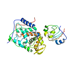
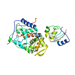
2AN2
DownloadVisualize
BU of 2an2 by Molmil
P332G, A333S Double mutant of the Bacillus subtilis Nitric Oxide Synthase
Descriptor: 5,6,7,8-TETRAHYDROBIOPTERIN, ARGININE, P332G A333S double mutant of Nitric Oxide Synthase from Bacillus subtilis, ...
Authors:Pant, K, Crane, B.R.
Deposit date:2005-08-11
Release date:2006-08-29
Last modified:2024-02-14
Method:X-RAY DIFFRACTION (2.6 Å)
Cite:Structure of a loose dimer: an intermediate in nitric oxide synthase assembly
J.Mol.Biol., 352, 2005
2B12
DownloadVisualize
BU of 2b12 by Molmil
Crystal structure of the protein-protein complex between F82Y cytochrome c and cytochrome c peroxidase
Descriptor: Cytochrome c iso-1, Cytochrome c peroxidase, mitochondrial, ...
Authors:Kang, S.A, Crane, B.R.
Deposit date:2005-09-15
Release date:2005-10-25
Last modified:2021-10-20
Method:X-RAY DIFFRACTION (3.02 Å)
Cite:Effects of interface mutations on association modes and electron-transfer rates between proteins
Proc.Natl.Acad.Sci.Usa, 102, 2005
2AN0
DownloadVisualize
BU of 2an0 by Molmil
Crystal Structure of the P332G mutant of the Bacillus subtilis NOS
Descriptor: Nitric Oxide Synthase, PROTOPORPHYRIN IX CONTAINING FE
Authors:Pant, K, Crane, B.R.
Deposit date:2005-08-10
Release date:2006-08-29
Last modified:2024-02-14
Method:X-RAY DIFFRACTION (2.6 Å)
Cite:Structure of a loose dimer: an intermediate in nitric oxide synthase assembly
J.Mol.Biol., 352, 2005
2B0Z
DownloadVisualize
BU of 2b0z by Molmil
Crystal structure of the protein-protein complex between F82I cytochrome c and cytochrome c peroxidase
Descriptor: Cytochrome c iso-1, Cytochrome c peroxidase, mitochondrial, ...
Authors:Kang, S.A, Crane, B.R.
Deposit date:2005-09-15
Release date:2005-10-25
Last modified:2021-10-20
Method:X-RAY DIFFRACTION (2.7 Å)
Cite:Effects of interface mutations on association modes and electron-transfer rates between proteins
Proc.Natl.Acad.Sci.Usa, 102, 2005
2AMO
DownloadVisualize
BU of 2amo by Molmil
Loose Dimer of a Bacillus subtilis Nitric Oxide Synthase
Descriptor: Nitric oxide synthase oxygenase, PROTOPORPHYRIN IX CONTAINING FE
Authors:Pant, K, Crane, B.R.
Deposit date:2005-08-09
Release date:2006-10-31
Last modified:2023-08-23
Method:X-RAY DIFFRACTION (2.6 Å)
Cite:Structure of a loose dimer: an intermediate in nitric oxide synthase assembly.
J.Mol.Biol., 352, 2005
2B10
DownloadVisualize
BU of 2b10 by Molmil
Crystal Structure of the Protein-Protein Complex between F82S cytochrome c and cytochrome c peroxidase
Descriptor: Cytochrome c iso-1, Cytochrome c peroxidase, mitochondrial, ...
Authors:Kang, S.A, Crane, B.R.
Deposit date:2005-09-15
Release date:2005-10-25
Last modified:2024-02-14
Method:X-RAY DIFFRACTION (2.8 Å)
Cite:Effects of interface mutations on association modes and electron-transfer rates between proteins
Proc.Natl.Acad.Sci.Usa, 102, 2005
2B11
DownloadVisualize
BU of 2b11 by Molmil
Crystal structure of the protein-protein complex between F82W cytochrome c and cytochrome c peroxidase
Descriptor: Cytochrome c iso-1, Cytochrome c peroxidase, mitochondrial, ...
Authors:Kang, S.A, Crane, B.R.
Deposit date:2005-09-15
Release date:2005-10-25
Last modified:2021-10-20
Method:X-RAY DIFFRACTION (2.3 Å)
Cite:Effects of interface mutations on association modes and electron-transfer rates between proteins
Proc.Natl.Acad.Sci.Usa, 102, 2005
2BCN
DownloadVisualize
BU of 2bcn by Molmil
Solvent isotope effects on interfacial protein electron transfer between cytochrome c and cytochrome c peroxidase
Descriptor: Cytochrome c iso-1, HEME C, PROTOPORPHYRIN IX CONTAINING ZN, ...
Authors:Kang, S.A, Crane, B.R.
Deposit date:2005-10-19
Release date:2006-02-28
Last modified:2021-10-20
Method:X-RAY DIFFRACTION (1.7 Å)
Cite:Solvent isotope effects on interfacial protein electron transfer in crystals and electrode films.
J.Am.Chem.Soc., 128, 2006
2CH7
DownloadVisualize
BU of 2ch7 by Molmil
Crystal structure of the cytoplasmic domain of a bacterial chemoreceptor from Thermotoga maritima
Descriptor: LEAD (II) ION, METHYL-ACCEPTING CHEMOTAXIS PROTEIN
Authors:Park, S.Y, Bilwes, A.M, Crane, B.R.
Deposit date:2006-03-13
Release date:2006-04-18
Last modified:2011-07-13
Method:X-RAY DIFFRACTION (2.5 Å)
Cite:Reconstruction of the Chemotaxis Receptor-Kinase Assembly
Nat.Struct.Mol.Biol., 13, 2006

219140

PDB entries from 2024-05-01

PDB statisticsPDBj update infoContact PDBjnumon