Loading
PDBj
MenuPDBj@FacebookPDBj@TwitterPDBj@YouTubewwPDB FoundationwwPDB
RCSB PDBPDBeBMRBAdv. SearchSearch help
Search by PDB author
3HN7
DownloadVisualize
BU of 3hn7 by Molmil
Crystal structure of a murein peptide ligase mpl (psyc_0032) from psychrobacter arcticus 273-4 at 1.65 A resolution
Descriptor: UDP-N-acetylmuramate-L-alanine ligase
Authors:Joint Center for Structural Genomics (JCSG)
Deposit date:2009-05-29
Release date:2009-06-16
Last modified:2023-02-01
Method:X-RAY DIFFRACTION (1.65 Å)
Cite:Structure and function of the first full-length murein Peptide ligase (mpl) cell wall recycling protein.
Plos One, 6, 2011
4H4J
DownloadVisualize
BU of 4h4j by Molmil
Crystal structure of a N-acetylmuramoyl-L-alanine amidase (BACUNI_02947) from Bacteroides uniformis ATCC 8492 at 1.15 A resolution
Descriptor: hypothetical protein
Authors:Joint Center for Structural Genomics (JCSG)
Deposit date:2012-09-17
Release date:2012-10-17
Last modified:2023-02-01
Method:X-RAY DIFFRACTION (1.15 Å)
Cite:Structure-guided functional characterization of DUF1460 reveals a highly specific NlpC/P60 amidase family.
Structure, 22, 2014
1G5P
DownloadVisualize
BU of 1g5p by Molmil
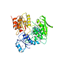
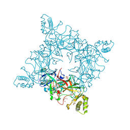
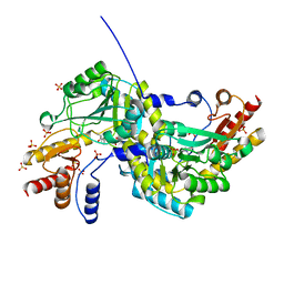
NITROGENASE IRON PROTEIN FROM AZOTOBACTER VINELANDII
Descriptor: IRON/SULFUR CLUSTER, NITROGENASE IRON PROTEIN
Authors:Strop, P, Takahara, P.M, Chiu, H.J, Angove, H.C, Burgess, B.K, Rees, D.C.
Deposit date:2000-11-01
Release date:2001-01-31
Last modified:2023-08-09
Method:X-RAY DIFFRACTION (2.2 Å)
Cite:Crystal structure of the all-ferrous [4Fe-4S]0 form of the nitrogenase iron protein from Azotobacter vinelandii.
Biochemistry, 40, 2001
4JG5
DownloadVisualize
BU of 4jg5 by Molmil
Crystal structure of a putative cell adhesion protein (BDI_3519) from Parabacteroides distasonis ATCC 8503 at 2.34 A resolution (PSI Community Target, Nakayama)
Descriptor: Putative cell adhesion protein
Authors:Joint Center for Structural Genomics (JCSG)
Deposit date:2013-02-28
Release date:2013-03-20
Last modified:2023-02-01
Method:X-RAY DIFFRACTION (2.34 Å)
Cite:A Distinct Type of Pilus from the Human Microbiome.
Cell, 165, 2016
4JRF
DownloadVisualize
BU of 4jrf by Molmil
Crystal structure of a putative cell adhesion protein (BACOVA_01548) from Bacteroides ovatus ATCC 8483 at 1.98 A resolution (PSI Community Target, Nakayama)
Descriptor: CALCIUM ION, CHLORIDE ION, SULFATE ION, ...
Authors:Joint Center for Structural Genomics (JCSG)
Deposit date:2013-03-21
Release date:2013-04-03
Last modified:2023-02-01
Method:X-RAY DIFFRACTION (1.98 Å)
Cite:A Distinct Type of Pilus from the Human Microbiome.
Cell, 165, 2016
4K4K
DownloadVisualize
BU of 4k4k by Molmil
Crystal structure of a putative cell adhesion protein (BACUNI_00621) from Bacteroides uniformis ATCC 8492 at 1.67 A resolution
Descriptor: CHLORIDE ION, Putative cell adhesion protein
Authors:Joint Center for Structural Genomics (JCSG)
Deposit date:2013-04-12
Release date:2013-05-01
Last modified:2023-02-01
Method:X-RAY DIFFRACTION (1.67 Å)
Cite:A Distinct Type of Pilus from the Human Microbiome.
Cell, 165, 2016
1O4U
DownloadVisualize
BU of 1o4u by Molmil
Crystal structure of a nicotinate nucleotide pyrophosphorylase (tm1645) from thermotoga maritima at 2.50 A resolution
Descriptor: Type II quinolic acid phosphoribosyltransferase
Authors:Joint Center for Structural Genomics (JCSG)
Deposit date:2003-07-09
Release date:2003-07-22
Last modified:2023-09-20
Method:X-RAY DIFFRACTION (2.5 Å)
Cite:Crystal structure of a type II quinolic acid phosphoribosyltransferase (TM1645) from Thermotoga maritima at 2.50 A resolution
Proteins, 55, 2004
1O4W
DownloadVisualize
BU of 1o4w by Molmil
CRYSTAL STRUCTURE OF a PIN (PILT N-TERMINUS) DOMAIN CONTAINING PROTEIN (AF0591) FROM ARCHAEOGLOBUS FULGIDUS AT 1.90 A RESOLUTION
Descriptor: PIN (PilT N-terminus) domain
Authors:Joint Center for Structural Genomics (JCSG)
Deposit date:2003-07-16
Release date:2003-07-29
Last modified:2023-01-25
Method:X-RAY DIFFRACTION (1.9 Å)
Cite:Crystal structure of a PIN (PilT N-terminus) domain (AF0591) from Archaeoglobus fulgidus at 1.90 A resolution
Proteins, 56, 2004
1O50
DownloadVisualize
BU of 1o50 by Molmil
Crystal structure of a cbs domain-containing protein (tm0935) from thermotoga maritima at 1.87 A resolution
Descriptor: CBS domain-containing predicted protein TM0935
Authors:Joint Center for Structural Genomics (JCSG)
Deposit date:2003-07-31
Release date:2003-08-12
Last modified:2023-01-25
Method:X-RAY DIFFRACTION (1.87 Å)
Cite:Crystal structure of a tandem cystathionine-beta-synthase (CBS) domain protein (TM0935) from Thermotoga maritima at 1.87 A resolution
Proteins, 57, 2004
1O5H
DownloadVisualize
BU of 1o5h by Molmil
Crystal structure of formiminotetrahydrofolate cyclodeaminase (TM1560) from Thermotoga maritima at 2.80 A resolution
Descriptor: Formiminotetrahydrofolate cyclodeaminase
Authors:Joint Center for Structural Genomics (JCSG)
Deposit date:2003-09-17
Release date:2003-09-30
Last modified:2023-01-25
Method:X-RAY DIFFRACTION (2.8 Å)
Cite:Crystal structure of a formiminotetrahydrofolate cyclodeaminase (TM1560) from Thermotoga maritima at 2.80 A resolution reveals a new fold
Proteins, 58, 2005
1O4V
DownloadVisualize
BU of 1o4v by Molmil
Crystal structure of the catalytic subunit of a phosphoribosylaminoimidazole mutase (tm0446) from thermotoga maritima at 1.77 A resolution
Descriptor: SULFATE ION, phosphoribosylaminoimidazole mutase PurE
Authors:Joint Center for Structural Genomics (JCSG)
Deposit date:2003-07-11
Release date:2003-07-22
Last modified:2023-09-20
Method:X-RAY DIFFRACTION (1.77 Å)
Cite:Crystal structure of a phosphoribosylaminoimidazole mutase PurE (TM0446) from Thermotoga maritima at 1.77 A resolution
Proteins, 55, 2004
1O4S
DownloadVisualize
BU of 1o4s by Molmil
Crystal structure of Aspartate aminotransferase (TM1255) from Thermotoga maritima at 1.90 A resolution
Descriptor: Aspartate aminotransferase, PYRIDOXAL-5'-PHOSPHATE, SULFATE ION
Authors:Joint Center for Structural Genomics (JCSG)
Deposit date:2003-06-26
Release date:2003-07-08
Last modified:2023-09-20
Method:X-RAY DIFFRACTION (1.9 Å)
Cite:Crystal structure of an aspartate aminotransferase (TM1255) from Thermotoga maritima at 1.90 A resolution
Proteins, 55, 2004
1O4T
DownloadVisualize
BU of 1o4t by Molmil
Crystal structure of a predicted oxalate decarboxylase (tm1287) from thermotoga maritima at 1.95 A resolution
Descriptor: MANGANESE (II) ION, OXALATE ION, putative oxalate decarboxylase
Authors:Joint Center for Structural Genomics (JCSG)
Deposit date:2003-07-03
Release date:2003-07-15
Last modified:2023-09-20
Method:X-RAY DIFFRACTION (1.95 Å)
Cite:Crystal structure of a putative oxalate decarboxylase (TM1287) from Thermotoga maritima at 1.95 A resolution
Proteins, 56, 2004
1O51
DownloadVisualize
BU of 1o51 by Molmil
Crystal structure of a putative PII-like signaling protein (TM0021) from Thermotoga maritima at 2.50 A resolution
Descriptor: ADENOSINE-5'-DIPHOSPHATE, Hypothetical protein TM0021, SULFATE ION
Authors:Joint Center for Structural Genomics (JCSG)
Deposit date:2003-08-01
Release date:2003-08-19
Last modified:2023-01-25
Method:X-RAY DIFFRACTION (2.5 Å)
Cite:Crystal structure of a putative PII-like signaling protein (TM0021) from Thermotoga maritima at 2.5 A resolution
Proteins, 54, 2004
1O59
DownloadVisualize
BU of 1o59 by Molmil
Crystal structure of Allantoicase (yir029w) from Saccharomyces cerevisiae at 2.40 A resolution
Descriptor: Allantoicase
Authors:Joint Center for Structural Genomics (JCSG)
Deposit date:2003-08-22
Release date:2003-09-02
Last modified:2023-01-25
Method:X-RAY DIFFRACTION (2.4 Å)
Cite:Crystal structure of an allantoicase (YIR029W) from Saccharomyces cerevisiae at 2.4 A resolution
Proteins, 56, 2004
2ETS
DownloadVisualize
BU of 2ets by Molmil
CRYSTAL STRUCTURE OF A BACTERIAL DOMAIN OF UNKNOWN FUNCTION FROM DUF1798 FAMILY (MW1337) FROM STAPHYLOCOCCUS AUREUS SUBSP. AUREUS AT 2.25 A RESOLUTION
Descriptor: CHLORIDE ION, PHOSPHATE ION, hypothetical protein
Authors:Joint Center for Structural Genomics (JCSG)
Deposit date:2005-10-27
Release date:2005-11-08
Last modified:2023-01-25
Method:X-RAY DIFFRACTION (2.25 Å)
Cite:Crystal structures of MW1337R and lin2004: representatives of a novel protein family that adopt a four-helical bundle fold.
Proteins, 71, 2008
2F46
DownloadVisualize
BU of 2f46 by Molmil
Crystal structure of a putative phosphatase (nma1982) from neisseria meningitidis z2491 at 1.41 A resolution
Descriptor: CHLORIDE ION, UNKNOWN LIGAND, hypothetical protein
Authors:Joint Center for Structural Genomics (JCSG)
Deposit date:2005-11-22
Release date:2006-02-07
Last modified:2023-01-25
Method:X-RAY DIFFRACTION (1.41 Å)
Cite:Crystal structure of NMA1982 from Neisseria meningitidis at 1.5 A resolution provides a structural scaffold for nonclassical, eukaryotic-like phosphatases.
Proteins, 69, 2007
2FG0
DownloadVisualize
BU of 2fg0 by Molmil
Crystal structure of a putative gamma-d-glutamyl-l-diamino acid endopeptidase (npun_r0659) from nostoc punctiforme pcc 73102 at 1.79 A resolution
Descriptor: COG0791: Cell wall-associated hydrolases (invasion-associated proteins), GLYCEROL
Authors:Joint Center for Structural Genomics (JCSG)
Deposit date:2005-12-20
Release date:2006-01-10
Last modified:2023-01-25
Method:X-RAY DIFFRACTION (1.79 Å)
Cite:Structural Basis of Murein Peptide Specificity of a gamma-D-Glutamyl-L-Diamino Acid Endopeptidase.
Structure, 17, 2009
2EVR
DownloadVisualize
BU of 2evr by Molmil
CRYSTAL STRUCTURE OF A PUTATIVE GAMMA-D-GLUTAMYL-L-DIAMINO ACID ENDOPEPTIDASE (NPUN_R0659) FROM NOSTOC PUNCTIFORME PCC 73102 AT 1.60 A RESOLUTION
Descriptor: 1,2-ETHANEDIOL, CHLORIDE ION, COG0791: Cell wall-associated hydrolases (invasion-associated proteins), ...
Authors:Joint Center for Structural Genomics (JCSG)
Deposit date:2005-10-31
Release date:2005-11-22
Last modified:2023-01-25
Method:X-RAY DIFFRACTION (1.6 Å)
Cite:Structural Basis of Murein Peptide Specificity of a gamma-D-Glutamyl-L-Diamino Acid Endopeptidase.
Structure, 17, 2009
2FEA
DownloadVisualize
BU of 2fea by Molmil
Crystal structure of MtnX phosphatase from Bacillus Subtilis at 2.00 A resolution
Descriptor: 1,2-ETHANEDIOL, 2-hydroxy-3-keto-5-methylthiopentenyl-1-phosphate phosphatase, MAGNESIUM ION, ...
Authors:Joint Center for Structural Genomics (JCSG)
Deposit date:2005-12-15
Release date:2005-12-27
Last modified:2023-01-25
Method:X-RAY DIFFRACTION (2 Å)
Cite:Crystal structure of MtnX phosphatase from Bacillus subtilis at 2.0 A resolution provides a structural basis for bipartite phosphomonoester hydrolysis of 2-hydroxy-3-keto-5-methylthiopentenyl-1-phosphate.
Proteins, 69, 2007
1J6U
DownloadVisualize
BU of 1j6u by Molmil
Crystal structure of UDP-N-acetylmuramate-alanine ligase MurC (TM0231) from Thermotoga maritima at 2.3 A resolution
Descriptor: UDP-N-acetylmuramate-alanine ligase MurC
Authors:Joint Center for Structural Genomics (JCSG)
Deposit date:2002-08-29
Release date:2002-11-06
Last modified:2023-01-25
Method:X-RAY DIFFRACTION (2.3 Å)
Cite:Crystal structure of an Udp-n-acetylmuramate-alanine ligase MurC (TM0231) from Thermotoga maritima at 2.3 A resolution.
Proteins, 55, 2004
2FNA
DownloadVisualize
BU of 2fna by Molmil
Crystal structure of an archaeal aaa+ atpase (sso1545) from sulfolobus solfataricus p2 at 2.00 A resolution
Descriptor: 1,2-ETHANEDIOL, ADENOSINE-5'-DIPHOSPHATE, Conserved hypothetical protein, ...
Authors:Joint Center for Structural Genomics (JCSG)
Deposit date:2006-01-10
Release date:2006-02-07
Last modified:2023-01-25
Method:X-RAY DIFFRACTION (2 Å)
Cite:Crystal structure of a novel archaeal AAA+ ATPase SSO1545 from Sulfolobus solfataricus.
Proteins, 74, 2009
2G36
DownloadVisualize
BU of 2g36 by Molmil
Crystal structure of Tryptophanyl-tRNA synthetase (EC 6.1.1.2) (Tryptophan-tRNA ligase)(TrpRS) (tm0492) from THERMOTOGA MARITIMA at 2.50 A resolution
Descriptor: IRON/SULFUR CLUSTER, TRYPTOPHAN, Tryptophanyl-tRNA synthetase
Authors:Joint Center for Structural Genomics (JCSG)
Deposit date:2006-02-17
Release date:2006-03-28
Last modified:2024-04-03
Method:X-RAY DIFFRACTION (2.5 Å)
Cite:Structure of a tryptophanyl-tRNA synthetase containing an iron-sulfur cluster.
Acta Crystallogr.,Sect.F, 66, 2010
2FNO
DownloadVisualize
BU of 2fno by Molmil
Crystal structure of a glutathione s-transferase (atu5508) from agrobacterium tumefaciens str. c58 at 2.00 A resolution
Descriptor: AGR_pAT_752p, THIOCYANATE ION
Authors:Joint Center for Structural Genomics (JCSG)
Deposit date:2006-01-11
Release date:2006-02-14
Last modified:2023-01-25
Method:X-RAY DIFFRACTION (2 Å)
Cite:Comparative structural analysis of a novel glutathioneS-transferase (ATU5508) from Agrobacterium tumefaciens at 2.0 A resolution.
Proteins, 65, 2006
2GHR
DownloadVisualize
BU of 2ghr by Molmil
Crystal structure of homoserine o-succinyltransferase (NP_981826.1) from Bacillus cereus ATCC 10987 at 2.40 A resolution
Descriptor: Homoserine O-succinyltransferase, SULFATE ION
Authors:Joint Center for Structural Genomics (JCSG)
Deposit date:2006-03-27
Release date:2006-04-11
Last modified:2023-01-25
Method:X-RAY DIFFRACTION (2.4 Å)
Cite:Crystal structure of homoserine O-succinyltransferase from Bacillus cereus at 2.4 A resolution
Proteins, 68, 2007

219869

数据于2024-05-15公开中

PDB statisticsPDBj update infoContact PDBjnumon