Loading
PDBj
MenuPDBj@FacebookPDBj@TwitterPDBj@YouTubewwPDB FoundationwwPDB
RCSB PDBPDBeBMRBAdv. SearchSearch help
Search by PDB author
1VR0
DownloadVisualize
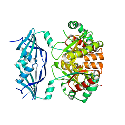
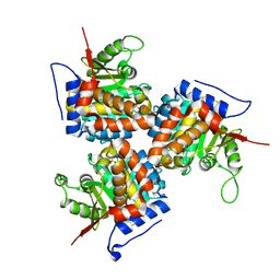
BU of 1vr0 by Molmil
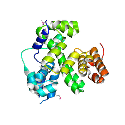
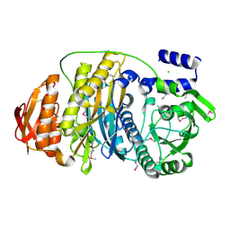
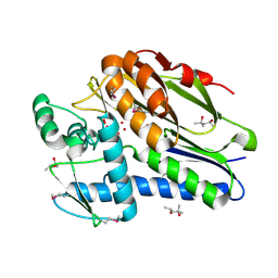
Crystal structure of putative 2-phosphosulfolactate phosphatase (15026306) from Clostridium acetobutylicum at 2.6 A resolution
Descriptor: (2R)-3-SULFOLACTIC ACID, MAGNESIUM ION, Probable 2-phosphosulfolactate phosphatase
Authors:Joint Center for Structural Genomics (JCSG)
Deposit date:2005-01-26
Release date:2005-02-15
Last modified:2023-01-25
Method:X-RAY DIFFRACTION (2.49 Å)
Cite:Crystal structure of 2-phosphosulfolactate phosphatase (ComB) from Clostridium acetobutylicum at 2.6 A resolution reveals a new fold with a novel active site.
Proteins, 65, 2006
1VK9
DownloadVisualize
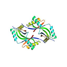
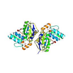
BU of 1vk9 by Molmil
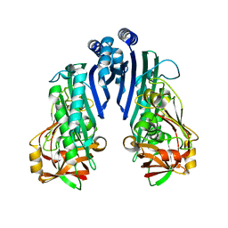
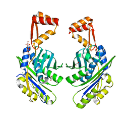
CRYSTAL STRUCTURE OF A DUF1893 family protein (TM1506) FROM THERMOTOGA MARITIMA AT 2.70 A RESOLUTION
Descriptor: UNKNOWN LIGAND, ZINC ION, conserved hypothetical protein TM1506
Authors:Joint Center for Structural Genomics (JCSG)
Deposit date:2004-05-06
Release date:2004-05-18
Last modified:2023-01-25
Method:X-RAY DIFFRACTION (2.7 Å)
Cite:Crystal structure of an ADP-ribosylated protein with a cytidine deaminase-like fold, but unknown function (TM1506), from Thermotoga maritima at 2.70 A resolution.
Proteins, 71, 2008
1VLR
DownloadVisualize
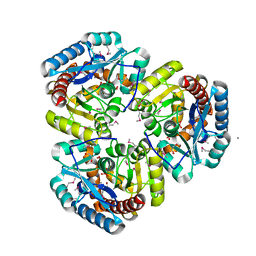
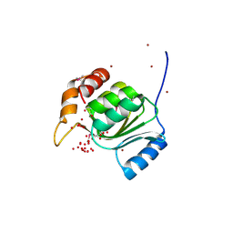
BU of 1vlr by Molmil
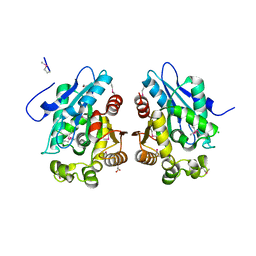
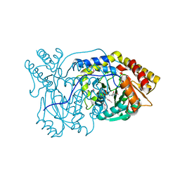
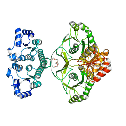
Crystal structure of mRNA decapping enzyme (DcpS) from Mus musculus at 1.83 A resolution
Descriptor: 1,2-ETHANEDIOL, mRNA decapping enzyme
Authors:Joint Center for Structural Genomics (JCSG)
Deposit date:2004-08-10
Release date:2004-08-24
Last modified:2023-09-20
Method:X-RAY DIFFRACTION (1.83 Å)
Cite:Crystal structure of an Apo mRNA decapping enzyme (DcpS) from Mouse at 1.83 A resolution.
Proteins, 60, 2005
1VQ3
DownloadVisualize
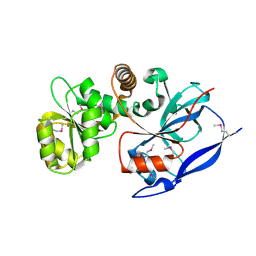
BU of 1vq3 by Molmil
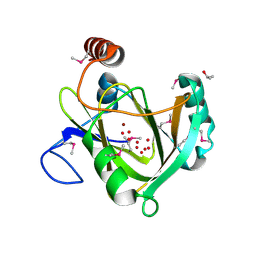
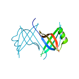
Crystal structure of Phosphoribosylformylglycinamidine synthase, purS subunit (EC 6.3.5.3) (TM1244) from Thermotoga maritima at 1.90 A resolution
Descriptor: Phosphoribosylformylglycinamidine synthase, purS subunit
Authors:Joint Center for Structural Genomics (JCSG)
Deposit date:2004-12-15
Release date:2004-12-28
Last modified:2023-09-20
Method:X-RAY DIFFRACTION (1.9 Å)
Cite:Crystal structure of phosphoribosylformyl-glycinamidine synthase II, PurS subunit (TM1244) from Thermotoga maritima at 1.90 A resolution.
Proteins, 65, 2006
1VKM
DownloadVisualize
BU of 1vkm by Molmil
Crystal structure of an indigoidine synthase a (idga)-like protein (tm1464) from thermotoga maritima msb8 at 1.90 A resolution
Descriptor: 1,2-ETHANEDIOL, MANGANESE (II) ION, UNKNOWN LIGAND, ...
Authors:Joint Center for Structural Genomics (JCSG)
Deposit date:2004-06-09
Release date:2004-06-29
Last modified:2023-01-25
Method:X-RAY DIFFRACTION (1.9 Å)
Cite:Crystal structure of an indigoidine synthase A (IndA)-like protein (TM1464) from Thermotoga maritima at 1.90 A resolution reveals a new fold.
Proteins, 59, 2005
1VJ1
DownloadVisualize
BU of 1vj1 by Molmil
Crystal structure of putative NADPH-dependent oxidoreductase from Mus musculus at 2.10 A resolution
Descriptor: putative NADPH-dependent oxidoreductase
Authors:Joint Center for Structural Genomics (JCSG)
Deposit date:2003-12-03
Release date:2003-12-09
Last modified:2023-01-25
Method:X-RAY DIFFRACTION (2.1 Å)
Cite:Crystal structure of a putative NADPH-dependent oxidoreductase (GI: 18204011) from mouse at 2.10 A resolution
Proteins, 56, 2004
1VQR
DownloadVisualize
BU of 1vqr by Molmil
Crystal structure of a virulence factor (cj0248) from campylobacter jejuni subsp. jejuni at 2.25 A resolution
Descriptor: hypothetical protein Cj0248
Authors:Joint Center for Structural Genomics (JCSG)
Deposit date:2004-12-17
Release date:2004-12-28
Last modified:2023-01-25
Method:X-RAY DIFFRACTION (2.25 Å)
Cite:Crystal structure of virulence factor CJ0248 from Campylobacter jejuni at 2.25 A resolution reveals a new fold.
Proteins, 62, 2006
1VR8
DownloadVisualize
BU of 1vr8 by Molmil
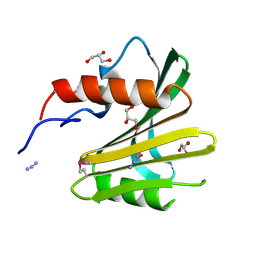
Crystal structure of GTP binding regulator (TM1622) from Thermotoga Maritima at 1.75 A resolution
Descriptor: AZIDE ION, GLYCEROL, GTP binding regulator
Authors:Joint Center for Structural Genomics (JCSG)
Deposit date:2005-02-16
Release date:2005-03-15
Last modified:2023-01-25
Method:X-RAY DIFFRACTION (1.75 Å)
Cite:Crystal structure of an ORFan protein (TM1622) from Thermotoga maritima at 1.75 A resolution reveals a fold similar to the Ran-binding protein Mog1p.
Proteins, 65, 2006
1VPZ
DownloadVisualize
BU of 1vpz by Molmil
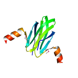
Crystal structure of a putative carbon storage regulator protein (csra, pa0905) from pseudomonas aeruginosa at 2.05 A resolution
Descriptor: Carbon storage regulator homolog
Authors:Joint Center for Structural Genomics (JCSG)
Deposit date:2004-11-24
Release date:2004-12-14
Last modified:2023-01-25
Method:X-RAY DIFFRACTION (2.05 Å)
Cite:Crystal structure of the global regulatory protein CsrA from Pseudomonas putida at 2.05 A resolution reveals a new fold.
Proteins, 61, 2005
1VKH
DownloadVisualize
BU of 1vkh by Molmil
CRYSTAL STRUCTURE OF A PUTATIVE SERINE HYDROLASE (YDR428C) FROM SACCHAROMYCES CEREVISIAE AT 1.85 A RESOLUTION
Descriptor: CHLORIDE ION, GLYCEROL, putative serine hydrolase
Authors:Joint Center for Structural Genomics (JCSG)
Deposit date:2004-05-20
Release date:2004-06-08
Last modified:2023-01-25
Method:X-RAY DIFFRACTION (1.85 Å)
Cite:Crystal structure of an alpha/beta serine hydrolase (YDR428C) from Saccharomyces cerevisiae at 1.85 A resolution
Proteins, 58, 2005
1VK3
DownloadVisualize
BU of 1vk3 by Molmil
Crystal structure of Phosphoribosylformylglycinamidine synthase II (TM1246) from Thermotoga maritima at 2.15 A resolution
Descriptor: CHLORIDE ION, Phosphoribosylformylglycinamidine synthase II
Authors:Joint Center for Structural Genomics (JCSG)
Deposit date:2004-04-23
Release date:2004-05-11
Last modified:2023-01-25
Method:X-RAY DIFFRACTION (2.15 Å)
Cite:Crystal structure of phosphoribosylformylglycinamidine synthase II (smPurL) from Thermotoga maritima at 2.15 A resolution.
Proteins, 63, 2006
1VL4
DownloadVisualize
BU of 1vl4 by Molmil
CRYSTAL STRUCTURE OF A PUTATIVE MODULATOR OF A DNA GYRASE (TM0727) FROM THERMOTOGA MARITIMA MSB8 AT 1.95 A RESOLUTION
Descriptor: pmbA-related protein
Authors:Joint Center for Structural Genomics (JCSG)
Deposit date:2004-07-09
Release date:2004-09-21
Last modified:2023-01-25
Method:X-RAY DIFFRACTION (1.95 Å)
Cite:Crystal structure of a putative modulator of DNA gyrase (pmbA) from Thermotoga maritima at 1.95 A resolution reveals a new fold.
Proteins, 61, 2005
1VJO
DownloadVisualize
BU of 1vjo by Molmil
Crystal structure of Alanine--glyoxylate aminotransferase (ALR1004) from Nostoc sp. at 1.70 A resolution
Descriptor: PYRIDOXAL-5'-PHOSPHATE, alanine--glyoxylate aminotransferase
Authors:Joint Center for Structural Genomics (JCSG)
Deposit date:2004-03-16
Release date:2004-03-23
Last modified:2023-01-25
Method:X-RAY DIFFRACTION (1.7 Å)
Cite:Crystal structure of an alanine-glyoxylate aminotransferase from Anabaena sp. at 1.70 A resolution reveals a noncovalently linked PLP cofactor
Proteins, 58, 2005
1VR3
DownloadVisualize
BU of 1vr3 by Molmil
Crystal structure of Acireductone dioxygenase (13543033) from Mus musculus at 2.06 A resolution
Descriptor: Acireductone dioxygenase, ISOPROPYL ALCOHOL, NICKEL (II) ION, ...
Authors:Joint Center for Structural Genomics (JCSG)
Deposit date:2005-01-28
Release date:2005-02-15
Last modified:2023-01-25
Method:X-RAY DIFFRACTION (2.06 Å)
Cite:Crystal structure of acireductone dioxygenase (ARD) from Mus musculus at 2.06 angstrom resolution.
Proteins, 64, 2006
1VRM
DownloadVisualize
BU of 1vrm by Molmil
Crystal structure of the apbe protein (tm1553) from thermotoga maritima msb8 at 1.58 A resolution
Descriptor: (4R)-2-METHYLPENTANE-2,4-DIOL, (4S)-2-METHYL-2,4-PENTANEDIOL, UNKNOWN LIGAND, ...
Authors:Joint Center for Structural Genomics (JCSG)
Deposit date:2005-03-10
Release date:2005-03-22
Last modified:2023-01-25
Method:X-RAY DIFFRACTION (1.58 Å)
Cite:Crystal structure of the ApbE protein (TM1553) from Thermotoga maritima at 1.58 A resolution.
Proteins, 64, 2006
1VQ0
DownloadVisualize
BU of 1vq0 by Molmil
Crystal structure of 33 kDa chaperonin (Heat shock protein 33 homolog) (HSP33) (TM1394) from Thermotoga maritima at 2.20 A resolution
Descriptor: 1,2-ETHANEDIOL, 33 kDa chaperonin, CHLORIDE ION, ...
Authors:Joint Center for Structural Genomics (JCSG)
Deposit date:2004-11-30
Release date:2004-12-14
Last modified:2023-09-20
Method:X-RAY DIFFRACTION (2.2 Å)
Cite:Crystal structure of Hsp33 chaperone (TM1394) from Thermotoga maritima at 2.20 A resolution.
Proteins, 61, 2005
1ZCZ
DownloadVisualize
BU of 1zcz by Molmil
Crystal structure of Phosphoribosylaminoimidazolecarboxamide formyltransferase / IMP cyclohydrolase (TM1249) from THERMOTOGA MARITIMA at 1.88 A resolution
Descriptor: Bifunctional purine biosynthesis protein purH, POTASSIUM ION, TETRAETHYLENE GLYCOL
Authors:Joint Center for Structural Genomics (JCSG)
Deposit date:2005-04-13
Release date:2005-04-26
Last modified:2023-09-20
Method:X-RAY DIFFRACTION (1.88 Å)
Cite:Crystal structure of AICAR transformylase IMP cyclohydrolase (TM1249) from Thermotoga maritima at 1.88 A resolution.
Proteins, 71, 2008
1Z9F
DownloadVisualize
BU of 1z9f by Molmil
Crystal structure of single stranded DNA-binding protein (TM0604) from Thermotoga maritima at 2.60 A resolution
Descriptor: Single-strand binding protein
Authors:Joint Center for Structural Genomics (JCSG)
Deposit date:2005-04-01
Release date:2005-04-12
Last modified:2023-09-20
Method:X-RAY DIFFRACTION (2.3 Å)
Cite:Crystal structure of a single-stranded DNA-binding protein (TM0604) from Thermotoga maritima at 2.60 A resolution.
Proteins, 63, 2006
1ZKG
DownloadVisualize
BU of 1zkg by Molmil
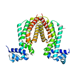
Crystal structure of Transcriptional regulator, TETR family (tm1030) from Thermotoga maritima at 2.30 A resolution
Descriptor: UNKNOWN LIGAND, transcriptional regulator, TetR family
Authors:Joint Center for Structural Genomics (JCSG)
Deposit date:2005-05-02
Release date:2005-07-26
Last modified:2023-01-25
Method:X-RAY DIFFRACTION (2.3 Å)
Cite:Crystal structure of TM1030 from Thermotoga maritima at 2.3 A resolution reveals molecular details of its transcription repressor function.
Proteins, 68, 2007
1ZX8
DownloadVisualize
BU of 1zx8 by Molmil
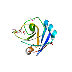
CRYSTAL STRUCTURE OF an atypical cyclophilin (peptidylprolyl cis-trans isomerase) (TM1367) FROM THERMOTOGA MARITIMA AT 1.90 A RESOLUTION
Descriptor: NICKEL (II) ION, PENTAETHYLENE GLYCOL, hypothetical protein TM1367
Authors:Joint Center for Structural Genomics (JCSG)
Deposit date:2005-06-07
Release date:2005-07-26
Last modified:2023-01-25
Method:X-RAY DIFFRACTION (1.9 Å)
Cite:Crystal structure of TM1367 from Thermotoga maritima at 1.90 A resolution reveals an atypical member of the cyclophilin (peptidylprolyl isomerase) fold.
Proteins, 63, 2006
7SUT
DownloadVisualize
BU of 7sut by Molmil
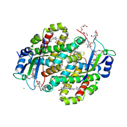
Light harvesting phycobiliprotein HaPE645 from the cryptophyte Hemiselmis andersenii CCMP644
Descriptor: (15,16)-DIHYDROBILIVERDIN (SINGLY LINKED), 2-[BIS-(2-HYDROXY-ETHYL)-AMINO]-2-HYDROXYMETHYL-PROPANE-1,3-DIOL, CHLORIDE ION, ...
Authors:Rathbone, H.W, Michie, K.A, Laos, A.L, Curmi, P.M.G.
Deposit date:2021-11-18
Release date:2023-10-25
Last modified:2023-11-29
Method:X-RAY DIFFRACTION (1.49 Å)
Cite:Molecular dissection of the soluble photosynthetic antenna from the cryptophyte alga Hemiselmis andersenii.
Commun Biol, 6, 2023
1ESQ
DownloadVisualize
BU of 1esq by Molmil
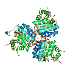
CRYSTAL STRUCTURE OF THIAZOLE KINASE MUTANT (C198S) WITH ATP AND THIAZOLE PHOSPHATE.
Descriptor: 4-METHYL-5-HYDROXYETHYLTHIAZOLE PHOSPHATE, ADENOSINE-5'-TRIPHOSPHATE, HYDROXYETHYLTHIAZOLE KINASE, ...
Authors:Campobasso, N, Mathews, I.I, Begley, T.P, Ealick, S.E.
Deposit date:2000-04-10
Release date:2000-08-09
Last modified:2024-02-07
Method:X-RAY DIFFRACTION (2.5 Å)
Cite:Crystal structure of 4-methyl-5-beta-hydroxyethylthiazole kinase from Bacillus subtilis at 1.5 A resolution.
Biochemistry, 39, 2000
1ESJ
DownloadVisualize
BU of 1esj by Molmil
CRYSTAL STRUCTURE OF THIAZOLE KINASE MUTANT (C198S)
Descriptor: HYDROXYETHYLTHIAZOLE KINASE, SULFATE ION
Authors:Campobasso, N, Mathews, I.I, Begley, T.P, Ealick, S.E.
Deposit date:2000-04-10
Release date:2000-08-09
Last modified:2024-02-07
Method:X-RAY DIFFRACTION (1.8 Å)
Cite:Crystal structure of 4-methyl-5-beta-hydroxyethylthiazole kinase from Bacillus subtilis at 1.5 A resolution.
Biochemistry, 39, 2000

219140

PDB entries from 2024-05-01

PDB statisticsPDBj update infoContact PDBjnumon