Loading
PDBj
MenuPDBj@FacebookPDBj@TwitterPDBj@YouTubewwPDB FoundationwwPDB
RCSB PDBPDBeBMRBAdv. SearchSearch help
Search by PDB author
3NL9
DownloadVisualize
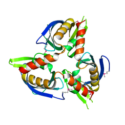
BU of 3nl9 by Molmil
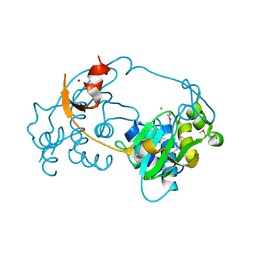
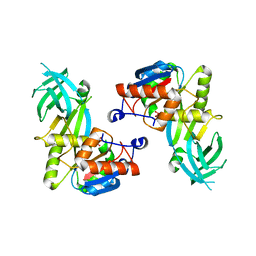
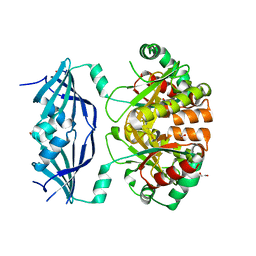
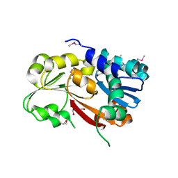
Crystal structure of a putative NTP pyrophosphohydrolase (Exig_1061) from EXIGUOBACTERIUM SP. 255-15 at 1.78 A resolution
Descriptor: 1,2-ETHANEDIOL, putative NTP pyrophosphohydrolase
Authors:Joint Center for Structural Genomics (JCSG)
Deposit date:2010-06-21
Release date:2010-07-21
Last modified:2023-02-01
Method:X-RAY DIFFRACTION (1.78 Å)
Cite:Structure of a putative NTP pyrophosphohydrolase: YP_001813558.1 from Exiguobacterium sibiricum 255-15.
Acta Crystallogr.,Sect.F, 66, 2010
3CM1
DownloadVisualize
BU of 3cm1 by Molmil
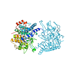
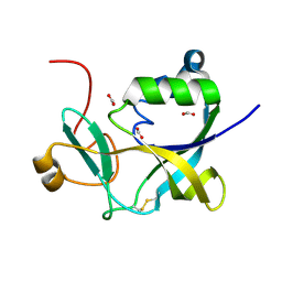
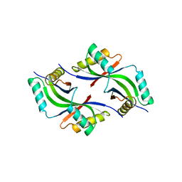
Crystal structure of SsgA-like sporulation-specific cell division protein (YP_290167.1) from Thermobifida fusca YX-ER1 at 2.60 A resolution
Descriptor: SsgA-like sporulation-specific cell division protein
Authors:Joint Center for Structural Genomics (JCSG), Chruszcz, M, Minor, W, Wang, S.
Deposit date:2008-03-20
Release date:2008-04-01
Last modified:2023-02-01
Method:X-RAY DIFFRACTION (2.6 Å)
Cite:Structural and functional characterizations of SsgB, a conserved activator of developmental cell division in morphologically complex actinomycetes.
J.Biol.Chem., 284, 2009
3D00
DownloadVisualize
BU of 3d00 by Molmil
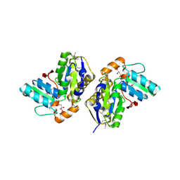
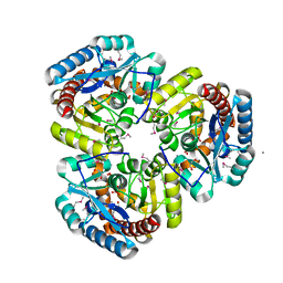
Crystal structure of a tungsten formylmethanofuran dehydrogenase subunit e (fmde)-like protein (syn_00638) from syntrophus aciditrophicus at 1.90 A resolution
Descriptor: CHLORIDE ION, Tungsten formylmethanofuran dehydrogenase subunit E, ZINC ION
Authors:Joint Center for Structural Genomics (JCSG)
Deposit date:2008-04-30
Release date:2008-05-20
Last modified:2023-02-01
Method:X-RAY DIFFRACTION (1.9 Å)
Cite:Structures of three members of Pfam PF02663 (FmdE) implicated in microbial methanogenesis reveal a conserved alpha+beta core domain and an auxiliary C-terminal treble-clef zinc finger.
Acta Crystallogr.,Sect.F, 66, 2010
3CGH
DownloadVisualize
BU of 3cgh by Molmil
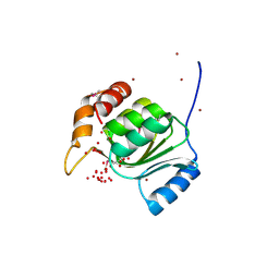
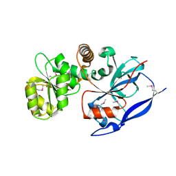
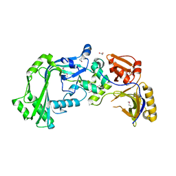
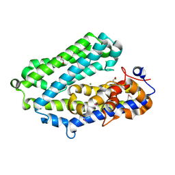
Crystal structure of a susd homolog (bt_3984) from bacteroides thetaiotaomicron vpi-5482 at 1.70 A resolution
Descriptor: ACETATE ION, SusD homolog, ZINC ION
Authors:Joint Center for Structural Genomics (JCSG)
Deposit date:2008-03-05
Release date:2008-03-18
Last modified:2023-02-01
Method:X-RAY DIFFRACTION (1.7 Å)
Cite:Structure of BT_3984, a member of the SusD/RagB family of nutrient-binding molecules.
Acta Crystallogr.,Sect.F, 66, 2010
5CAG
DownloadVisualize
BU of 5cag by Molmil
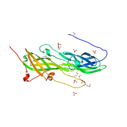
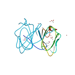
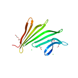
Crystal structure of a putative adhesin (BACOVA_02677) from Bacteroides ovatus ATCC 8483 at 3.00 A resolution (PSI Community Target, Nakayama)
Descriptor: SULFATE ION, Uncharacterized protein
Authors:Joint Center for Structural Genomics (JCSG), Nakayama, K.
Deposit date:2015-06-29
Release date:2015-10-14
Last modified:2020-04-22
Method:X-RAY DIFFRACTION (3 Å)
Cite:A Distinct Type of Pilus from the Human Microbiome.
Cell, 165, 2016
3B77
DownloadVisualize
BU of 3b77 by Molmil
Crystal structure of a ph domain containing bacterial protein (exig_2160) from exiguobacterium sibiricum 255-15 at 2.42 A resolution
Descriptor: Uncharacterized protein
Authors:Joint Center for Structural Genomics (JCSG)
Deposit date:2007-10-30
Release date:2007-11-13
Last modified:2023-01-25
Method:X-RAY DIFFRACTION (2.42 Å)
Cite:Bacterial pleckstrin homology domains: a prokaryotic origin for the PH domain.
J.Mol.Biol., 396, 2010
1VKY
DownloadVisualize
BU of 1vky by Molmil
Crystal structure of S-adenosylmethionine tRNA ribosyltransferase (TM0574) from Thermotoga maritima at 2.00 A resolution
Descriptor: S-adenosylmethionine:tRNA ribosyltransferase-isomerase, UNKNOWN LIGAND
Authors:Joint Center for Structural Genomics (JCSG)
Deposit date:2004-06-28
Release date:2004-08-17
Last modified:2023-01-25
Method:X-RAY DIFFRACTION (2 Å)
Cite:Crystal structure of S-adenosylmethionine:tRNA ribosyltransferase-isomerase (QueA) from Thermotoga maritima at 2.0 A resolution reveals a new fold.
Proteins, 59, 2005
1VKB
DownloadVisualize
BU of 1vkb by Molmil
Crystal structure of an aig2-like protein (a2ld1, ggact, mgc7867) from mus musculus at 1.90 A resolution
Descriptor: FORMIC ACID, hypothetical protein
Authors:Joint Center for Structural Genomics (JCSG)
Deposit date:2004-05-07
Release date:2004-05-18
Last modified:2023-01-25
Method:X-RAY DIFFRACTION (1.9 Å)
Cite:Crystal structure of a conserved hypothetical protein (gi: 13879369) from Mouse at 1.90 A resolution reveals a new fold.
Proteins, 61, 2005
1VR0
DownloadVisualize
BU of 1vr0 by Molmil
Crystal structure of putative 2-phosphosulfolactate phosphatase (15026306) from Clostridium acetobutylicum at 2.6 A resolution
Descriptor: (2R)-3-SULFOLACTIC ACID, MAGNESIUM ION, Probable 2-phosphosulfolactate phosphatase
Authors:Joint Center for Structural Genomics (JCSG)
Deposit date:2005-01-26
Release date:2005-02-15
Last modified:2023-01-25
Method:X-RAY DIFFRACTION (2.49 Å)
Cite:Crystal structure of 2-phosphosulfolactate phosphatase (ComB) from Clostridium acetobutylicum at 2.6 A resolution reveals a new fold with a novel active site.
Proteins, 65, 2006
1VK9
DownloadVisualize
BU of 1vk9 by Molmil
CRYSTAL STRUCTURE OF A DUF1893 family protein (TM1506) FROM THERMOTOGA MARITIMA AT 2.70 A RESOLUTION
Descriptor: UNKNOWN LIGAND, ZINC ION, conserved hypothetical protein TM1506
Authors:Joint Center for Structural Genomics (JCSG)
Deposit date:2004-05-06
Release date:2004-05-18
Last modified:2023-01-25
Method:X-RAY DIFFRACTION (2.7 Å)
Cite:Crystal structure of an ADP-ribosylated protein with a cytidine deaminase-like fold, but unknown function (TM1506), from Thermotoga maritima at 2.70 A resolution.
Proteins, 71, 2008
1VLR
DownloadVisualize
BU of 1vlr by Molmil
Crystal structure of mRNA decapping enzyme (DcpS) from Mus musculus at 1.83 A resolution
Descriptor: 1,2-ETHANEDIOL, mRNA decapping enzyme
Authors:Joint Center for Structural Genomics (JCSG)
Deposit date:2004-08-10
Release date:2004-08-24
Last modified:2023-09-20
Method:X-RAY DIFFRACTION (1.83 Å)
Cite:Crystal structure of an Apo mRNA decapping enzyme (DcpS) from Mouse at 1.83 A resolution.
Proteins, 60, 2005
1VQ3
DownloadVisualize
BU of 1vq3 by Molmil
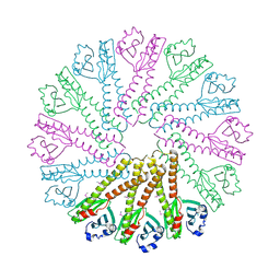
Crystal structure of Phosphoribosylformylglycinamidine synthase, purS subunit (EC 6.3.5.3) (TM1244) from Thermotoga maritima at 1.90 A resolution
Descriptor: Phosphoribosylformylglycinamidine synthase, purS subunit
Authors:Joint Center for Structural Genomics (JCSG)
Deposit date:2004-12-15
Release date:2004-12-28
Last modified:2023-09-20
Method:X-RAY DIFFRACTION (1.9 Å)
Cite:Crystal structure of phosphoribosylformyl-glycinamidine synthase II, PurS subunit (TM1244) from Thermotoga maritima at 1.90 A resolution.
Proteins, 65, 2006
1VKM
DownloadVisualize
BU of 1vkm by Molmil
Crystal structure of an indigoidine synthase a (idga)-like protein (tm1464) from thermotoga maritima msb8 at 1.90 A resolution
Descriptor: 1,2-ETHANEDIOL, MANGANESE (II) ION, UNKNOWN LIGAND, ...
Authors:Joint Center for Structural Genomics (JCSG)
Deposit date:2004-06-09
Release date:2004-06-29
Last modified:2023-01-25
Method:X-RAY DIFFRACTION (1.9 Å)
Cite:Crystal structure of an indigoidine synthase A (IndA)-like protein (TM1464) from Thermotoga maritima at 1.90 A resolution reveals a new fold.
Proteins, 59, 2005
1VJ1
DownloadVisualize
BU of 1vj1 by Molmil
Crystal structure of putative NADPH-dependent oxidoreductase from Mus musculus at 2.10 A resolution
Descriptor: putative NADPH-dependent oxidoreductase
Authors:Joint Center for Structural Genomics (JCSG)
Deposit date:2003-12-03
Release date:2003-12-09
Last modified:2023-01-25
Method:X-RAY DIFFRACTION (2.1 Å)
Cite:Crystal structure of a putative NADPH-dependent oxidoreductase (GI: 18204011) from mouse at 2.10 A resolution
Proteins, 56, 2004
3H50
DownloadVisualize
BU of 3h50 by Molmil
CRYSTAL STRUCTURE OF A TETRACENOMYCIN POLYKETIDE SYNTHESIS PROTEIN (TCMJ) FROM XANTHOMONAS CAMPESTRIS PV. CAMPESTRIS AT 1.60 A RESOLUTION
Descriptor: ACETATE ION, Tetracenomycin polyketide synthesis protein, ZINC ION
Authors:Joint Center for Structural Genomics (JCSG)
Deposit date:2009-04-21
Release date:2009-05-05
Last modified:2023-02-01
Method:X-RAY DIFFRACTION (1.6 Å)
Cite:Conformational changes associated with the binding of zinc acetate at the putative active site of XcTcmJ, a cupin from Xanthomonas campestris pv. campestris.
Acta Crystallogr.,Sect.F, 66, 2010
3KK7
DownloadVisualize
BU of 3kk7 by Molmil
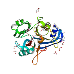
Crystal structure of Putative cell invasion protein with MAC/Perforin domain (NP_812351.1) from BACTERIODES THETAIOTAOMICRON VPI-5482 at 2.46 A resolution
Descriptor: (4S)-2-METHYL-2,4-PENTANEDIOL, 1,2-ETHANEDIOL, CHLORIDE ION, ...
Authors:Joint Center for Structural Genomics (JCSG)
Deposit date:2009-11-04
Release date:2009-11-24
Last modified:2023-02-01
Method:X-RAY DIFFRACTION (2.46 Å)
Cite:Structure of a membrane-attack complex/perforin (MACPF) family protein from the human gut symbiont Bacteroides thetaiotaomicron.
Acta Crystallogr.,Sect.F, 66, 2010
3L5O
DownloadVisualize
BU of 3l5o by Molmil
Crystal structure of protein with unknown function from DUF364 family (ZP_00559375.1) from Desulfitobacterium hafniense DCB-2 at 2.01 A resolution
Descriptor: 1,2-ETHANEDIOL, CHLORIDE ION, IMIDAZOLE, ...
Authors:Joint Center for Structural Genomics, Joint Center for Structural Genomics (JCSG)
Deposit date:2009-12-22
Release date:2010-02-02
Last modified:2023-02-01
Method:X-RAY DIFFRACTION (2.01 Å)
Cite:Structure of the first representative of Pfam family PF04016 (DUF364) reveals enolase and Rossmann-like folds that combine to form a unique active site with a possible role in heavy-metal chelation.
Acta Crystallogr.,Sect.F, 66, 2010
3LIU
DownloadVisualize
BU of 3liu by Molmil
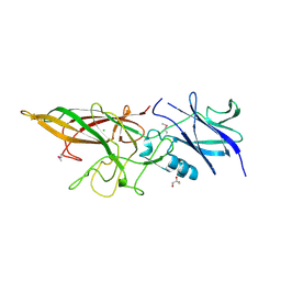
Crystal structure of Putative cell adhesion protein (YP_001304840.1) from Parabacteroides distasonis ATCC 8503 at 2.05 A resolution
Descriptor: 1,2-ETHANEDIOL, CHLORIDE ION, GLYCEROL, ...
Authors:Joint Center for Structural Genomics (JCSG)
Deposit date:2010-01-25
Release date:2010-02-09
Last modified:2023-02-01
Method:X-RAY DIFFRACTION (2.05 Å)
Cite:A Distinct Type of Pilus from the Human Microbiome.
Cell, 165, 2016
3N8U
DownloadVisualize
BU of 3n8u by Molmil
Crystal structure of an imelysin peptidase (BACOVA_03801) from Bacteroides ovatus at 1.44 A resolution
Descriptor: 1,2-ETHANEDIOL, CHLORIDE ION, MAGNESIUM ION, ...
Authors:Joint Center for Structural Genomics (JCSG)
Deposit date:2010-05-28
Release date:2010-07-28
Last modified:2023-02-01
Method:X-RAY DIFFRACTION (1.44 Å)
Cite:Structural and sequence analysis of imelysin-like proteins implicated in bacterial iron uptake.
Plos One, 6, 2011
3MSW
DownloadVisualize
BU of 3msw by Molmil
Crystal structure of a Protein with unknown function (BF3112) from Bacteroides fragilis NCTC 9343 at 1.90 A resolution
Descriptor: CHLORIDE ION, R-1,2-PROPANEDIOL, uncharacterized protein
Authors:Joint Center for Structural Genomics (JCSG)
Deposit date:2010-04-29
Release date:2010-06-09
Last modified:2023-02-01
Method:X-RAY DIFFRACTION (1.9 Å)
Cite:Structures of single-layer beta-sheet proteins evolved from beta-hairpin repeats.
Protein Sci., 28, 2019
3O0F
DownloadVisualize
BU of 3o0f by Molmil
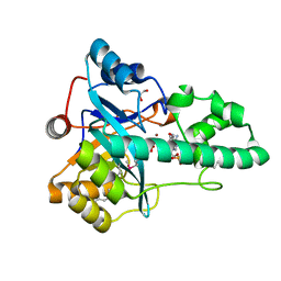
Crystal structure of a putative metal-dependent phosphoesterase (BAD_1165) from bifidobacterium adolescentis atcc 15703 at 1.94 A resolution
Descriptor: 1,2-ETHANEDIOL, ADENOSINE MONOPHOSPHATE, FE (III) ION, ...
Authors:Joint Center for Structural Genomics (JCSG)
Deposit date:2010-07-19
Release date:2010-08-25
Last modified:2023-02-01
Method:X-RAY DIFFRACTION (1.94 Å)
Cite:Crystal structure of a metal-dependent phosphoesterase (YP_910028.1) from Bifidobacterium adolescentis: Computational prediction and experimental validation of phosphoesterase activity.
Proteins, 79, 2011
3OHG
DownloadVisualize
BU of 3ohg by Molmil
Crystal structure of a protein with unknown function from DUF2233 family (BACOVA_00430) from Bacteroides ovatus at 1.80 A resolution
Descriptor: 1,2-ETHANEDIOL, CHLORIDE ION, SULFATE ION, ...
Authors:Joint Center for Structural Genomics (JCSG)
Deposit date:2010-08-17
Release date:2010-09-08
Last modified:2023-02-01
Method:X-RAY DIFFRACTION (1.8 Å)
Cite:Structure and function of the DUF2233 domain in bacteria and in the human mannose 6-phosphate uncovering enzyme.
J.Biol.Chem., 288, 2013
3OYV
DownloadVisualize
BU of 3oyv by Molmil
Crystal structure of an imelysin peptidase (BACOVA_03801) from Bacteroides ovatus ATCC 8483 at 1.25 A resolution
Descriptor: CHLORIDE ION, GLYCEROL, Imelysin, ...
Authors:Joint Center for Structural Genomics (JCSG)
Deposit date:2010-09-23
Release date:2010-10-20
Last modified:2023-02-01
Method:X-RAY DIFFRACTION (1.25 Å)
Cite:Structural and sequence analysis of imelysin-like proteins implicated in bacterial iron uptake.
Plos One, 6, 2011
1G69
DownloadVisualize
BU of 1g69 by Molmil
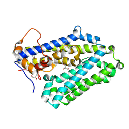
THIAMIN PHOSPHATE SYNTHASE
Descriptor: 2-METHYL-5-METHYLENE-5H-PYRIMIDIN-4-YLIDENEAMINE, 4-METHYL-5-HYDROXYETHYLTHIAZOLE PHOSPHATE, MAGNESIUM ION, ...
Authors:Peapus, D.H, Chiu, H.-J, Campobasso, N, Reddick, J.J, Begley, T.P, Ealick, S.E.
Deposit date:2000-11-03
Release date:2001-09-26
Last modified:2023-08-09
Method:X-RAY DIFFRACTION (1.5 Å)
Cite:Structural characterization of the enzyme-substrate, enzyme-intermediate, and enzyme-product complexes of thiamin phosphate synthase.
Biochemistry, 40, 2001
1G4P
DownloadVisualize
BU of 1g4p by Molmil
THIAMIN PHOSPHATE SYNTHASE
Descriptor: 4-AMINO-2-TRIFLUOROMETHYL-5-HYDROXYMETHYLPYRIMIDINE PYROPHOSPHATE, MAGNESIUM ION, THIAMIN PHOSPHATE SYNTHASE
Authors:Peapus, D.H, Chiu, H.-J, Campobasso, N, Reddick, J.J, Begley, T.P, Ealick, S.E.
Deposit date:2000-10-27
Release date:2001-09-26
Last modified:2023-08-09
Method:X-RAY DIFFRACTION (2.5 Å)
Cite:Structural characterization of the enzyme-substrate, enzyme-intermediate, and enzyme-product complexes of thiamin phosphate synthase.
Biochemistry, 40, 2001

219140

PDB entries from 2024-05-01

PDB statisticsPDBj update infoContact PDBjnumon