Loading
PDBj
MenuPDBj@FacebookPDBj@TwitterPDBj@YouTubewwPDB FoundationwwPDB
RCSB PDBPDBeBMRBAdv. SearchSearch help
Search by PDB author
1JGX
DownloadVisualize
BU of 1jgx by Molmil
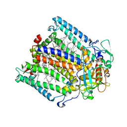
Photosynthetic Reaction Center Mutant With Thr M 21 Replaced With Asp
Descriptor: BACTERIOCHLOROPHYLL A, BACTERIOPHEOPHYTIN A, FE (III) ION, ...
Authors:Camara-Artigas, A, Magee, C.L, Williams, J.C, Allen, J.P.
Deposit date:2001-06-27
Release date:2001-09-05
Last modified:2023-08-16
Method:X-RAY DIFFRACTION (3.01 Å)
Cite:Individual interactions influence the crystalline order for membrane proteins.
Acta Crystallogr.,Sect.D, 57, 2001
6QJI
DownloadVisualize
BU of 6qji by Molmil
Crystal Structure of the third PDZ domain of PSD-95 protein: space group P3112
Descriptor: Disks large homolog 4, SULFATE ION
Authors:Camara-Artigas, A.
Deposit date:2019-01-24
Release date:2019-04-17
Last modified:2024-01-24
Method:X-RAY DIFFRACTION (1.5 Å)
Cite:Conformational changes in the third PDZ domain of the neuronal postsynaptic density protein 95.
Acta Crystallogr D Struct Biol, 75, 2019
6QJG
DownloadVisualize
BU of 6qjg by Molmil
Crystal Structure of the third PDZ domain of PSD-95 protein D332P mutant: space group C121, structure 2
Descriptor: Disks large homolog 4
Authors:Camara-Artigas, A.
Deposit date:2019-01-24
Release date:2019-04-17
Last modified:2024-01-24
Method:X-RAY DIFFRACTION (2.003 Å)
Cite:Conformational changes in the third PDZ domain of the neuronal postsynaptic density protein 95.
Acta Crystallogr D Struct Biol, 75, 2019
6QJK
DownloadVisualize
BU of 6qjk by Molmil
Crystal Structure of the third PDZ domain of PSD-95 protein D332G mutant: space group P43
Descriptor: Disks large homolog 4, GLYCEROL, SULFATE ION
Authors:Camara-Artigas, A.
Deposit date:2019-01-24
Release date:2019-04-17
Last modified:2024-01-24
Method:X-RAY DIFFRACTION (1.046 Å)
Cite:Conformational changes in the third PDZ domain of the neuronal postsynaptic density protein 95.
Acta Crystallogr D Struct Biol, 75, 2019
6QJF
DownloadVisualize
BU of 6qjf by Molmil
Crystal Structure of the third PDZ domain of PSD-95 protein D332P mutant: space group C121, structure 1
Descriptor: Disks large homolog 4
Authors:Camara-Artigas, A.
Deposit date:2019-01-24
Release date:2019-04-17
Last modified:2024-01-24
Method:X-RAY DIFFRACTION (1.5 Å)
Cite:Conformational changes in the third PDZ domain of the neuronal postsynaptic density protein 95.
Acta Crystallogr D Struct Biol, 75, 2019
6QJL
DownloadVisualize
BU of 6qjl by Molmil
Crystal Structure of the third PDZ domain of PSD-95 protein D332G mutant: space group P21
Descriptor: Disks large homolog 4, GLYCEROL, SULFATE ION
Authors:Camara-Artigas, A.
Deposit date:2019-01-24
Release date:2019-04-17
Last modified:2024-01-24
Method:X-RAY DIFFRACTION (1.043 Å)
Cite:Conformational changes in the third PDZ domain of the neuronal postsynaptic density protein 95.
Acta Crystallogr D Struct Biol, 75, 2019
6TG7
DownloadVisualize
BU of 6tg7 by Molmil
Crystal structure of the CheY in presence of magnesium
Descriptor: Chemotaxis protein CheY, MAGNESIUM ION
Authors:Camara-Artigas, A, Salinas-Garcia, M.C, Alba-Elena, D.
Deposit date:2019-11-15
Release date:2019-12-04
Last modified:2024-01-24
Method:X-RAY DIFFRACTION (1.65 Å)
Cite:Crystal structure of the CheY in presence of magnesium
To be published
4F16
DownloadVisualize
BU of 4f16 by Molmil
Crystal Structure of the alpha spectrin SH3 domain at pH 5
Descriptor: Spectrin alpha chain, brain
Authors:Camara-Artigas, A, Gavira, J.A.
Deposit date:2012-05-06
Release date:2013-05-08
Last modified:2023-09-13
Method:X-RAY DIFFRACTION (1.93 Å)
Cite:Crystal Structure of the alpha spectrin SH3 domain at pH 5
To be Published
4F17
DownloadVisualize
BU of 4f17 by Molmil
Crystal Structure of the alpha spectrin SH3 domain at pH 9
Descriptor: Spectrin alpha chain, brain
Authors:Camara-Artigas, A, Gavira, J.A.
Deposit date:2012-05-06
Release date:2013-05-08
Last modified:2023-09-13
Method:X-RAY DIFFRACTION (1.55 Å)
Cite:Crystal Structure of the alpha spectrin SH3 domain at pH 9
To be Published
7A2O
DownloadVisualize
BU of 7a2o by Molmil
Crystal structure of the Fyn SH3 domain L112V-S114N-S115T-E121L-R123H mutant at pH 4.5
Descriptor: TRIETHYLENE GLYCOL, Tyrosine-protein kinase Fyn
Authors:Camara-Artigas, A, Plaza-Garrido, M, Salinas-Garcia, M.C.
Deposit date:2020-08-18
Release date:2021-08-25
Last modified:2024-01-31
Method:X-RAY DIFFRACTION (0.94 Å)
Cite:Crystal structure of the Fyn SH3 domain L112V-S114N-S115T-E121L-R123H mutant at pH 4.5
To be published
7A2X
DownloadVisualize
BU of 7a2x by Molmil
Crystal structure of the Fyn SH3 domain L112V-S114N-S115T-E121L-R123H mutant in complex with VSL12 at pH 5.0
Descriptor: Tyrosine-protein kinase Fyn, VSL12 high affinity synthetic peptide acetylated in the amino-terminus
Authors:Camara-Artigas, A, Plaza-Garrido, M, Salinas-Garcia, M.C.
Deposit date:2020-08-18
Release date:2021-08-25
Last modified:2024-01-31
Method:X-RAY DIFFRACTION (0.92 Å)
Cite:Crystal structure of the Fyn SH3 domain L112V-S114N-S115T-E121L-R123H mutant in complex with VSL12 at pH 5.0
To be published
7A3D
DownloadVisualize
BU of 7a3d by Molmil
Intertwined dimer of the c-Src SH3 domain mutant T125S
Descriptor: DI(HYDROXYETHYL)ETHER, GLYCEROL, Proto-oncogene tyrosine-protein kinase Src, ...
Authors:Camara-Artigas, A, Plaza-Garrido, M, Salinas-Garcia, M.C.
Deposit date:2020-08-18
Release date:2021-08-25
Last modified:2024-01-31
Method:X-RAY DIFFRACTION (2.2 Å)
Cite:Intertwined dimer of the c-Src SH3 domain mutant T125S
To be published
7A2P
DownloadVisualize
BU of 7a2p by Molmil
Crystal structure of the Fyn SH3 domain L112V-S114N-S115T-E121L-R123H mutant at pH 5.0 with PEG
Descriptor: TRIETHYLENE GLYCOL, Tyrosine-protein kinase Fyn
Authors:Camara-Artigas, A, Plaza-Garrido, M, Salinas-Garcia, M.C.
Deposit date:2020-08-18
Release date:2021-08-25
Last modified:2024-01-31
Method:X-RAY DIFFRACTION (0.9 Å)
Cite:Crystal structure of the Fyn SH3 domain L112V-S114N-S115T-E121L-R123H mutant at pH 5.0 with PEG
To be published
7A2Z
DownloadVisualize
BU of 7a2z by Molmil
Crystal structure of the Fyn SH3 domain L112V-S114N-S115T-E121L-R123H mutant in complex with VSL12 at pH 6.0
Descriptor: Tyrosine-protein kinase Fyn, VSL12
Authors:Camara-Artigas, A, Plaza-Garrido, M, Salinas-Garcia, M.C.
Deposit date:2020-08-18
Release date:2021-08-25
Last modified:2024-01-31
Method:X-RAY DIFFRACTION (1.14 Å)
Cite:Crystal structure of the Fyn SH3 domain L112V-S114N-S115T-E121L-R123H mutant in complex with VSL12 at pH 6.0
To be published
7A2J
DownloadVisualize
BU of 7a2j by Molmil
Crystal structure of the Fyn SH3 domain in space group C21 at pH 7.5
Descriptor: SODIUM ION, Tyrosine-protein kinase Fyn
Authors:Camara-Artigas, A, Plaza-Garrido, M, Salinas-Garcia, M.C.
Deposit date:2020-08-18
Release date:2021-08-25
Last modified:2024-01-31
Method:X-RAY DIFFRACTION (1.5 Å)
Cite:Crystal structure of the Fyn SH3 domain in space group C21 at pH 7.5
To be published
7A30
DownloadVisualize
BU of 7a30 by Molmil
Intertwined dimer of the c-Src SH3 domain mutant Q128E
Descriptor: DI(HYDROXYETHYL)ETHER, Proto-oncogene tyrosine-protein kinase Src, SULFATE ION, ...
Authors:Camara-Artigas, A, Plaza-Garrido, M, Salinas-Garcia, M.C.
Deposit date:2020-08-18
Release date:2021-08-25
Last modified:2024-01-31
Method:X-RAY DIFFRACTION (1.67 Å)
Cite:Intertwined dimer of the c-Src SH3 domain mutant Q128E
To be published
7A39
DownloadVisualize
BU of 7a39 by Molmil
Crystal structure of the c-Src SH3 domain mutant V111L-N113S-T114S-Q128E at pH 7.0
Descriptor: Proto-oncogene tyrosine-protein kinase Src, SULFATE ION
Authors:Camara-Artigas, A, Plaza-Garrido, M, Salinas-Garcia, M.C.
Deposit date:2020-08-18
Release date:2021-08-25
Last modified:2024-01-31
Method:X-RAY DIFFRACTION (1.65 Å)
Cite:Crystal structure of the c-Src SH3 domain mutant V111L-N113S-T114S-Q128E at pH 7.0
To be published
7A2Q
DownloadVisualize
BU of 7a2q by Molmil
Crystal structure of the Fyn SH3 domain L112V-S114N-S115T-E121L-R123H mutant at pH 3.0 with PEG
Descriptor: SULFATE ION, TETRAETHYLENE GLYCOL, Tyrosine-protein kinase Fyn
Authors:Camara-Artigas, A, Plaza-Garrido, M, Salinas-Garcia, M.C.
Deposit date:2020-08-18
Release date:2021-08-25
Last modified:2024-01-31
Method:X-RAY DIFFRACTION (0.94 Å)
Cite:Crystal structure of the Fyn SH3 domain L112V-S114N-S115T-E121L-R123H mutant at pH 3.0 with PEG
To be published
7A2Y
DownloadVisualize
BU of 7a2y by Molmil
Crystal structure of the Fyn SH3 domain L112V-S114N-S115T-E121L-R123H mutant in complex with VSL12 at pH 4.0
Descriptor: Tyrosine-protein kinase Fyn, VSL12
Authors:Camara-Artigas, A, Plaza-Garrido, M, Salinas-Garcia, M.C.
Deposit date:2020-08-18
Release date:2021-08-25
Last modified:2024-01-31
Method:X-RAY DIFFRACTION (0.97 Å)
Cite:Crystal structure of the Fyn SH3 domain L112V-S114N-S115T-E121L-R123H mutant in complex with VSL12 at pH 4.0
To be published
7A3C
DownloadVisualize
BU of 7a3c by Molmil
Crystal structure of the c-Src SH3 domain mutant L100I at pH 3.0
Descriptor: GLYCEROL, Proto-oncogene tyrosine-protein kinase Src, SULFATE ION
Authors:Camara-Artigas, A, Plaza-Garrido, M, Salinas-Garcia, M.C.
Deposit date:2020-08-18
Release date:2021-08-25
Last modified:2024-01-31
Method:X-RAY DIFFRACTION (1.8 Å)
Cite:Crystal structure of the c-Src SH3 domain mutant L100I at pH 3.0
To be published
7A2R
DownloadVisualize
BU of 7a2r by Molmil
Crystal structure of the Fyn SH3 domain L112V-S114N-S115T-E121L-R123H mutant at pH 6.0
Descriptor: Tyrosine-protein kinase Fyn
Authors:Camara-Artigas, A, Plaza-Garrido, M, Salinas-Garcia, M.C.
Deposit date:2020-08-18
Release date:2021-08-25
Last modified:2024-01-31
Method:X-RAY DIFFRACTION (1.05 Å)
Cite:Crystal structure of the Fyn SH3 domain L112V-S114N-S115T-E121L-R123H mutant at pH 6.0
To be published
7A31
DownloadVisualize
BU of 7a31 by Molmil
Crystal structure of the c-Src SH3 domain mutant S94A-T98D-V111L-N113S-T114S at pH 4.5
Descriptor: Proto-oncogene tyrosine-protein kinase Src
Authors:Camara-Artigas, A, Plaza-Garrido, M, Salinas-Garcia, M.C.
Deposit date:2020-08-18
Release date:2021-08-25
Last modified:2024-01-31
Method:X-RAY DIFFRACTION (0.94 Å)
Cite:Crystal structure of the c-Src SH3 domain mutant S94A-T98D-V111L-N113S-T114S at pH 4.5
To be published
7A3B
DownloadVisualize
BU of 7a3b by Molmil
Intertwined dimer of the c-Src SH3 domain mutant E106D
Descriptor: GLYCEROL, Proto-oncogene tyrosine-protein kinase Src, TRIETHYLENE GLYCOL
Authors:Camara-Artigas, A, Plaza-Garrido, M, Salinas-Garcia, M.C.
Deposit date:2020-08-18
Release date:2021-08-25
Last modified:2024-01-31
Method:X-RAY DIFFRACTION (1.91 Å)
Cite:Intertwined dimer of the c-Src SH3 domain mutant E106D
To be published
7A2L
DownloadVisualize
BU of 7a2l by Molmil
Crystal structure of the Fyn SH3 domain mutant E129Q in space group C21 at pH 4.0
Descriptor: FORMIC ACID, SODIUM ION, Tyrosine-protein kinase Fyn
Authors:Camara-Artigas, A, Plaza-Garrido, M, Salinas-Garcia, M.C.
Deposit date:2020-08-18
Release date:2021-08-25
Last modified:2024-01-31
Method:X-RAY DIFFRACTION (1.9 Å)
Cite:Crystal structure of the Fyn SH3 domain mutant E129Q in space group C21 at pH 4.0
To be published
7A2V
DownloadVisualize
BU of 7a2v by Molmil
Crystal structure of the Fyn SH3 domain L112V-S114N-S115T-E129Q at pH 4.0
Descriptor: Tyrosine-protein kinase Fyn
Authors:Camara-Artigas, A, Plaza-Garrido, M, Salinas-Garcia, M.C.
Deposit date:2020-08-18
Release date:2021-08-25
Last modified:2024-01-31
Method:X-RAY DIFFRACTION (1.81 Å)
Cite:Crystal structure of the Fyn SH3 domain L112V-S114N-S115T-E129Q at pH 4.0
To be published

218853

PDB entries from 2024-04-24

PDB statisticsPDBj update infoContact PDBjnumon