Loading
PDBj
MenuPDBj@FacebookPDBj@TwitterPDBj@YouTubewwPDB FoundationwwPDB
RCSB PDBPDBeBMRBAdv. SearchSearch help
Search by PDB author
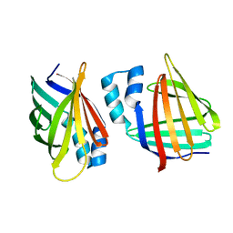
6MOQ
DownloadVisualize
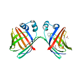
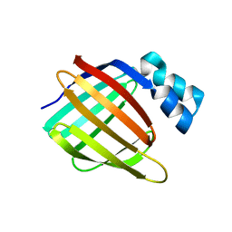
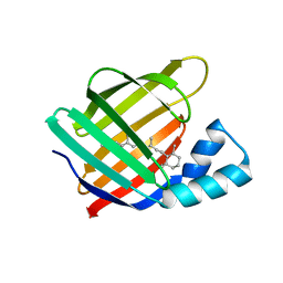
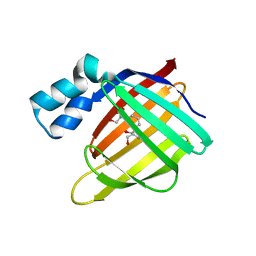
BU of 6moq by Molmil
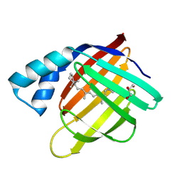
Crystal Structure of the All-trans Retinal-Bound R111K:Y134F:T54V:R132Q:P39Y:R59Y:L121W Mutant of Human Cellular Retinoic Acid Binding Protein II in the Dark at 2.2 Angstrom Resolution
Descriptor: Cellular retinoic acid-binding protein 2, RETINAL
Authors:Ghanbarpour, A, Geiger, J.
Deposit date:2018-10-04
Release date:2019-01-09
Last modified:2023-10-11
Method:X-RAY DIFFRACTION (2.198 Å)
Cite:Mimicking Microbial Rhodopsin Isomerization in a Single Crystal.
J. Am. Chem. Soc., 141, 2019
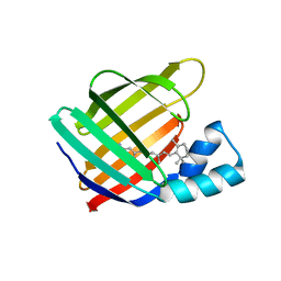
6MOP
DownloadVisualize
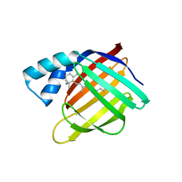
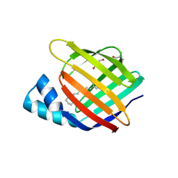
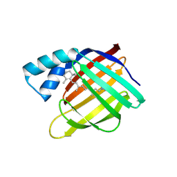
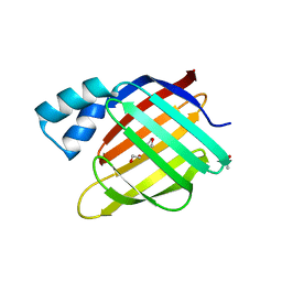
BU of 6mop by Molmil
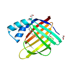
Crystal Structure of the All-trans Retinal-Bound R111K:Y134F:T54V:R132Q:P39Y:R59Y:L121E Mutant of Human Cellular Retinoic Acid Binding Protein II in the Dark at 1.9 Angstrom Resolution
Descriptor: Cellular retinoic acid-binding protein 2, RETINAL
Authors:Ghanbarpour, A, Geiger, J.
Deposit date:2018-10-04
Release date:2019-01-09
Last modified:2023-10-11
Method:X-RAY DIFFRACTION (1.9 Å)
Cite:Mimicking Microbial Rhodopsin Isomerization in a Single Crystal.
J. Am. Chem. Soc., 141, 2019
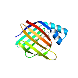
6MPK
DownloadVisualize
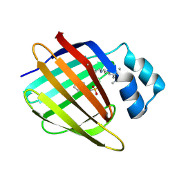
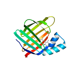
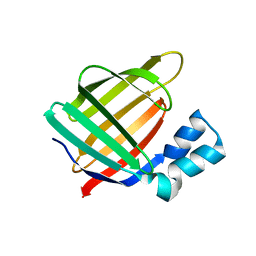
BU of 6mpk by Molmil
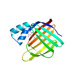
Crystal Structure of the 13-cis Retinal-Bound R111K:Y134F:T54V:R132Q:P39Y:R59Y:L121E:A32Y Mutant of Human Cellular Retinoic Acid Binding Protein II in the Dark at 1.58 Angstrom Resolution
Descriptor: Cellular retinoic acid-binding protein 2, GLYCEROL, RETINAL
Authors:Ghanbarpour, A, Geiger, J.
Deposit date:2018-10-07
Release date:2019-01-09
Last modified:2023-10-11
Method:X-RAY DIFFRACTION (1.58 Å)
Cite:Mimicking Microbial Rhodopsin Isomerization in a Single Crystal.
J. Am. Chem. Soc., 141, 2019
6MQY
DownloadVisualize
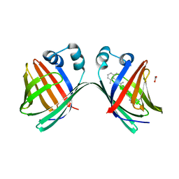
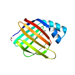
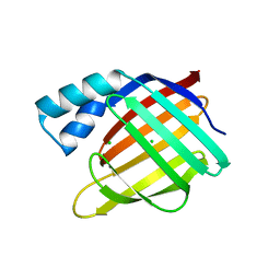
BU of 6mqy by Molmil
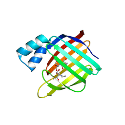
Crystal Structure of the 13-cis Product of All-trans Retinal-Bound R111K:Y134F:T54V:R132Q:P39Y:R59Y:L121E Mutant of Human Cellular Retinoic Acid Binding Protein II Irradiated with 400 nm Laser (30 seconds) at 1.9 Angstrom
Descriptor: Cellular retinoic acid-binding protein 2, GLYCEROL, RETINAL
Authors:Ghanbarpour, A, Geiger, J.
Deposit date:2018-10-11
Release date:2019-01-09
Last modified:2023-10-11
Method:X-RAY DIFFRACTION (1.9 Å)
Cite:Mimicking Microbial Rhodopsin Isomerization in a Single Crystal.
J. Am. Chem. Soc., 141, 2019
6MOV
DownloadVisualize
BU of 6mov by Molmil
Crystal Structure of the All-trans Retinal bound R111K:Y134F:T54V:R132Q:P39Y:R59Y:L121Q Human Cellular Retinoic Acid Binding Protein II in the Dark at 1.75 Angstrom Resolution
Descriptor: Cellular retinoic acid-binding protein 2, RETINAL
Authors:Ghanbarpour, A, Geiger, J.
Deposit date:2018-10-04
Release date:2019-01-09
Last modified:2023-10-11
Method:X-RAY DIFFRACTION (1.752 Å)
Cite:Mimicking Microbial Rhodopsin Isomerization in a Single Crystal.
J. Am. Chem. Soc., 141, 2019
3FEL
DownloadVisualize
BU of 3fel by Molmil
Crystal structure of the R132K:R111L:T54E mutant of cellular retinoic acid-binding protein II at 1.85 angstrom resolution
Descriptor: 2-[3-(2-HYDROXY-1,1-DIHYDROXYMETHYL-ETHYLAMINO)-PROPYLAMINO]-2-HYDROXYMETHYL-PROPANE-1,3-DIOL, Cellular retinoic acid-binding protein 2
Authors:Jia, X, Geiger, J.H.
Deposit date:2008-11-30
Release date:2009-11-10
Last modified:2023-09-06
Method:X-RAY DIFFRACTION (1.85 Å)
Cite:Crystal structures of apo cellular retinoic acid-binding protein II mutants: Structural integrity investigated through multiple site mutations
To be Published
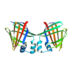
6MLB
DownloadVisualize
BU of 6mlb by Molmil
Crystal structure of the holo retinal-bound domain-swapped dimer Q108K:K40L:T51F mutant of human cellular retinol binding protein II
Descriptor: ACETATE ION, GLYCEROL, RETINAL, ...
Authors:Ghanbarpour, A, Geiger, J.
Deposit date:2018-09-27
Release date:2019-10-16
Last modified:2023-10-11
Method:X-RAY DIFFRACTION (2.15 Å)
Cite:Engineering the hCRBPII Domain-Swapped Dimer into a New Class of Protein Switches.
J.Am.Chem.Soc., 141, 2019
6MR0
DownloadVisualize
BU of 6mr0 by Molmil
Crystal Structure of the All-trans Retinal Bound R111K:Y134F:T54V:R132Q:P39Q:R59Y:L121E Human Cellular Retinoic Acid Binding Protein II Mutant After 5 Minutes UV irradiation at 2.6 Angstrom Resolution
Descriptor: Cellular retinoic acid-binding protein 2, RETINAL
Authors:Ghanbarpour, A, Geiger, J.
Deposit date:2018-10-11
Release date:2019-01-09
Last modified:2023-10-11
Method:X-RAY DIFFRACTION (2.649 Å)
Cite:Mimicking Microbial Rhodopsin Isomerization in a Single Crystal.
J. Am. Chem. Soc., 141, 2019
6MOX
DownloadVisualize
BU of 6mox by Molmil
Crystal Structure of the All-trans Retinal-Bound R111K:Y134F:T54V:R132Q:P39Q:R59Y:L121E Human Cellular Retinoic Acid Binding Protein II Mutant in the Dark at 2.18 Angstrom Resolution
Descriptor: Cellular retinoic acid-binding protein 2, RETINAL
Authors:Ghanbarpour, A, Geiger, J.
Deposit date:2018-10-04
Release date:2019-01-09
Last modified:2023-10-11
Method:X-RAY DIFFRACTION (2.18 Å)
Cite:Mimicking Microbial Rhodopsin Isomerization in a Single Crystal.
J. Am. Chem. Soc., 141, 2019
3FEP
DownloadVisualize
BU of 3fep by Molmil
Crystal structure of the R132K:R111L:L121E:R59W-CRABPII mutant complexed with a synthetic ligand (merocyanin) at 2.60 angstrom resolution.
Descriptor: (2E,4E,6E)-3-methyl-6-(1,3,3-trimethyl-1,3-dihydro-2H-indol-2-ylidene)hexa-2,4-dienal, 2-(N-MORPHOLINO)-ETHANESULFONIC ACID, Cellular retinoic acid-binding protein 2
Authors:Jia, X, Geiger, J.H.
Deposit date:2008-11-30
Release date:2009-11-10
Last modified:2023-09-06
Method:X-RAY DIFFRACTION (2.6 Å)
Cite:"Turn-on" protein fluorescence: in situ formation of cyanine dyes.
J.Am.Chem.Soc., 137, 2015
6MKV
DownloadVisualize
BU of 6mkv by Molmil
Crystal structure of Retinal-bound holo Q108K:K40L:T51W domain-swapped dimer of human cellular retinol binding protein 2
Descriptor: ACETATE ION, GLYCEROL, RETINAL, ...
Authors:Ghanbarpour, A, Geiger, J.
Deposit date:2018-09-26
Release date:2019-10-16
Last modified:2023-10-11
Method:X-RAY DIFFRACTION (2.107 Å)
Cite:Engineering the hCRBPII Domain-Swapped Dimer into a New Class of Protein Switches.
J.Am.Chem.Soc., 141, 2019
3FA9
DownloadVisualize
BU of 3fa9 by Molmil
Crystal structure of the apo R132K:Y134F:R111L:L121D mutant of cellular retinoic acid-binding protein II at 1.94 angstrom resolution
Descriptor: ACETATE ION, Cellular retinoic acid-binding protein 2
Authors:Jia, X, Geiger, J.H.
Deposit date:2008-11-16
Release date:2009-10-27
Last modified:2023-12-27
Method:X-RAY DIFFRACTION (1.94 Å)
Cite:Crystal structures of apo cellular retinoic acid-binding protein II mutants: Structural integrity investigated through multiple site mutations
To be Published
6MQX
DownloadVisualize
BU of 6mqx by Molmil
Crystal Structure of All-trans Retinal-Bound R111K:Y134F:T54V:R132Q:P39Y:R59Y:L121E Human Cellular Retinoic Acid Binding Protein II Irradiated with 400 nm Laser (30 seconds) and Subsequently Dark Adapted (25 minutes) at 2.0 Angstrom Resolution
Descriptor: Cellular retinoic acid-binding protein 2, GLYCEROL, RETINAL
Authors:Ghanbarpour, A, Geiger, J.
Deposit date:2018-10-11
Release date:2019-01-09
Last modified:2023-10-11
Method:X-RAY DIFFRACTION (2.01 Å)
Cite:Mimicking Microbial Rhodopsin Isomerization in a Single Crystal.
J. Am. Chem. Soc., 141, 2019
6MQI
DownloadVisualize
BU of 6mqi by Molmil
Crystal Structure of the All-trans Retinal bound R111K:Y134F:T54V:R132Q:P39Y:R59Y:L121Q mutant of Human Cellular Retinoic Acid Binding Protein II Irradiated with 400 nm Laser for 5 minutes at 1.87 Angstrom Resolution
Descriptor: Cellular retinoic acid-binding protein 2, RETINAL
Authors:Ghanbarpour, A, Geiger, J.
Deposit date:2018-10-09
Release date:2019-01-09
Last modified:2023-10-11
Method:X-RAY DIFFRACTION (1.87 Å)
Cite:Mimicking Microbial Rhodopsin Isomerization in a Single Crystal.
J. Am. Chem. Soc., 141, 2019
6MQZ
DownloadVisualize
BU of 6mqz by Molmil
Crystal Structure of the 13-cis Product of the All-trans Retinal-Bound R111K:Y134F:T54V:R132Q:P39Y:R59Y:L121E Mutant of Human Cellular Retinoic Acid Binding Protein II Irradiated with 400 nm Laser (5 minutes) at 2.07 Angstrom
Descriptor: Cellular retinoic acid-binding protein 2, GLYCEROL, RETINAL
Authors:Ghanbarpour, A, Geiger, J.
Deposit date:2018-10-11
Release date:2019-01-09
Last modified:2023-10-11
Method:X-RAY DIFFRACTION (2.07 Å)
Cite:Mimicking Microbial Rhodopsin Isomerization in a Single Crystal.
J. Am. Chem. Soc., 141, 2019
6MOR
DownloadVisualize
BU of 6mor by Molmil
Crystal Structure of the All-Trans Retinal-Bound R111K:Y134F:T54V:R132Q:P39Y:R59Y:L121Y Mutant of Human Cellular Retinoic Acid Binding Protein II in the Dark at 1.79 Angstrom Resolution
Descriptor: Cellular retinoic acid-binding protein 2, RETINAL
Authors:Ghanbarpour, A, Geiger, J.
Deposit date:2018-10-04
Release date:2019-01-09
Last modified:2023-10-11
Method:X-RAY DIFFRACTION (1.79 Å)
Cite:Mimicking Microbial Rhodopsin Isomerization in a Single Crystal.
J. Am. Chem. Soc., 141, 2019
6MQW
DownloadVisualize
BU of 6mqw by Molmil
Crystal Structure of All-trans Retinal-Bound R111K:Y134F:T54V:R132Q:P39Y:R59Y:L121E Human Cellular Retinoic Acid Binding Protein II Irradiated with 400 nm laser (30 seconds) and subsequently dark adapted (10 minutes) at 2.1 Angstrom Resolution
Descriptor: Cellular retinoic acid-binding protein 2, RETINAL
Authors:Ghanbarpour, A, Geiger, J.
Deposit date:2018-10-11
Release date:2019-01-09
Last modified:2023-10-11
Method:X-RAY DIFFRACTION (2.09 Å)
Cite:Mimicking Microbial Rhodopsin Isomerization in a Single Crystal.
J. Am. Chem. Soc., 141, 2019
3FA8
DownloadVisualize
BU of 3fa8 by Molmil
Crystal structure of the apo R132K:Y134F:R111L:L121E mutant of cellular retinoic acid-binding protein II at 1.78 angstrom resolution
Descriptor: Cellular retinoic acid-binding protein 2
Authors:Jia, X, Geiger, J.H.
Deposit date:2008-11-16
Release date:2009-10-27
Last modified:2023-09-06
Method:X-RAY DIFFRACTION (1.78 Å)
Cite:Crystal structures of apo cellular retinoic acid-binding protein II mutants: Structural integrity investigated through multiple site mutations
To be Published
3FEN
DownloadVisualize
BU of 3fen by Molmil
Crystal structure of the R132K:R111L:A32E mutant of cellular retinoic acid-binding protein II at 1.56 angstrom resolution
Descriptor: CHLORIDE ION, Cellular retinoic acid-binding protein 2
Authors:Jia, X, Geiger, J.H.
Deposit date:2008-11-30
Release date:2009-11-10
Last modified:2023-09-06
Method:X-RAY DIFFRACTION (1.56 Å)
Cite:Crystal structures of apo cellular retinoic acid-binding protein II mutants: Structural integrity investigated through multiple site mutations
To be Published
3FA7
DownloadVisualize
BU of 3fa7 by Molmil
Crystal structure of the apo R132K:R111L:L121E:R59E mutant of cellular retinoic acid-binding protein II at 1.90 angstrom resolution
Descriptor: 2-[3-(2-HYDROXY-1,1-DIHYDROXYMETHYL-ETHYLAMINO)-PROPYLAMINO]-2-HYDROXYMETHYL-PROPANE-1,3-DIOL, Cellular retinoic acid-binding protein 2
Authors:Jia, X, Geiger, J.H.
Deposit date:2008-11-15
Release date:2009-10-27
Last modified:2023-12-27
Method:X-RAY DIFFRACTION (1.9 Å)
Cite:Crystal structures of apo cellular retinoic acid-binding protein II mutants: Structural integrity investigated through multiple site mutations
To be Published
3FEK
DownloadVisualize
BU of 3fek by Molmil
Crystal structure of the R132K:Y134F:R111L:L121D:T54V mutant of cellular retinoic acid-binding protein II at 1.51 angstrom resolution
Descriptor: ACETATE ION, Cellular retinoic acid-binding protein 2, DI(HYDROXYETHYL)ETHER
Authors:Jia, X, Geiger, J.H.
Deposit date:2008-11-30
Release date:2009-11-10
Last modified:2023-09-06
Method:X-RAY DIFFRACTION (1.51 Å)
Cite:Crystal structures of apo cellular retinoic acid-binding protein II mutants: Structural integrity investigated through multiple site mutations
To be Published
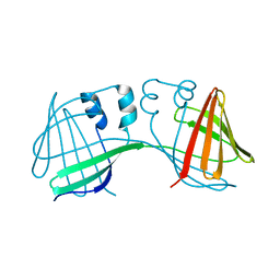
6ON5
DownloadVisualize
BU of 6on5 by Molmil
Crystal Structure of the Zn-bound Domain-Swapped Dimer Q108K:T51D:A28C:L36C:F57H Mutant of Human Cellular Retinol Binding Protein II
Descriptor: Retinol-binding protein 2, ZINC ION
Authors:Ghanbarpour, A, Geiger, J.
Deposit date:2019-04-20
Release date:2019-10-16
Last modified:2023-10-11
Method:X-RAY DIFFRACTION (1.638 Å)
Cite:Engineering the hCRBPII Domain-Swapped Dimer into a New Class of Protein Switches.
J.Am.Chem.Soc., 141, 2019
6ON8
DownloadVisualize
BU of 6on8 by Molmil
Crystal Structure of the Reduced Form of Apo Domain-Swapped Dimer Q108K:T51D:A28C:L36C:F57H Mutant of Human Cellular Retinol Binding Protein II
Descriptor: Retinol-binding protein 2
Authors:Ghanbarpour, A, Geiger, J.
Deposit date:2019-04-20
Release date:2019-10-16
Last modified:2023-10-11
Method:X-RAY DIFFRACTION (2.395 Å)
Cite:Engineering the hCRBPII Domain-Swapped Dimer into a New Class of Protein Switches.
J.Am.Chem.Soc., 141, 2019
3I17
DownloadVisualize
BU of 3i17 by Molmil
Crystal structure of the apo R132K:L121E mutant of cellular retinoic acid-binding protein II at 1.68 angstrom resolution
Descriptor: Cellular retinoic acid-binding protein 2
Authors:Jia, X, Watson, C.T, Geiger, J.H.
Deposit date:2009-06-25
Release date:2009-07-28
Last modified:2023-09-06
Method:X-RAY DIFFRACTION (1.68 Å)
Cite:Elucidating the exact role of engineered CRABPII residues for the formation of a retinal protonated Schiff base.
Proteins, 77, 2009

219869

数据于2024-05-15公开中

PDB statisticsPDBj update infoContact PDBjnumon