Loading
PDBj
MenuPDBj@FacebookPDBj@TwitterPDBj@YouTubewwPDB FoundationwwPDB
RCSB PDBPDBeBMRBAdv. SearchSearch help
Search by PDB author
4ZR2
DownloadVisualize
BU of 4zr2 by Molmil
Crystal Structure of the Domain-Swapped Dimer K40L:Q108K:Y60W mutant of Human Cellular Retinol Binding Protein II
Descriptor: ACETATE ION, RETINAL, Retinol-binding protein 2
Authors:Assar, Z, Nossoni, Z, Geiger, J.H.
Deposit date:2015-05-11
Release date:2016-06-08
Last modified:2023-09-27
Method:X-RAY DIFFRACTION (1.8024 Å)
Cite:Domain-Swapped Dimers of Intracellular Lipid-Binding Proteins: Evidence for Ordered Folding Intermediates.
Structure, 24, 2016
6VIT
DownloadVisualize
BU of 6vit by Molmil
The Crystal Structure of Apo Domain-Swapped dimer Q108K:T51D:I32C Variant of HCRBPII with an Engineered Disulfide Bond
Descriptor: Retinol-binding protein 2
Authors:Ghanbarpour, A, Geiger, J.
Deposit date:2020-01-13
Release date:2020-08-19
Last modified:2023-10-11
Method:X-RAY DIFFRACTION (3.2 Å)
Cite:Human Cellular Retinol Binding Protein II Forms a Domain-Swapped Trimer Representing a Novel Fold and a New Template for Protein Engineering.
Chembiochem, 21, 2020
6VIS
DownloadVisualize
BU of 6vis by Molmil
The Crystal Structure of Domain-Swapped Trimer Q108K:K40D:T53A:R58L:Q38F:Q4F:V62E Variant of HCRBPII
Descriptor: GLYCEROL, Retinol-binding protein 2
Authors:Ghanbarpour, A, Geiger, J.
Deposit date:2020-01-13
Release date:2020-09-09
Last modified:2023-10-11
Method:X-RAY DIFFRACTION (2.79 Å)
Cite:Human Cellular Retinol Binding Protein II Forms a Domain-Swapped Trimer Representing a Novel Fold and a New Template for Protein Engineering.
Chembiochem, 21, 2020
4ZGU
DownloadVisualize
BU of 4zgu by Molmil
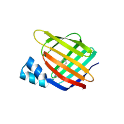
Crystal structure of monomer Y60W hCRBPII
Descriptor: ACETATE ION, Retinol-binding protein 2
Authors:Nossoni, Z, Geiger, J.
Deposit date:2015-04-24
Release date:2016-06-08
Last modified:2024-03-06
Method:X-RAY DIFFRACTION (1.49 Å)
Cite:Domain-Swapped Dimers of Intracellular Lipid-Binding Proteins: Evidence for Ordered Folding Intermediates.
Structure, 24, 2016
6WNF
DownloadVisualize
BU of 6wnf by Molmil
The Crystal Structure of Apo Domain-Swapped Dimer Q108K:K40D:T53A:R58L:Q38F:Q4F:F57H Variant of HCRBPII
Descriptor: ACETATE ION, GLYCEROL, Retinol-binding protein 2
Authors:Ghanbarpour, A, Geiger, J.
Deposit date:2020-04-22
Release date:2020-08-19
Last modified:2023-10-18
Method:X-RAY DIFFRACTION (1.67 Å)
Cite:Human Cellular Retinol Binding Protein II Forms a Domain-Swapped Trimer Representing a Novel Fold and a New Template for Protein Engineering.
Chembiochem, 21, 2020
6WNJ
DownloadVisualize
BU of 6wnj by Molmil
The Crystal Structure of Apo Domain-Swapped Trimer Q108K:T51D:A28C:I32C of HCRBPII
Descriptor: GLYCEROL, Retinol-binding protein 2, SULFATE ION, ...
Authors:Ghanbarpour, A, Geiger, J.
Deposit date:2020-04-22
Release date:2020-08-19
Last modified:2023-10-18
Method:X-RAY DIFFRACTION (2.1 Å)
Cite:Human Cellular Retinol Binding Protein II Forms a Domain-Swapped Trimer Representing a Novel Fold and a New Template for Protein Engineering.
Chembiochem, 21, 2020
6WP1
DownloadVisualize
BU of 6wp1 by Molmil
The Crystal Structure of Apo Domain-Swapped Trimer Q108K:K40L:T51K Variant of HCRBPII
Descriptor: ACETATE ION, GLYCEROL, Retinol-binding protein 2
Authors:Ghanbarpour, A, Geiger, J.
Deposit date:2020-04-26
Release date:2020-08-19
Last modified:2023-10-18
Method:X-RAY DIFFRACTION (2.99 Å)
Cite:Human Cellular Retinol Binding Protein II Forms a Domain-Swapped Trimer Representing a Novel Fold and a New Template for Protein Engineering.
Chembiochem, 21, 2020
6WP0
DownloadVisualize
BU of 6wp0 by Molmil
The Crystal Structure of Domain-Swapped Trimer Q108K:T51D variant of HCRBPII
Descriptor: ACETATE ION, GLYCEROL, Retinol-binding protein 2
Authors:Ghanbarpour, A, Geiger, J.
Deposit date:2020-04-26
Release date:2020-08-19
Last modified:2023-10-18
Method:X-RAY DIFFRACTION (2.78 Å)
Cite:Human Cellular Retinol Binding Protein II Forms a Domain-Swapped Trimer Representing a Novel Fold and a New Template for Protein Engineering.
Chembiochem, 21, 2020
6WP2
DownloadVisualize
BU of 6wp2 by Molmil
The Crystal Structure of Apo Zinc-Bound Domain Swapped-Trimer Q108K:K40D:T53A:R58L:Q38F:Q4F:F57H Variant of HCRBPII
Descriptor: ACETATE ION, GLYCEROL, Retinol-binding protein 2, ...
Authors:Ghanbarpour, A, Geiger, J.
Deposit date:2020-04-26
Release date:2020-08-19
Last modified:2023-10-18
Method:X-RAY DIFFRACTION (2.48 Å)
Cite:Human Cellular Retinol Binding Protein II Forms a Domain-Swapped Trimer Representing a Novel Fold and a New Template for Protein Engineering.
Chembiochem, 21, 2020
4I9S
DownloadVisualize
BU of 4i9s by Molmil
Crystal Structure of the R111K:R132L:Y134F:T54V:R59W Mutant of the Cellular Retinoic Acid Binding Protein Type II in Complex with All-Trans Retinal at 2.58 Angstrom Resolution
Descriptor: Cellular retinoic acid-binding protein 2, RETINAL
Authors:Nosrati, M, Geiger, J.H.
Deposit date:2012-12-05
Release date:2013-10-09
Last modified:2023-09-20
Method:X-RAY DIFFRACTION (2.58 Å)
Cite:Rational Design of a Colorimetric pH Sensor from a Soluble Retinoic Acid Chaperone.
J.Am.Chem.Soc., 135, 2013
4I9R
DownloadVisualize
BU of 4i9r by Molmil
Crystal Structure of the R111K:R132L:Y134F:T54V:R59W:A32W Mutant of the Cellular Retinoic Acid Binding Protein Type II in Complex with All-Trans Retinal at 2.6 Angstrom Resolution
Descriptor: Cellular retinoic acid-binding protein 2, RETINAL
Authors:Nosrati, M, Geiger, J.H.
Deposit date:2012-12-05
Release date:2013-10-09
Last modified:2023-09-20
Method:X-RAY DIFFRACTION (2.6 Å)
Cite:Rational Design of a Colorimetric pH Sensor from a Soluble Retinoic Acid Chaperone.
J.Am.Chem.Soc., 135, 2013
4M6S
DownloadVisualize
BU of 4m6s by Molmil
Crystal structure of the R111K:R132Y:Y134F:T54V:R59W:A32W mutant of the Cellular Retinoic Acid Binding Protein Type II in complex with All-Trans Retinal at 2.38 Angstrom Resolution
Descriptor: Cellular retinoic acid-binding protein 2, RETINAL
Authors:Nosrati, M, Geiger, J.H.
Deposit date:2013-08-10
Release date:2013-10-09
Last modified:2023-09-20
Method:X-RAY DIFFRACTION (2.47 Å)
Cite:Rational Design of a Colorimetric pH Sensor from a Soluble Retinoic Acid Chaperone.
J.Am.Chem.Soc., 135, 2013
4M7M
DownloadVisualize
BU of 4m7m by Molmil
Crystal structure of the R111K:R132Q:Y134F:T54V:R59W:A32W mutant of the Cellular Retinoic Acid Binding Protein Type II in complex with All-Trans Retinal at 2.57 Angstrom Resolution
Descriptor: Cellular retinoic acid-binding protein 2, RETINAL
Authors:Nosrati, M, Geiger, J.H.
Deposit date:2013-08-12
Release date:2013-10-09
Last modified:2014-02-19
Method:X-RAY DIFFRACTION (2.571 Å)
Cite:Rational Design of a Colorimetric pH Sensor from a Soluble Retinoic Acid Chaperone.
J.Am.Chem.Soc., 135, 2013
3CR6
DownloadVisualize
BU of 3cr6 by Molmil
Crystal Structure of the R132K:R111L:A32E Mutant of Cellular Retinoic Acid Binding Protein Type II Complexed with C15-aldehyde (a retinal analog) at 1.22 Angstrom resolution.
Descriptor: 1,3,3-trimethyl-2-[(1E,3E)-3-methylpenta-1,3-dien-1-yl]cyclohexene, Cellular retinoic acid-binding protein 2
Authors:Jia, X, Geiger, J.H.
Deposit date:2008-04-04
Release date:2009-03-17
Last modified:2023-08-30
Method:X-RAY DIFFRACTION (1.22 Å)
Cite:Two distinctive orientations of binding determined by a single mutation in the CRABPII mutant-C15-aldehyde complexes
To be Published
3F8A
DownloadVisualize
BU of 3f8a by Molmil
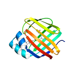
Crystal Structure of the R132K:R111L:L121E:R59W Mutant of Cellular Retinoic Acid-Binding Protein Type II Complexed with C15-aldehyde (a retinal analog) at 1.95 Angstrom resolution.
Descriptor: 1,3,3-trimethyl-2-[(1E,3E)-3-methylpenta-1,3-dien-1-yl]cyclohexene, 2-[3-(2-HYDROXY-1,1-DIHYDROXYMETHYL-ETHYLAMINO)-PROPYLAMINO]-2-HYDROXYMETHYL-PROPANE-1,3-DIOL, Cellular retinoic acid-binding protein 2
Authors:Jia, X, Geiger, J.H.
Deposit date:2008-11-12
Release date:2009-11-10
Last modified:2023-09-06
Method:X-RAY DIFFRACTION (1.95 Å)
Cite:Probing Wavelength Regulation with an Engineered Rhodopsin Mimic and a C15-Retinal Analogue
Chempluschem, 77, 2012
3FA6
DownloadVisualize
BU of 3fa6 by Molmil
Crystal structure of the R132K:Y134F:R111L:L121D:T54V mutant of cellular retinoic acid-binding protein II complexed with C15-aldehyde (a retinal analog) at 1.54 angstrom resolution
Descriptor: 1,3,3-trimethyl-2-[(1E,3E)-3-methylpenta-1,3-dien-1-yl]cyclohexene, Cellular retinoic acid-binding protein 2
Authors:Jia, X, Geiger, J.H.
Deposit date:2008-11-15
Release date:2009-10-27
Last modified:2023-12-27
Method:X-RAY DIFFRACTION (1.54 Å)
Cite:Two distinctive orientations of binding determined by a single mutation in the CRABPII mutant-C15-aldehyde complexes
To be Published
3F9D
DownloadVisualize
BU of 3f9d by Molmil
Crystal structure of the R132K:R111L:T54E mutant of cellular retinoic acid-binding protein II complexed with C15-aldehyde (a retinal analog) at 2.00 angstrom resolution
Descriptor: 1,3,3-trimethyl-2-[(1E,3E)-3-methylpenta-1,3-dien-1-yl]cyclohexene, Cellular retinoic acid-binding protein 2
Authors:Jia, X, Geiger, J.H.
Deposit date:2008-11-13
Release date:2009-10-27
Last modified:2023-09-06
Method:X-RAY DIFFRACTION (2 Å)
Cite:Two distinctive orientations of binding determined by a single mutation in the CRABPII mutant-C15-aldehyde complexes
To be Published
5F58
DownloadVisualize
BU of 5f58 by Molmil
Crystal structure of the Q108K:K40L:T51V:R58F mutant of human Cellular Retinol Binding Protein II in complex with All-trans-Retinal after 24 hours of incubation at 1.54 Angstrom Resolution
Descriptor: ACETATE ION, RETINAL, Retinol-binding protein 2
Authors:Nosrati, M, Nossoni, Z, Geiger, J.H.
Deposit date:2015-12-04
Release date:2016-12-07
Last modified:2023-09-27
Method:X-RAY DIFFRACTION (1.54 Å)
Cite:Crystal structure of the Q108K:K40L:T51V:R58F mutant of human Cellular Retinol Binding Protein II in complex with All-trans-Retinal after 24 hours of incubation at 1.54 Angstrom Resolution
To Be Published
5F6B
DownloadVisualize
BU of 5f6b by Molmil
Crystal structure of the Q108K:K40L:T51V:R58Y:Y19W mutant of human Cellular Retinol Binding Protein II in complex with All-trans-Retinal at 1.3 Angstrom Resolution
Descriptor: ACETATE ION, RETINAL, Retinol-binding protein 2
Authors:Nosrati, M, Nossoni, Z, Geiger, J.H.
Deposit date:2015-12-05
Release date:2016-12-07
Last modified:2023-09-27
Method:X-RAY DIFFRACTION (1.31 Å)
Cite:Crystal structure of the Q108K:K40L:T51V:R58Y:Y19W mutant of human Cellular Retinol Binding ProteinII in complex with All-trans-Retinal at 1.31 Angstrom Resolution
To Be Published
5FAZ
DownloadVisualize
BU of 5faz by Molmil
Crystal structure of the Q108K:K40L:T51V mutant of human Cellular Retinol Binding Protein II in complex with All-trans-Retinal after 24 hours of incubation at 1.4 Angstrom Resolution
Descriptor: ACETATE ION, RETINAL, Retinol-binding protein 2
Authors:Nosrati, M, Nossoni, Z, Geiger, J.H.
Deposit date:2015-12-13
Release date:2016-12-14
Last modified:2023-09-27
Method:X-RAY DIFFRACTION (1.4 Å)
Cite:Crystal structure of the Q108K:K40L:T51V mutant of human Cellular Retinol Binding Protein II in complex with All-trans-Retinal after 24 hours of incubation at 1.54 Angstrom Resolution
To Be Published
5FEN
DownloadVisualize
BU of 5fen by Molmil
Crystal structure of the Q108K:K40L:T53C mutant of human Cellular Retinol Binding Protein II in complex with All-trans-Retinal after 24 hours of incubation at 1.55 Angstrom Resolution
Descriptor: ACETATE ION, RETINAL, Retinol-binding protein 2
Authors:Nossoni, Z, Nosrati, M, Geiger, J.H.
Deposit date:2015-12-17
Release date:2016-12-21
Last modified:2023-09-27
Method:X-RAY DIFFRACTION (1.55 Å)
Cite:Crystal structure of the Q108K:K40L:T53C mutant of human Cellular Retinol Binding Protein II in complex with All-trans-Retinal after 24 hours of incubation at 1.55 Angstrom Resolution
To Be Published
5FFH
DownloadVisualize
BU of 5ffh by Molmil
Crystal structure of the Q108K:K40L:T51V:R58W:Y19W mutant of human Cellular Retinol Binding Protein II in complex with All-trans-Retinal at 1.68 Angstrom Resolution
Descriptor: ACETATE ION, RETINAL, Retinol-binding protein 2
Authors:Nossoni, Z, Nosrati, M, Geiger, J.H.
Deposit date:2015-12-18
Release date:2016-12-21
Last modified:2023-09-27
Method:X-RAY DIFFRACTION (1.68 Å)
Cite:Crystal structure of the Q108K:K40L:T51V:R58W:Y19W mutant of human Cellular Retinol Binding ProteinII in complex with All-trans-Retinal at 1.68 Angstrom Resolution
To Be Published
5F7G
DownloadVisualize
BU of 5f7g by Molmil
Crystal structure of the Q108K:K40L:T51V:R58Y:Y19W:Q38L mutant of human Cellular Retinol Binding Protein II in complex with All-trans-Retinal at 1.48 Angstrom Resolution
Descriptor: ACETATE ION, RETINAL, Retinol-binding protein 2
Authors:Nosrati, M, Nossoni, Z, Geiger, J.H.
Deposit date:2015-12-08
Release date:2016-12-14
Last modified:2023-09-27
Method:X-RAY DIFFRACTION (1.48 Å)
Cite:Crystal structure of the Q108K:K40L:T51V:R58F mutant of human Cellular Retinol Binding Protein II in complex with All-trans-Retinal after 24 hours of incubation at 1.54 Angstrom Resolution
To Be Published
4YGH
DownloadVisualize
BU of 4ygh by Molmil
Crystal Structure of R111K:Y134F:T54V:R132Q:P39Y:R59Y mutant of human Cellular Retinoic Acid Binding Protein II with Retinal at 2.1 Angstrom resolution - UV irradiated crystal - 2nd cycle
Descriptor: Cellular retinoic acid-binding protein 2, RETINAL
Authors:Nosrati, M, Geiger, J.H.
Deposit date:2015-02-26
Release date:2016-03-02
Last modified:2023-09-27
Method:X-RAY DIFFRACTION (2.1 Å)
Cite:A Photoisomerizing Rhodopsin Mimic Observed at Atomic Resolution.
J.Am.Chem.Soc., 138, 2016
4YH0
DownloadVisualize
BU of 4yh0 by Molmil
Crystal structure of the R111K:Y134F:T54V:R132Q:P39Y:R59Y mutant of human Cellular Retinoic Acid Binding Protein II in complex with Retinal at 2.14 angstrom resolution - UV irradiated crystal - 3rd cycle
Descriptor: Cellular retinoic acid-binding protein 2, RETINAL
Authors:Nosrati, M, Geiger, J.H.
Deposit date:2015-02-26
Release date:2016-03-02
Last modified:2023-09-27
Method:X-RAY DIFFRACTION (2.144 Å)
Cite:A Photoisomerizing Rhodopsin Mimic Observed at Atomic Resolution.
J.Am.Chem.Soc., 138, 2016

219869

数据于2024-05-15公开中

PDB statisticsPDBj update infoContact PDBjnumon