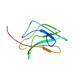
Loading
PDBj
メニューPDBj@FacebookPDBj@TwitterPDBj@YouTubewwPDB FoundationwwPDB
RCSB PDBPDBeBMRBAdv. SearchSearch help
Search by PDB author
1CO1
DownloadVisualize
BU of 1co1 by Molmil
FOLD OF THE CBFA
分子名称: CORE BINDING FACTOR ALPHA
著者Berardi, M.J, Bushweller, J.H.
登録日1999-05-31
公開日2000-06-07
最終更新日2023-12-27
実験手法SOLUTION NMR
主引用文献The Ig fold of the core binding factor alpha Runt domain is a member of a family of structurally and functionally related Ig-fold DNA-binding domains.
Structure Fold.Des., 7, 1999
1QFN
DownloadVisualize
BU of 1qfn by Molmil
GLUTAREDOXIN-1-RIBONUCLEOTIDE REDUCTASE B1 MIXED DISULFIDE BOND
分子名称: PROTEIN (GLUTAREDOXIN 1), PROTEIN (RIBONUCLEOSIDE-DIPHOSPHATE REDUCTASE 1)
著者Berardi, M.J, Bushweller, J.H.
登録日1999-04-12
公開日2000-01-01
最終更新日2023-12-27
実験手法SOLUTION NMR
主引用文献Binding specificity and mechanistic insight into glutaredoxin-catalyzed protein disulfide reduction.
J.Mol.Biol., 292, 1999
2LCK
DownloadVisualize
BU of 2lck by Molmil
Structure of the mitochondrial uncoupling protein 2 determined by NMR molecular fragment replacement
分子名称: Mitochondrial uncoupling protein 2
著者Berardi, M.J, Chou, J.J, Membrane Protein Structures by Solution NMR (MPSbyNMR)
登録日2011-04-29
公開日2011-08-03
最終更新日2011-08-17
実験手法SOLUTION NMR
主引用文献Mitochondrial uncoupling protein 2 structure determined by NMR molecular fragment searching.
Nature, 476, 2011
1JHB
DownloadVisualize
BU of 1jhb by Molmil
HUMAN GLUTAREDOXIN IN FULLY REDUCED FORM, NMR, 20 STRUCTURES
分子名称: GLUTAREDOXIN
著者Sun, C, Bushweller, J.H.
登録日1998-02-17
公開日1998-08-26
最終更新日2022-02-23
実験手法SOLUTION NMR
主引用文献The NMR solution structure of human glutaredoxin in the fully reduced form.
J.Mol.Biol., 280, 1998
2M6X
DownloadVisualize
BU of 2m6x by Molmil
Structure of the p7 channel of Hepatitis C virus, genotype 5a
分子名称: p7
著者OuYang, B, Chou, J.J.
登録日2013-04-12
公開日2013-06-12
最終更新日2014-01-29
実験手法SOLUTION NMR
主引用文献Unusual architecture of the p7 channel from hepatitis C virus
Nature, 498, 2013

218853

件を2024-04-24に公開中

PDB statisticsPDBj update infoContact PDBjnumon