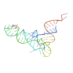
1FIR
 
 | |
4NEB
 
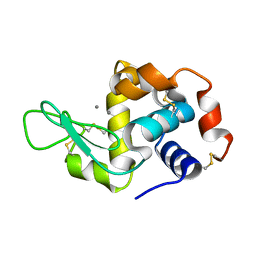 | | Previously de-ionized HEW lysozyme batch crystallized in 0.5 M MnCl2 | | 分子名称: | CHLORIDE ION, Lysozyme C, MANGANESE (II) ION | | 著者 | Benas, P, Legrand, L, Ries-Kautt, M. | | 登録日 | 2013-10-29 | | 公開日 | 2014-05-28 | | 最終更新日 | 2024-10-09 | | 実験手法 | X-RAY DIFFRACTION (1.48 Å) | | 主引用文献 | Weak protein-cationic co-ion interactions addressed by X-ray crystallography and mass spectrometry.
Acta Crystallogr.,Sect.D, 70, 2014
|
|
4NGZ
 
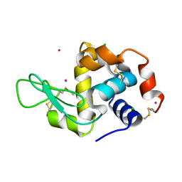 | | Previously de-ionized HEW lysozyme crystallized in 0.5 M YbCl3/30% (v/v) glycerol and collected at 125K | | 分子名称: | CHLORIDE ION, Lysozyme C, YTTERBIUM (III) ION | | 著者 | Benas, P, Legrand, L, Ries-Kautt, M. | | 登録日 | 2013-11-03 | | 公開日 | 2014-05-28 | | 最終更新日 | 2024-11-20 | | 実験手法 | X-RAY DIFFRACTION (1.7 Å) | | 主引用文献 | Weak protein-cationic co-ion interactions addressed by X-ray crystallography and mass spectrometry.
Acta Crystallogr.,Sect.D, 70, 2014
|
|
4NGO
 
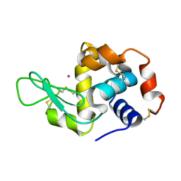 | | Previously de-ionized HEW lysozyme batch crystallized in 1.0 M CoCl2 | | 分子名称: | CHLORIDE ION, COBALT (II) ION, Lysozyme C | | 著者 | Benas, P, Legrand, L, Ries-Kautt, M. | | 登録日 | 2013-11-02 | | 公開日 | 2014-05-28 | | 最終更新日 | 2024-11-20 | | 実験手法 | X-RAY DIFFRACTION (1.58 Å) | | 主引用文献 | Weak protein-cationic co-ion interactions addressed by X-ray crystallography and mass spectrometry.
Acta Crystallogr.,Sect.D, 70, 2014
|
|
4NGW
 
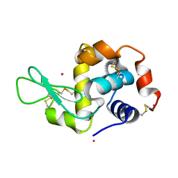 | | Dialyzed HEW lysozyme batch crystallized in 0.5 M YbCl3 and collected at 100 K | | 分子名称: | CHLORIDE ION, Lysozyme C, YTTERBIUM (III) ION | | 著者 | Benas, P, Legrand, L, Ries-Kautt, M. | | 登録日 | 2013-11-03 | | 公開日 | 2014-05-28 | | 最終更新日 | 2024-11-06 | | 実験手法 | X-RAY DIFFRACTION (1.37 Å) | | 主引用文献 | Weak protein-cationic co-ion interactions addressed by X-ray crystallography and mass spectrometry.
Acta Crystallogr.,Sect.D, 70, 2014
|
|
4NGK
 
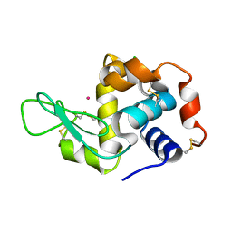 | | Previously de-ionized HEW lysozyme batch crystallized in 0.2 M CoCl2 | | 分子名称: | CHLORIDE ION, COBALT (II) ION, Lysozyme C | | 著者 | Benas, P, Legrand, L, Ries-Kautt, M. | | 登録日 | 2013-11-02 | | 公開日 | 2014-05-28 | | 最終更新日 | 2024-10-30 | | 実験手法 | X-RAY DIFFRACTION (1.5 Å) | | 主引用文献 | Weak protein-cationic co-ion interactions addressed by X-ray crystallography and mass spectrometry.
Acta Crystallogr.,Sect.D, 70, 2014
|
|
4NGJ
 
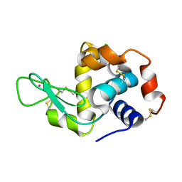 | | Dialyzed HEW lysozyme batch crystallized in 1.0 M RbCl and collected at 100 K | | 分子名称: | CHLORIDE ION, Lysozyme C, RUBIDIUM ION | | 著者 | Benas, P, Legrand, L, Ries-Kautt, M. | | 登録日 | 2013-11-02 | | 公開日 | 2014-05-28 | | 最終更新日 | 2024-10-09 | | 実験手法 | X-RAY DIFFRACTION (1.1 Å) | | 主引用文献 | Weak protein-cationic co-ion interactions addressed by X-ray crystallography and mass spectrometry.
Acta Crystallogr.,Sect.D, 70, 2014
|
|
4NFV
 
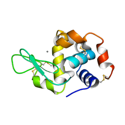 | | Previously de-ionized HEW lysozyme batch crystallized in 1.1 M MnCl2 | | 分子名称: | CHLORIDE ION, Lysozyme C, MANGANESE (II) ION | | 著者 | Benas, P, Legrand, L, Ries-Kautt, M. | | 登録日 | 2013-11-01 | | 公開日 | 2014-05-28 | | 最終更新日 | 2024-11-20 | | 実験手法 | X-RAY DIFFRACTION (1.63 Å) | | 主引用文献 | Weak protein-cationic co-ion interactions addressed by X-ray crystallography and mass spectrometry.
Acta Crystallogr.,Sect.D, 70, 2014
|
|
4NG1
 
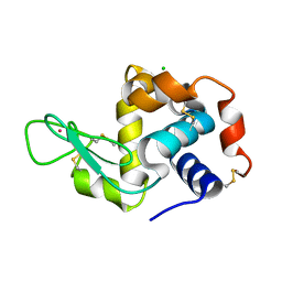 | | Previously de-ionized HEW lysozyme batch crystallized in 1.9 M CsCl | | 分子名称: | CESIUM ION, CHLORIDE ION, Lysozyme C | | 著者 | Benas, P, Legrand, L, Ries-Kautt, M. | | 登録日 | 2013-11-01 | | 公開日 | 2014-05-28 | | 最終更新日 | 2024-11-20 | | 実験手法 | X-RAY DIFFRACTION (1.82 Å) | | 主引用文献 | Weak protein-cationic co-ion interactions addressed by X-ray crystallography and mass spectrometry.
Acta Crystallogr.,Sect.D, 70, 2014
|
|
4NGI
 
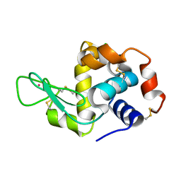 | | Previously de-ionized HEW lysozyme crystallized in 1.0 M RbCl and collected at 125K | | 分子名称: | CHLORIDE ION, Lysozyme C, RUBIDIUM ION | | 著者 | Benas, P, Legrand, L, Ries-Kautt, M. | | 登録日 | 2013-11-02 | | 公開日 | 2014-05-28 | | 最終更新日 | 2024-11-20 | | 実験手法 | X-RAY DIFFRACTION (1.7 Å) | | 主引用文献 | Weak protein-cationic co-ion interactions addressed by X-ray crystallography and mass spectrometry.
Acta Crystallogr.,Sect.D, 70, 2014
|
|
4NGL
 
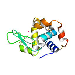 | | Previously de-ionized HEW lysozyme batch crystallized in 0.6 M CoCl2 | | 分子名称: | CHLORIDE ION, COBALT (II) ION, Lysozyme C | | 著者 | Benas, P, Legrand, L, Ries-Kautt, M. | | 登録日 | 2013-11-02 | | 公開日 | 2014-05-28 | | 最終更新日 | 2024-11-20 | | 実験手法 | X-RAY DIFFRACTION (1.52 Å) | | 主引用文献 | Weak protein-cationic co-ion interactions addressed by X-ray crystallography and mass spectrometry.
Acta Crystallogr.,Sect.D, 70, 2014
|
|
4NGV
 
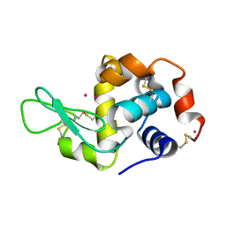 | | Previously de-ionized HEW lysozyme batch crystallized in 0.5 M YbCl3 | | 分子名称: | CHLORIDE ION, Lysozyme C, YTTERBIUM (III) ION | | 著者 | Benas, P, Legrand, L, Ries-Kautt, M. | | 登録日 | 2013-11-03 | | 公開日 | 2014-05-28 | | 最終更新日 | 2024-11-20 | | 実験手法 | X-RAY DIFFRACTION (1.64 Å) | | 主引用文献 | Weak protein-cationic co-ion interactions addressed by X-ray crystallography and mass spectrometry.
Acta Crystallogr.,Sect.D, 70, 2014
|
|
4NGY
 
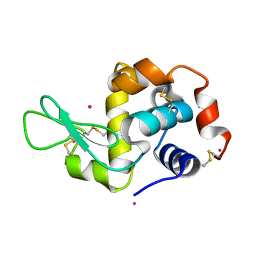 | | Dialyzed HEW lysozyme batch crystallized in 0.75 M YbCl3 and collected at 100 K | | 分子名称: | CHLORIDE ION, Lysozyme C, YTTERBIUM (III) ION | | 著者 | Benas, P, Legrand, L, Ries-Kautt, M. | | 登録日 | 2013-11-03 | | 公開日 | 2014-05-28 | | 最終更新日 | 2024-10-16 | | 実験手法 | X-RAY DIFFRACTION (1.35 Å) | | 主引用文献 | Weak protein-cationic co-ion interactions addressed by X-ray crystallography and mass spectrometry.
Acta Crystallogr.,Sect.D, 70, 2014
|
|
4NG8
 
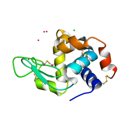 | | Dialyzed HEW lysozyme batch crystallized in 1.9 M CsCl and collected at 100 K. | | 分子名称: | CESIUM ION, CHLORIDE ION, Lysozyme C | | 著者 | Benas, P, Legrand, L, Ries-Kautt, M. | | 登録日 | 2013-11-01 | | 公開日 | 2014-05-28 | | 最終更新日 | 2024-11-27 | | 実験手法 | X-RAY DIFFRACTION (1.09 Å) | | 主引用文献 | Weak protein-cationic co-ion interactions addressed by X-ray crystallography and mass spectrometry.
Acta Crystallogr.,Sect.D, 70, 2014
|
|
8BHD
 
 | | N-terminal domain of Plasmodium berghei glutamyl-tRNA synthetase (Tbxo4 derivative crystal structure) | | 分子名称: | GLYCEROL, Glutamate--tRNA ligase, SULFATE ION, ... | | 著者 | Benas, P, Jaramillo Ponce, J.R, Legrand, P, Frugier, M, Sauter, C. | | 登録日 | 2022-10-31 | | 公開日 | 2023-01-25 | | 最終更新日 | 2024-06-19 | | 実験手法 | X-RAY DIFFRACTION (3.17 Å) | | 主引用文献 | Solution X-ray scattering highlights discrepancies in Plasmodium multi-aminoacyl-tRNA synthetase complexes.
Protein Sci., 32, 2023
|
|
8BCQ
 
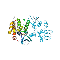 | | N-terminal domain of Plasmodium berghei glutamyl-tRNA synthetase (native crystal structure) | | 分子名称: | GLYCEROL, Glutamate--tRNA ligase, SULFATE ION | | 著者 | Benas, P, Jaramillo Ponce, J.R, Frugier, M, Sauter, C. | | 登録日 | 2022-10-17 | | 公開日 | 2023-01-25 | | 最終更新日 | 2024-02-07 | | 実験手法 | X-RAY DIFFRACTION (2.7 Å) | | 主引用文献 | Solution X-ray scattering highlights discrepancies in Plasmodium multi-aminoacyl-tRNA synthetase complexes.
Protein Sci., 32, 2023
|
|
2CLB
 
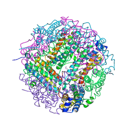 | | The structure of the DPS-like protein from Sulfolobus solfataricus reveals a bacterioferritin-like di-metal binding site within a Dps- like dodecameric assembly | | 分子名称: | DPS-LIKE PROTEIN, FE (III) ION, ZINC ION | | 著者 | Gauss, G.H, Benas, P, Wiedenheft, B, Young, M, Douglas, T, Lawrence, C.M. | | 登録日 | 2006-04-26 | | 公開日 | 2006-07-17 | | 最終更新日 | 2024-11-20 | | 実験手法 | X-RAY DIFFRACTION (2.4 Å) | | 主引用文献 | Structure of the Dps-Like Protein from Sulfolobus Solfataricus Reveals a Bacterioferritin-Like Dimetal Binding Site within a Dps-Like Dodecameric Assembly.
Biochemistry, 45, 2006
|
|
2QIR
 
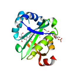 | | Crystal structure of aminoglycoside acetyltransferase AAC(6')-Ib in complex whith coenzyme A and kanamycin | | 分子名称: | (1R,2S,3S,4R,6S)-4,6-DIAMINO-3-[(3-AMINO-3-DEOXY-ALPHA-D-GLUCOPYRANOSYL)OXY]-2-HYDROXYCYCLOHEXYL 2,6-DIAMINO-2,6-DIDEOXY-ALPHA-D-GLUCOPYRANOSIDE, Aminoglycoside 6-N-acetyltransferase type Ib11, COENZYME A | | 著者 | Maurice, F, Broutin, I, Podglajen, I, Benas, P, Collatz, E, Dardel, F. | | 登録日 | 2007-07-05 | | 公開日 | 2008-04-08 | | 最終更新日 | 2023-08-30 | | 実験手法 | X-RAY DIFFRACTION (2.4 Å) | | 主引用文献 | Enzyme structural plasticity and the emergence of broad-spectrum antibiotic resistance.
Embo Rep., 9, 2008
|
|
2PRB
 
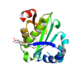 | | crystal structure of aminoglycoside acetyltransferase AAC(6')-Ib in complex whith coenzyme A | | 分子名称: | Aminoglycoside 6-N-acetyltransferase type Ib11, COENZYME A | | 著者 | Maurice, F, Broutin, I, Podglajen, I, Benas, P, Collatz, E, Dardel, F. | | 登録日 | 2007-05-04 | | 公開日 | 2008-04-08 | | 最終更新日 | 2023-08-30 | | 実験手法 | X-RAY DIFFRACTION (1.8 Å) | | 主引用文献 | Enzyme structural plasticity and the emergence of broad-spectrum antibiotic resistance.
Embo Rep., 9, 2008
|
|
2PR8
 
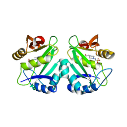 | | crystal structure of aminoglycoside N-acetyltransferase AAC(6')-Ib11 | | 分子名称: | 4-(2-HYDROXYETHYL)-1-PIPERAZINE ETHANESULFONIC ACID, Aminoglycoside 6-N-acetyltransferase type Ib11 | | 著者 | Maurice, F, Broutin, I, Podglajen, I, Benas, P, Collatz, E, Dardel, F. | | 登録日 | 2007-05-04 | | 公開日 | 2008-04-08 | | 最終更新日 | 2024-02-21 | | 実験手法 | X-RAY DIFFRACTION (2.1 Å) | | 主引用文献 | Enzyme structural plasticity and the emergence of broad-spectrum antibiotic resistance.
Embo Rep., 9, 2008
|
|
3D5K
 
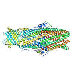 | |
6TVZ
 
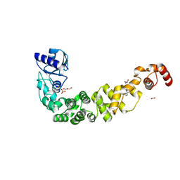 | | Structure of a psychrophilic CCA-adding enzyme crystallized in the XtalController device | | 分子名称: | ACETATE ION, CCA-adding enzyme, GLYCEROL, ... | | 著者 | de Wijn, R, Rollet, K, Coudray, L, Hennig, O, Betat, H, Moerl, M, Lorber, B, Sauter, C. | | 登録日 | 2020-01-10 | | 公開日 | 2020-12-16 | | 最終更新日 | 2024-01-24 | | 実験手法 | X-RAY DIFFRACTION (2.28 Å) | | 主引用文献 | Monitoring the Production of High Diffraction-Quality Crystals of Two Enzymes in Real Time Using In Situ Dynamic Light Scattering
Crystals, 2020
|
|
6TVY
 
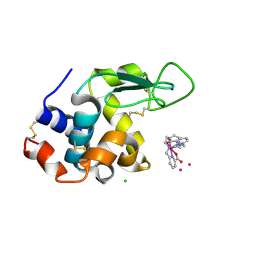 | | Structure of hen egg white lysozyme crystallized in the presence of Tb-Xo4 crystallophore in the XtalController device | | 分子名称: | CHLORIDE ION, Lysozyme C, SODIUM ION, ... | | 著者 | de Wijn, R, Rollet, K, Coudray, L, McEwen, A.G, Lorber, B, Sauter, C. | | 登録日 | 2020-01-10 | | 公開日 | 2020-12-16 | | 最終更新日 | 2024-10-23 | | 実験手法 | X-RAY DIFFRACTION (1.51 Å) | | 主引用文献 | Monitoring the Production of High Diffraction-Quality Crystals of Two Enzymes in Real Time Using In Situ Dynamic Light Scattering
Crystals, 2020
|
|
5IUY
 
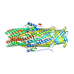 | | Structural insights of the outer-membrane channel OprN | | 分子名称: | CHLORIDE ION, FORMIC ACID, Multidrug efflux outer membrane protein OprN, ... | | 著者 | Ntsogo, Y, Garnier, C, Phan, G, Monlezun, L, Benas, P, Broutin, I. | | 登録日 | 2016-03-18 | | 公開日 | 2016-07-06 | | 最終更新日 | 2024-11-20 | | 実験手法 | X-RAY DIFFRACTION (2.29 Å) | | 主引用文献 | Xenon for tunnelling analysis of the efflux pump component OprN.
PLoS ONE, 12, 2017
|
|
