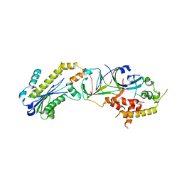
5V4B
 
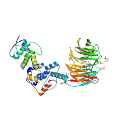 | | Crystal structure of the Skp1-FBXW7-DISC1 complex | | 分子名称: | DISC1 peptide, F-box/WD repeat-containing protein 7, IMIDAZOLE, ... | | 著者 | Li, Y, Baillie, G.S, Hao, B. | | 登録日 | 2017-03-08 | | 公開日 | 2017-09-20 | | 最終更新日 | 2023-10-04 | | 実験手法 | X-RAY DIFFRACTION (2.6 Å) | | 主引用文献 | FBXW7 regulates DISC1 stability via the ubiquitin-proteosome system.
Mol. Psychiatry, 23, 2018
|
|
8E1D
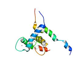 | |
2MH0
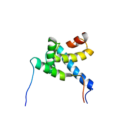 | |
1E9K
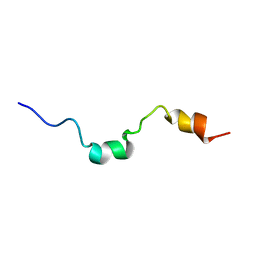 | | The structure of the RACK1 interaction sites located within the unique N-terminal region of the cAMP-specific phosphodiesterase, PDE4D5. | | 分子名称: | cAMP-specific 3',5'-cyclic phosphodiesterase 4D | | 著者 | Bolger, G.B, Smith, K.J, McCahill, A, Hyde, E.I, Steele, M.R, Houslay, M.D. | | 登録日 | 2000-10-20 | | 公開日 | 2001-10-18 | | 最終更新日 | 2018-06-20 | | 実験手法 | SOLUTION NMR | | 主引用文献 | 1H NMR structural and functional characterisation of a cAMP-specific phosphodiesterase-4D5 (PDE4D5) N-terminal region peptide that disrupts PDE4D5 interaction with the signalling scaffold proteins, beta-arrestin and RACK1.
Cell. Signal., 19, 2007
|
|
3P56
 | |
3P5J
 | |


