1QVE
 
 | |
1RG0
 | |
1JXN
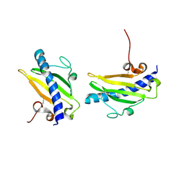
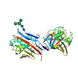
 | | Crystal Structure of the Lectin I from Ulex europaeus in complex with the methyl glycoside of alpha-L-fucose | | 分子名称: | (4R)-2-METHYLPENTANE-2,4-DIOL, CALCIUM ION, MANGANESE (II) ION, ... | | 著者 | Audette, G.F, Olson, D.J.H, Ross, A.R.S, Quail, J.W, Delbaere, L.T.J. | | 登録日 | 2001-09-07 | | 公開日 | 2002-12-06 | | 最終更新日 | 2023-08-16 | | 実験手法 | X-RAY DIFFRACTION (2.3 Å) | | 主引用文献 | Examination of the Structural Basis for O(H) Blood Group Specificity by Ulex europaeus Lectin I
Can.J.Chem., 80, 2002
|
|
1FU0
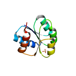
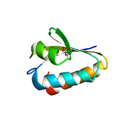 | | CRYSTAL STRUCTURE ANALYSIS OF THE PHOSPHO-SERINE 46 HPR FROM ENTEROCOCCUS FAECALIS | | 分子名称: | PHOSPHOCARRIER PROTEIN HPR | | 著者 | Audette, G.F, Engelmann, R, Hengstenberg, W, Deutscher, J, Hayakawa, K, Quail, J.W, Delbaere, L.T.J. | | 登録日 | 2000-09-13 | | 公開日 | 2000-11-22 | | 最終更新日 | 2022-12-21 | | 実験手法 | X-RAY DIFFRACTION (1.9 Å) | | 主引用文献 | The 1.9 A resolution structure of phospho-serine 46 HPr from Enterococcus faecalis.
J.Mol.Biol., 303, 2000
|
|
1FX5
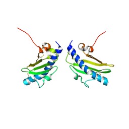
 | | CRYSTAL STRUCTURE ANALYSIS OF ULEX EUROPAEUS LECTIN I | | 分子名称: | (4R)-2-METHYLPENTANE-2,4-DIOL, ANTI-H(O) LECTIN I, CALCIUM ION, ... | | 著者 | Audette, G.F, Vandonselaar, M, Delbaere, L.T.J. | | 登録日 | 2000-09-25 | | 公開日 | 2001-01-17 | | 最終更新日 | 2023-08-09 | | 実験手法 | X-RAY DIFFRACTION (2.2 Å) | | 主引用文献 | The 2.2 A resolution structure of the O(H) blood-group-specific lectin I from Ulex europaeus.
J.Mol.Biol., 304, 2000
|
|
6BHF
 | |
6M8O
 | |
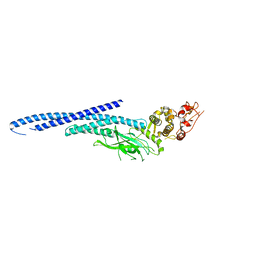
6MBZ
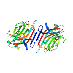
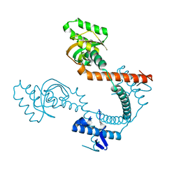
 | | Structure of Transcription Factor | | 分子名称: | Signal transducer and activator of transcription 5B | | 著者 | Seo, H.-S, Dhe-Paganon, S. | | 登録日 | 2018-08-30 | | 公開日 | 2019-06-19 | | 最終更新日 | 2023-10-11 | | 実験手法 | X-RAY DIFFRACTION (3.21 Å) | | 主引用文献 | Structural and functional consequences of the STAT5BN642Hdriver mutation.
Nat Commun, 10, 2019
|
|
6MBW
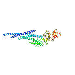
 | | Structure of Transcription Factor | | 分子名称: | Signal transducer and activator of transcription 5B | | 著者 | Seo, H.-S, Dhe-Paganon, S. | | 登録日 | 2018-08-30 | | 公開日 | 2019-06-19 | | 最終更新日 | 2023-10-11 | | 実験手法 | X-RAY DIFFRACTION (3.29 Å) | | 主引用文献 | Structural and functional consequences of the STAT5BN642H driver mutation.
Nat Commun, 10, 2019
|
|


