Loading
PDBj
MenuPDBj@FacebookPDBj@TwitterPDBj@YouTubewwPDB FoundationwwPDB
RCSB PDBPDBeBMRBAdv. SearchSearch help
Search by PDB author
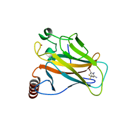
5AOL
DownloadVisualize
BU of 5aol by Molmil
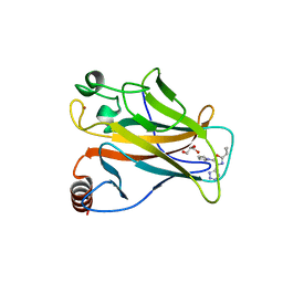
Structure of the p53 cancer mutant Y220C with bound 3-bromo-5-(trifluoromethyl)benzene-1,2-diamine
Descriptor: 3-BROMO-5-(TRIFLUOROMETHYL)BENZENE-1,2-DIAMINE, CELLULAR TUMOR ANTIGEN P53, ZINC ION
Authors:Joerger, A.C, Harbrecht, H.
Deposit date:2015-09-10
Release date:2015-12-16
Last modified:2024-01-10
Method:X-RAY DIFFRACTION (1.5 Å)
Cite:Exploiting Transient Protein States for the Design of Small-Molecule Stabilizers of Mutant P53.
Structure, 23, 2015
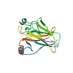
5AOJ
DownloadVisualize
BU of 5aoj by Molmil
Structure of the p53 cancer mutant Y220C in complex with 2-hydroxy-3, 5-diiodo-4-(1H-pyrrol-1-yl)benzoic acid
Descriptor: 2-hydroxy-3,5-diiodo-4-(1H-pyrrol-1-yl)benzoic acid, CELLULAR TUMOR ANTIGEN P53, DI(HYDROXYETHYL)ETHER, ...
Authors:Joerger, A.C, Baud, M.G, Bauer, M.R, Fersht, A.R.
Deposit date:2015-09-10
Release date:2015-12-16
Last modified:2024-01-10
Method:X-RAY DIFFRACTION (1.47 Å)
Cite:Exploiting Transient Protein States for the Design of Small-Molecule Stabilizers of Mutant P53.
Structure, 23, 2015
5AOK
DownloadVisualize
BU of 5aok by Molmil
Structure of the p53 cancer mutant Y220C with bound small molecule PhiKan7099
Descriptor: 5-[2-cyclopropyl-5-(1H-pyrrol-1-yl)-1,3-oxazol-4-yl]-1H-1,2,3,4-tetrazole, CELLULAR TUMOR ANTIGEN P53, DI(HYDROXYETHYL)ETHER, ...
Authors:Joerger, A.C.
Deposit date:2015-09-10
Release date:2015-12-16
Last modified:2024-01-10
Method:X-RAY DIFFRACTION (1.35 Å)
Cite:Exploiting Transient Protein States for the Design of Small-Molecule Stabilizers of Mutant P53.
Structure, 23, 2015
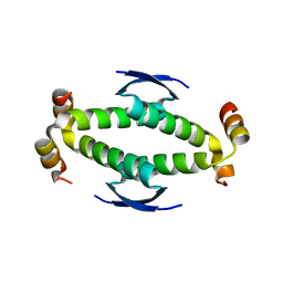
4CKM
DownloadVisualize
BU of 4ckm by Molmil
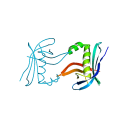
Structure of the N-terminal domain of Leishmania SAS-6
Descriptor: GLYCEROL, MAGNESIUM ION, SAS-6
Authors:van Breugel, M.
Deposit date:2014-01-07
Release date:2014-03-05
Last modified:2024-05-08
Method:X-RAY DIFFRACTION (2.15 Å)
Cite:Structure of the SAS-6 cartwheel hub from Leishmania major.
Elife, 3, 2014
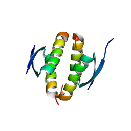
4CKP
DownloadVisualize
BU of 4ckp by Molmil
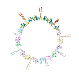
Structure of an N-terminal fragment of Leishmania SAS-6 that contains part of its coiled coil domain
Descriptor: SAS-6
Authors:van Breugel, M.
Deposit date:2014-01-07
Release date:2014-03-05
Last modified:2023-12-20
Method:X-RAY DIFFRACTION (3.45 Å)
Cite:Structure of the SAS-6 cartwheel hub from Leishmania major.
Elife, 3, 2014
4CZ7
DownloadVisualize
BU of 4cz7 by Molmil
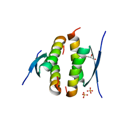
Truncated tetramerization domain of zebrafish p53 (crystal form III)
Descriptor: CELLULAR TUMOR ANTIGEN P53, GLYCEROL, PHOSPHATE ION
Authors:Joerger, A.C.
Deposit date:2014-04-16
Release date:2014-08-27
Last modified:2024-05-08
Method:X-RAY DIFFRACTION (1.1 Å)
Cite:Tracing the Evolution of the P53 Tetramerization Domain
Structure, 22, 2014
4D1L
DownloadVisualize
BU of 4d1l by Molmil
Tetramerization domain of zebrafish p53 (crystal form I)
Descriptor: CELLULAR TUMOR ANTIGEN P53, ZINC ION
Authors:Joerger, A.C.
Deposit date:2014-05-02
Release date:2014-08-27
Last modified:2023-12-20
Method:X-RAY DIFFRACTION (1.97 Å)
Cite:Tracing the Evolution of the P53 Tetramerization Domain
Structure, 22, 2014
4CZ5
DownloadVisualize
BU of 4cz5 by Molmil
Truncated tetramerization domain of zebrafish p53 (crystal form I)
Descriptor: CELLULAR TUMOR ANTIGEN P53
Authors:Joerger, A.C.
Deposit date:2014-04-16
Release date:2014-08-27
Last modified:2024-05-08
Method:X-RAY DIFFRACTION (1.02 Å)
Cite:Tracing the Evolution of the P53 Tetramerization Domain
Structure, 22, 2014
<12


220113

數據於2024-05-22公開中

PDB statisticsPDBj update infoContact PDBjnumon