3AYA
 
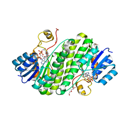
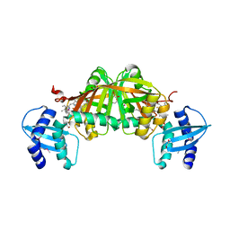
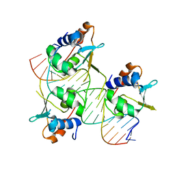
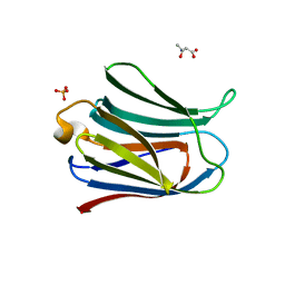 | | Crystal structure of galectin-3 CRD domian complexed with Thomsen-Friedenreich antigen | | 分子名称: | Galectin-3, SULFATE ION, THREONINE, ... | | 著者 | Bian, C.F, Li, D.F, Wang, D.C. | | 登録日 | 2011-05-04 | | 公開日 | 2011-10-12 | | 最終更新日 | 2023-11-01 | | 実験手法 | X-RAY DIFFRACTION (2 Å) | | 主引用文献 | Structural basis for distinct binding properties of the human galectins to thomsen-friedenreich antigen
Plos One, 6, 2011
|
|
3AYD
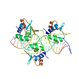
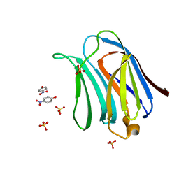 | | Crystal structure of galectin-3 CRD domian complexed with TFN | | 分子名称: | Galectin-3, P-NITROPHENOL, SULFATE ION, ... | | 著者 | Bian, C.F, Li, D.F, Wang, D.C. | | 登録日 | 2011-05-04 | | 公開日 | 2011-10-12 | | 最終更新日 | 2023-11-01 | | 実験手法 | X-RAY DIFFRACTION (1.9 Å) | | 主引用文献 | Structural basis for distinct binding properties of the human galectins to thomsen-friedenreich antigen
Plos One, 6, 2011
|
|
1P9G
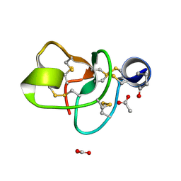 | | Crystal structure of a novel antifungal protein distinct with five disulfide bridges from Ecommia ulmoides Oliver at atomic resolution | | 分子名称: | ACETATE ION, EAFP 2 | | 著者 | Xiang, Y, Huang, R.H, Liu, X.Z, Wang, D.C. | | 登録日 | 2003-05-12 | | 公開日 | 2004-06-01 | | 最終更新日 | 2019-12-25 | | 実験手法 | X-RAY DIFFRACTION (0.84 Å) | | 主引用文献 | Crystal structure of a novel antifungal protein distinct with five disulfide bridges from Eucommia ulmoides Oliver at an atomic resolution.
J.Struct.Biol., 148, 2004
|
|
2P1W
 | |
5X80
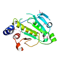
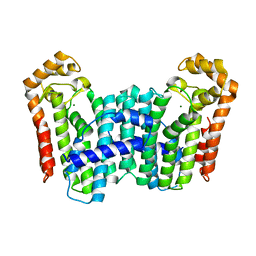
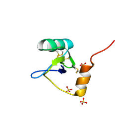
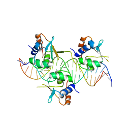
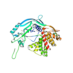
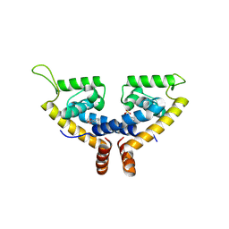 | | CRYSTAL STRUCTURE OF MYCOBACTERIUM TUBERCULOSIS MARR FAMILY PROTEIN RV2887 COMPLEX WITH SALICYLIC ACID | | 分子名称: | 2-HYDROXYBENZOIC ACID, SULFATE ION, Uncharacterized HTH-type transcriptional regulator Rv2887 | | 著者 | Gao, Y.R, Li, D.F, Wang, D.C, Bi, L.J. | | 登録日 | 2017-02-28 | | 公開日 | 2017-08-09 | | 最終更新日 | 2023-11-22 | | 実験手法 | X-RAY DIFFRACTION (2.4 Å) | | 主引用文献 | Structural analysis of the regulatory mechanism of MarR protein Rv2887 in M. tuberculosis
Sci Rep, 7, 2017
|
|
5X7Z
 | | CRYSTAL STRUCTURE OF MYCOBACTERIUM TUBERCULOSIS MARR FAMILY PROTEIN RV2887 COMPLEX WITH P-AMINOSALICYLIC ACID | | 分子名称: | 2-HYDROXY-4-AMINOBENZOIC ACID, Uncharacterized HTH-type transcriptional regulator Rv2887 | | 著者 | Gao, Y.R, Li, D.F, Wang, D.C, Bi, L.J. | | 登録日 | 2017-02-28 | | 公開日 | 2017-08-09 | | 最終更新日 | 2023-11-22 | | 実験手法 | X-RAY DIFFRACTION (2.2 Å) | | 主引用文献 | Structural analysis of the regulatory mechanism of MarR protein Rv2887 in M. tuberculosis
Sci Rep, 7, 2017
|
|
5XUA
 | |
5XUB
 | |
5Y6G
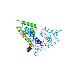
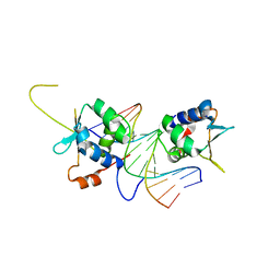
 | | PilZ domain with c-di-GMP of YcgR from Escherichia coli | | 分子名称: | 9,9'-[(2R,3R,3aS,5S,7aR,9R,10R,10aS,12S,14aR)-3,5,10,12-tetrahydroxy-5,12-dioxidooctahydro-2H,7H-difuro[3,2-d:3',2'-j][1,3,7,9,2,8]tetraoxadiphosphacyclododecine-2,9-diyl]bis(2-amino-1,9-dihydro-6H-purin-6-one), Flagellar brake protein YcgR, SULFATE ION | | 著者 | Hou, Y.J, Wang, D.C, Li, D.F. | | 登録日 | 2017-08-11 | | 公開日 | 2018-07-18 | | 最終更新日 | 2023-11-22 | | 実験手法 | X-RAY DIFFRACTION (2.3 Å) | | 主引用文献 | Structural insights into the mechanism of c-di-GMP-bound YcgR regulating flagellar motility inEscherichia coli.
J.Biol.Chem., 295, 2020
|
|
5Y6H
 | |
5Y6F
 | | Crystal structure of YcgR in complex with c-di-GMP from Escherichia coli | | 分子名称: | 9,9'-[(2R,3R,3aS,5S,7aR,9R,10R,10aS,12S,14aR)-3,5,10,12-tetrahydroxy-5,12-dioxidooctahydro-2H,7H-difuro[3,2-d:3',2'-j][1,3,7,9,2,8]tetraoxadiphosphacyclododecine-2,9-diyl]bis(2-amino-1,9-dihydro-6H-purin-6-one), Flagellar brake protein YcgR, SULFATE ION | | 著者 | Hou, Y.J, Wang, D.C, Li, D.F. | | 登録日 | 2017-08-11 | | 公開日 | 2018-07-18 | | 最終更新日 | 2024-03-27 | | 実験手法 | X-RAY DIFFRACTION (2.3 Å) | | 主引用文献 | Structural insights into the mechanism of c-di-GMP-bound YcgR regulating flagellar motility inEscherichia coli.
J.Biol.Chem., 295, 2020
|
|
5YZ2
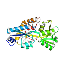
 | | the cystathionine-beta-synthase (CBS) domain of magnesium and cobalt efflux protein CorC in complex with both C2'- and C3'-endo AMP | | 分子名称: | ADENOSINE MONOPHOSPHATE, Magnesium and cobalt efflux protein CorC | | 著者 | Feng, N, Qi, C, Li, D.F, Wang, D.C. | | 登録日 | 2017-12-12 | | 公開日 | 2018-05-30 | | 最終更新日 | 2023-11-22 | | 実験手法 | X-RAY DIFFRACTION (1.75 Å) | | 主引用文献 | The C2'- and C3'-endo equilibrium for AMP molecules bound in the cystathionine-beta-synthase domain.
Biochem. Biophys. Res. Commun., 497, 2018
|
|
5Z2L
 | | Crystal structure of BdcA in complex with NADPH | | 分子名称: | 1,2-ETHANEDIOL, 2-(2-METHOXYETHOXY)ETHANOL, Cyclic-di-GMP-binding biofilm dispersal mediator protein, ... | | 著者 | Yang, W.S, Hou, Y.J, Li, D.F, Wang, D.C. | | 登録日 | 2018-01-03 | | 公開日 | 2018-03-07 | | 最終更新日 | 2023-11-22 | | 実験手法 | X-RAY DIFFRACTION (1.7 Å) | | 主引用文献 | A potential substrate binding pocket of BdcA plays a critical role in NADPH recognition and biofilm dispersal
Biochem. Biophys. Res. Commun., 497, 2018
|
|
3TC1
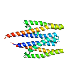
 | | Crystal Structure of Octaprenyl Pyrophosphate Synthase from Helicobacter pylori | | 分子名称: | MAGNESIUM ION, Octaprenyl Pyrophosphate Synthase | | 著者 | Zhang, J.Y, Zhang, X.L, Li, D.F, Zou, Q.M, Wang, D.C. | | 登録日 | 2011-08-08 | | 公開日 | 2011-08-31 | | 最終更新日 | 2024-03-20 | | 実験手法 | X-RAY DIFFRACTION (2 Å) | | 主引用文献 | Crystal Structure of Octaprenyl Pyrophosphate Synthase from Helicobacter pylori
To be Published
|
|
2IA4
 | |
4TQK
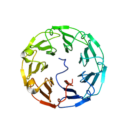
 | | Structural basis of specific recognition of non-reducing terminal N-acetylglucosamine by an Agrocybe aegerita lection | | 分子名称: | 2-acetamido-2-deoxy-beta-D-glucopyranose, Lectin 2 | | 著者 | Hu, Y.L, Ren, X.M, Li, D.F, Jiang, S, Lan, X.Q, Sun, H, Wang, D.C. | | 登録日 | 2014-06-11 | | 公開日 | 2015-06-03 | | 最終更新日 | 2024-03-20 | | 実験手法 | X-RAY DIFFRACTION (2.1 Å) | | 主引用文献 | Structural Basis of Specific Recognition of Non-Reducing Terminal N-Acetylglucosamine by an Agrocybe aegerita Lectin.
Plos One, 10, 2015
|
|
4TQJ
 | | Structural basis of specific recognition of non-reducing terminal N-acetylglucosamine by an Agrocybe aegerita lection | | 分子名称: | Lectin 2 | | 著者 | Hu, Y.L, Ren, X.M, Li, D.F, Jiang, S, Lan, X.Q, Sun, H, Wang, D.C. | | 登録日 | 2014-06-11 | | 公開日 | 2015-06-03 | | 最終更新日 | 2024-03-20 | | 実験手法 | X-RAY DIFFRACTION (2 Å) | | 主引用文献 | Structural Basis of Specific Recognition of Non-Reducing Terminal N-Acetylglucosamine by an Agrocybe aegerita Lectin.
Plos One, 10, 2015
|
|
4TQM
 | | Structural basis of specific recognition of non-reducing terminal N-acetylglucosamine by an Agrocybe aegerita lection | | 分子名称: | 2-acetamido-2-deoxy-beta-D-glucopyranose-(1-3)-beta-D-galactopyranose, 2-acetamido-2-deoxy-beta-D-glucopyranose-(1-3)-beta-D-galactopyranose-(1-4)-2-acetamido-2-deoxy-beta-D-glucopyranose, Lectin 2 | | 著者 | Hu, Y.L, Ren, X.M, Li, D.F, Jiang, S, Lan, X.Q, Sun, H, Wang, D.C. | | 登録日 | 2014-06-11 | | 公開日 | 2015-06-03 | | 最終更新日 | 2024-03-20 | | 実験手法 | X-RAY DIFFRACTION (2 Å) | | 主引用文献 | Structural Basis of Specific Recognition of Non-Reducing Terminal N-Acetylglucosamine by an Agrocybe aegerita Lectin.
Plos One, 10, 2015
|
|
3GAS
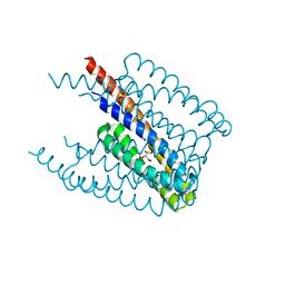
 | | Crystal Structure of Helicobacter pylori Heme Oxygenase Hugz in Complex with Heme | | 分子名称: | 1,2-ETHANEDIOL, AZIDE ION, PROTOPORPHYRIN IX CONTAINING FE, ... | | 著者 | Jiang, F, Hu, Y.L, Guo, Y, Guo, G, Zou, Q.M, Wang, D.C. | | 登録日 | 2009-02-18 | | 公開日 | 2010-02-23 | | 最終更新日 | 2011-07-13 | | 実験手法 | X-RAY DIFFRACTION (1.8 Å) | | 主引用文献 | Crystal Structure of Helicobacter pylori Heme Oxygenase Hugz in Complex with Heme
To be Published
|
|
1T0Z
 | | Structure of an Excitatory Insect-specific Toxin with an Analgesic Effect on Mammalian from Scorpion Buthus martensii Karsch | | 分子名称: | SULFATE ION, insect neurotoxin | | 著者 | Li, C, Guan, R.-J, Xiang, Y, Zhang, Y, Wang, D.-C. | | 登録日 | 2004-04-14 | | 公開日 | 2004-12-28 | | 最終更新日 | 2011-07-13 | | 実験手法 | X-RAY DIFFRACTION (2.6 Å) | | 主引用文献 | Structure of an excitatory insect-specific toxin with an analgesic effect on mammals from the scorpion Buthus martensii Karsch.
Acta Crystallogr.,Sect.D, 61, 2005
|
|
7DCT
 | |
7DCJ
 | |
7DCU
 | |
7DCS
 | |
5B5R
 | | Crystal structure of GSDMA3 | | 分子名称: | Gasdermin-A3 | | 著者 | Ding, J, Shao, F. | | 登録日 | 2016-05-14 | | 公開日 | 2016-06-15 | | 最終更新日 | 2017-09-27 | | 実験手法 | X-RAY DIFFRACTION (1.902 Å) | | 主引用文献 | Pore-forming activity and structural autoinhibition of the gasdermin family.
Nature, 535, 2016
|
|


