3QFP
 
 | |
3QML
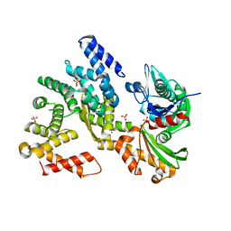 | | The structural analysis of Sil1-Bip complex reveals the mechanism for Sil1 to function as a novel nucleotide exchange factor | | 分子名称: | 78 kDa glucose-regulated protein homolog, MAGNESIUM ION, Nucleotide exchange factor SIL1, ... | | 著者 | Yan, M, Li, J.Z, Sha, B.D. | | 登録日 | 2011-02-04 | | 公開日 | 2011-06-29 | | 最終更新日 | 2024-02-21 | | 実験手法 | X-RAY DIFFRACTION (2.31 Å) | | 主引用文献 | Structural analysis of the Sil1-Bip complex reveals the mechanism for Sil1 to function as a nucleotide-exchange factor.
Biochem.J., 438, 2011
|
|
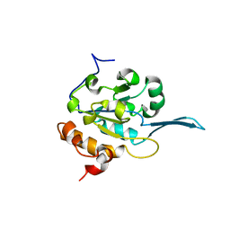
3QFU
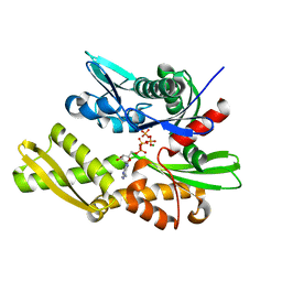 | | Crystal structure of Yeast Hsp70 (Bip/kar2) complexed with ADP | | 分子名称: | 78 kDa glucose-regulated protein homolog, ADENOSINE-5'-DIPHOSPHATE, MAGNESIUM ION, ... | | 著者 | Yan, M, Li, J.Z, Sha, B.D. | | 登録日 | 2011-01-22 | | 公開日 | 2011-06-29 | | 最終更新日 | 2023-09-13 | | 実験手法 | X-RAY DIFFRACTION (1.8 Å) | | 主引用文献 | Structural analysis of the Sil1-Bip complex reveals the mechanism for Sil1 to function as a nucleotide-exchange factor.
Biochem.J., 438, 2011
|
|
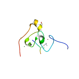
3QLE
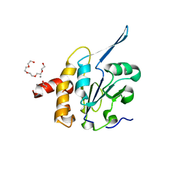 | | Structural Basis for the Function of Tim50 in the Mitochondrial Presequence Translocase | | 分子名称: | ACETATE ION, CALCIUM ION, PENTAETHYLENE GLYCOL, ... | | 著者 | Qian, X.G, Gebert, M, Hpker, J, Yan, M, Li, J.Z, Wiedemann, N, Laan, M.V.D, Pfanner, N, Sha, B.D. | | 登録日 | 2011-02-02 | | 公開日 | 2011-03-02 | | 最終更新日 | 2023-09-13 | | 実験手法 | X-RAY DIFFRACTION (1.831 Å) | | 主引用文献 | Structural basis for the function of tim50 in the mitochondrial presequence translocase.
J.Mol.Biol., 411, 2011
|
|
1JBK
 | |
4QQF
 | |
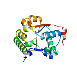
3KQI
 | | crystal structure of PHF2 PHD domain complexed with H3K4Me3 peptide | | 分子名称: | CHLORIDE ION, GLYCEROL, H3K4Me3 peptide, ... | | 著者 | Wen, H, Li, J.Z, Song, T, Lu, M, Lee, M. | | 登録日 | 2009-11-17 | | 公開日 | 2010-02-02 | | 最終更新日 | 2019-02-13 | | 実験手法 | X-RAY DIFFRACTION (1.78 Å) | | 主引用文献 | Recognition of histone H3K4 trimethylation by the plant homeodomain of PHF2 modulates histone demethylation.
J.Biol.Chem., 285, 2010
|
|
3QD2
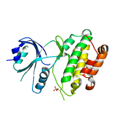 | | Crystal structure of mouse PERK kinase domain | | 分子名称: | Eukaryotic translation initiation factor 2-alpha kinase 3 | | 著者 | Wenjun, C, Jingzhi, L, David, R, Bingdong, S. | | 登録日 | 2011-01-17 | | 公開日 | 2011-04-27 | | 最終更新日 | 2019-01-16 | | 実験手法 | X-RAY DIFFRACTION (2.81 Å) | | 主引用文献 | The structure of the PERK kinase domain suggests the mechanism for its activation.
Acta Crystallogr.,Sect.D, 67, 2011
|
|


