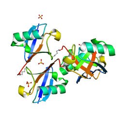
2ZCB
 
 | | Crystal Structure of ubiquitin P37A/P38A | | 分子名称: | Ubiquitin, ZINC ION | | 著者 | Kitahara, R, Tanaka, T, Sakata, E, Yamaguchi, Y, Kato, K, Yokoyama, S. | | 登録日 | 2007-11-08 | | 公開日 | 2007-11-20 | | 最終更新日 | 2023-11-01 | | 実験手法 | X-RAY DIFFRACTION (1.6 Å) | | 主引用文献 | Crystal Structure of ubiquitin P37A/P38A
To be published
|
|
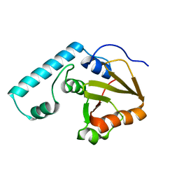
3ALB
 | | Cyclic Lys48-linked tetraubiquitin | | 分子名称: | SULFATE ION, ubiquitin | | 著者 | Satoh, T, Sakata, E, Yamamoto, S, Yamaguchi, Y, Sumiyoshi, A, Wakatsuki, S, Kato, K. | | 登録日 | 2010-07-29 | | 公開日 | 2010-08-25 | | 最終更新日 | 2023-11-01 | | 実験手法 | X-RAY DIFFRACTION (1.85 Å) | | 主引用文献 | Crystal structure of cyclic Lys48-linked tetraubiquitin
Biochem.Biophys.Res.Commun., 2010
|
|
3J47
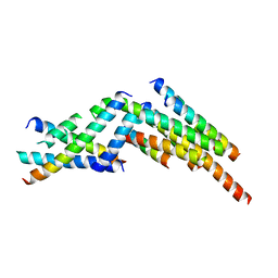 | | Formation of an intricate helical bundle dictates the assembly of the 26S proteasome lid | | 分子名称: | 26S proteasome regulatory subunit RPN11, 26S proteasome regulatory subunit RPN12, 26S proteasome regulatory subunit RPN3, ... | | 著者 | Estrin, E, Lopez-Blanco, J.R, Chacon, P, Martin, A. | | 登録日 | 2013-06-27 | | 公開日 | 2013-08-28 | | 最終更新日 | 2024-02-21 | | 実験手法 | ELECTRON MICROSCOPY (7.4 Å) | | 主引用文献 | Formation of an Intricate Helical Bundle Dictates the Assembly of the 26S Proteasome Lid.
Structure, 21, 2013
|
|
2DOS
 | |
5L4K
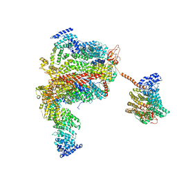 | | The human 26S proteasome lid | | 分子名称: | 26S proteasome complex subunit DSS1, 26S proteasome non-ATPase regulatory subunit 1, 26S proteasome non-ATPase regulatory subunit 11, ... | | 著者 | Schweitzer, A, Aufderheide, A, Rudack, T, Beck, F. | | 登録日 | 2016-05-25 | | 公開日 | 2016-09-07 | | 最終更新日 | 2024-05-08 | | 実験手法 | ELECTRON MICROSCOPY (3.9 Å) | | 主引用文献 | Structure of the human 26S proteasome at a resolution of 3.9 angstrom.
Proc.Natl.Acad.Sci.USA, 113, 2016
|
|
5L4G
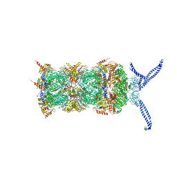 | | The human 26S proteasome at 3.9 A | | 分子名称: | 26S protease regulatory subunit 10B, 26S protease regulatory subunit 4, 26S protease regulatory subunit 6A, ... | | 著者 | Schweitzer, A, Aufderheide, A, Rudack, T, Beck, F. | | 登録日 | 2016-05-25 | | 公開日 | 2016-09-07 | | 最終更新日 | 2024-05-08 | | 実験手法 | ELECTRON MICROSCOPY (3.9 Å) | | 主引用文献 | Structure of the human 26S proteasome at a resolution of 3.9 angstrom.
Proc.Natl.Acad.Sci.USA, 113, 2016
|
|


