Loading
PDBj
MenuPDBj@FacebookPDBj@TwitterPDBj@YouTubewwPDB FoundationwwPDB
RCSB PDBPDBeBMRBAdv. SearchSearch help
Search by PDB author
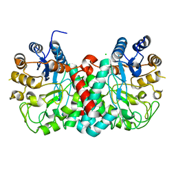
4EGJ
DownloadVisualize
BU of 4egj by Molmil
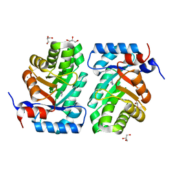
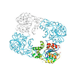
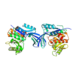
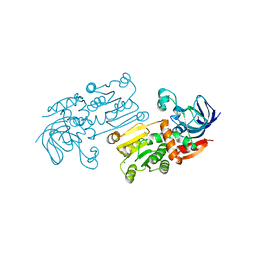
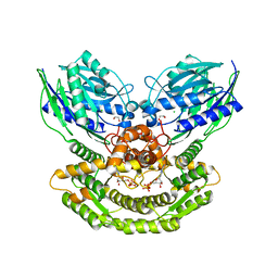
Crystal structure of D-alanine-D-alanine ligase from Burkholderia xenovorans
Descriptor: D-alanine--D-alanine ligase
Authors:SSGCID, Seattle Structural Genomics Center for Infectious Disease (SSGCID)
Deposit date:2012-03-31
Release date:2012-07-18
Last modified:2023-09-13
Method:X-RAY DIFFRACTION (2.3 Å)
Cite:Combining functional and structural genomics to sample the essential Burkholderia structome.
Plos One, 8, 2013
4GNV
DownloadVisualize
BU of 4gnv by Molmil
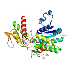
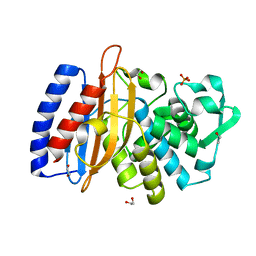
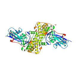
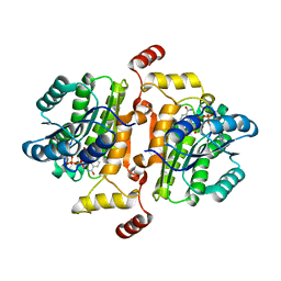
Crystal structure of beta-hexosaminidase 1 from Burkholderia cenocepacia J2315 with bound N-Acetyl-D-Glucosamine
Descriptor: 2-acetamido-2-deoxy-beta-D-glucopyranose, Beta-hexosaminidase 1, CHLORIDE ION
Authors:SSGCID, Seattle Structural Genomics Center for Infectious Disease (SSGCID)
Deposit date:2012-08-17
Release date:2012-08-29
Last modified:2024-02-28
Method:X-RAY DIFFRACTION (1.5 Å)
Cite:Crystal structure of beta-hexosaminidase 1 from Burkholderia cenocepacia J2315 with bound N-Acetyl-D-Glucosamine
TO BE PUBLISHED
3OMF
DownloadVisualize
BU of 3omf by Molmil
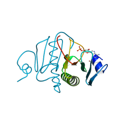
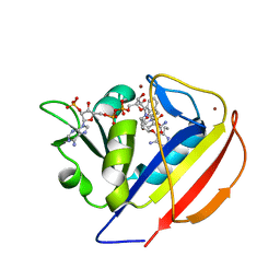
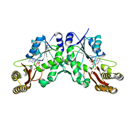
Crystal structure of a histidine triad family protein from Entamoeba histolytica, bound to AMP
Descriptor: ADENOSINE MONOPHOSPHATE, Putative histidine triad family protein, ZINC ION
Authors:SSGCID, Seattle Structural Genomics Center for Infectious Disease (SSGCID)
Deposit date:2010-08-26
Release date:2010-09-08
Last modified:2023-09-06
Method:X-RAY DIFFRACTION (1.8 Å)
Cite:Structures of a histidine triad family protein from Entamoeba histolytica bound to sulfate, AMP and GMP.
Acta Crystallogr F Struct Biol Commun, 71, 2015
5UZH
DownloadVisualize
BU of 5uzh by Molmil
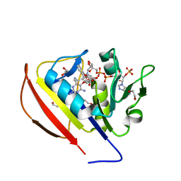
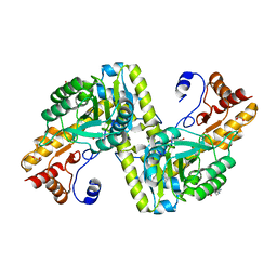
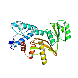
Crystal structure of a GDP-mannose dehydratase from Naegleria fowleri
Descriptor: (4S)-2-METHYL-2,4-PENTANEDIOL, CHLORIDE ION, GUANOSINE-5'-DIPHOSPHATE, ...
Authors:SSGCID, Seattle Structural Genomics Center for Infectious Disease (SSGCID)
Deposit date:2017-02-26
Release date:2017-04-05
Last modified:2023-10-04
Method:X-RAY DIFFRACTION (2.25 Å)
Cite:Crystal structure of a GDP-mannose dehydratase from Naegleria fowleri
to be published
3NDO
DownloadVisualize
BU of 3ndo by Molmil
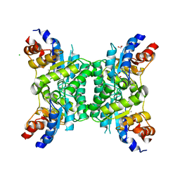
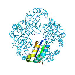
Crystal structure of deoxyribose phosphate aldolase from mycobacterium smegmatis
Descriptor: AMMONIUM ION, Deoxyribose-phosphate aldolase, GLYCEROL, ...
Authors:SSGCID, Seattle Structural Genomics Center for Infectious Disease (SSGCID)
Deposit date:2010-06-07
Release date:2010-07-21
Last modified:2023-09-06
Method:X-RAY DIFFRACTION (1.25 Å)
Cite:Increasing the structural coverage of tuberculosis drug targets.
Tuberculosis (Edinb), 95, 2015
3NG3
DownloadVisualize
BU of 3ng3 by Molmil
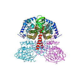
Crystal structure of deoxyribose phosphate aldolase from mycobacterium avium 104 in a schiff base with an unknown aldehyde
Descriptor: 1,2-ETHANEDIOL, CHLORIDE ION, Deoxyribose-phosphate aldolase, ...
Authors:SSGCID, Seattle Structural Genomics Center for Infectious Disease (SSGCID)
Deposit date:2010-06-10
Release date:2010-06-23
Last modified:2023-09-06
Method:X-RAY DIFFRACTION (2.15 Å)
Cite:Increasing the structural coverage of tuberculosis drug targets.
Tuberculosis (Edinb), 95, 2015
5W15
DownloadVisualize
BU of 5w15 by Molmil
Crystal structure of an alpha/beta hydrolase fold protein from Burkholderia ambifaria.
Descriptor: 1,2-ETHANEDIOL, Alpha/beta hydrolase fold protein, CHLORIDE ION, ...
Authors:SSGCID, Seattle Structural Genomics Center for Infectious Disease (SSGCID)
Deposit date:2017-06-01
Release date:2017-06-14
Last modified:2023-10-04
Method:X-RAY DIFFRACTION (1.75 Å)
Cite:Crystal structure of an alpha/beta hydrolase fold protein from Burkholderia ambifaria.
TO BE PUBLISHED
5VPQ
DownloadVisualize
BU of 5vpq by Molmil
Crystal structure of beta-lactamase from Burkholderia phymatum
Descriptor: 1,2-ETHANEDIOL, Beta-lactamase, PHOSPHATE ION
Authors:SSGCID, Seattle Structural Genomics Center for Infectious Disease (SSGCID)
Deposit date:2017-05-05
Release date:2017-05-17
Last modified:2023-10-04
Method:X-RAY DIFFRACTION (1.4 Å)
Cite:Crystal structure of beta-lactamase from Burkholderia phymatum
to be published
5SCO
DownloadVisualize
BU of 5sco by Molmil
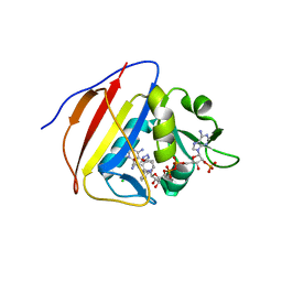
Crystal Structure of Dihydrofolate Reductase from Mycobacterium tuberculosis bound to NADP and SDDC Inhibitor SDDC-M7
Descriptor: 2-(2-{3-[(2,4-diamino-6-ethylpyrimidin-5-yl)oxy]propoxy}phenyl)-N,N-dimethylethane-1-sulfonamide, BROMIDE ION, Dihydrofolate reductase, ...
Authors:Seattle Structural Genomics Center for Infectious Disease (SSGCID)
Deposit date:2021-12-20
Release date:2022-02-09
Last modified:2022-03-02
Method:X-RAY DIFFRACTION (1.3 Å)
Cite:Crystal Structure of Dihydrofolate Reductase from Mycobacterium tuberculosis bound to NADP and SDDC Inhibitor SDDC-M7
to be published
5SD3
DownloadVisualize
BU of 5sd3 by Molmil
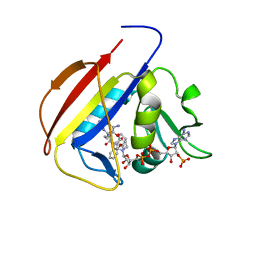
Crystal Structure of Dihydrofolate Reductase from Mycobacterium tuberculosis bound to NADP and SDDC Inhibitor SDDC-1088
Descriptor: 1,2-ETHANEDIOL, 3-{2-[(3-{[(2,4-diamino-6-ethylpyrimidin-5-yl)oxy]methyl}oxetan-3-yl)methoxy]phenyl}propanoic acid, Dihydrofolate reductase, ...
Authors:Seattle Structural Genomics Center for Infectious Disease (SSGCID)
Deposit date:2021-12-20
Release date:2022-02-09
Last modified:2022-03-02
Method:X-RAY DIFFRACTION (1.3 Å)
Cite:Crystal Structure of Dihydrofolate Reductase from Mycobacterium tuberculosis bound to NADP and SDDC Inhibitor SDDC-1088
to be published
5SCV
DownloadVisualize
BU of 5scv by Molmil
Crystal Structure of Dihydrofolate Reductase from Mycobacterium tuberculosis bound to NADP and SDDC Inhibitor SDDC-889
Descriptor: 6-ethyl-5-(4-{3-[3-(1H-tetrazol-5-yl)propyl]phenoxy}butoxy)pyrimidine-2,4-diamine, CHLORIDE ION, Dihydrofolate reductase, ...
Authors:Seattle Structural Genomics Center for Infectious Disease (SSGCID)
Deposit date:2021-12-20
Release date:2022-02-09
Last modified:2022-03-02
Method:X-RAY DIFFRACTION (1.55 Å)
Cite:Crystal Structure of Dihydrofolate Reductase from Mycobacterium tuberculosis bound to NADP and SDDC Inhibitor SDDC-889
to be published
5SCR
DownloadVisualize
BU of 5scr by Molmil
Crystal Structure of Dihydrofolate Reductase from Mycobacterium tuberculosis bound to NADP and SDDC Inhibitor SDDC-735
Descriptor: (2R)-3-(2-{3-[(2,4-diamino-6-ethylpyrimidin-5-yl)oxy]propoxy}phenyl)-2-methylpropanoic acid, Dihydrofolate reductase, NADP NICOTINAMIDE-ADENINE-DINUCLEOTIDE PHOSPHATE
Authors:Seattle Structural Genomics Center for Infectious Disease (SSGCID)
Deposit date:2021-12-20
Release date:2022-02-09
Last modified:2022-03-02
Method:X-RAY DIFFRACTION (1.95 Å)
Cite:Crystal Structure of Dihydrofolate Reductase from Mycobacterium tuberculosis bound to NADP and SDDC Inhibitor SDDC-735
to be published
6PZJ
DownloadVisualize
BU of 6pzj by Molmil
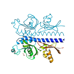
Structure of the N-terminal domain (residues 43-304) of Methyl-accepting chemotaxis protein from Leptospira interrogans serogroup Icterohaemorrhagiae serovar Copenhageni (strain Fiocruz L1-130)
Descriptor: 1,2-ETHANEDIOL, CHLORIDE ION, Methyl-accepting chemotaxis protein
Authors:Seattle Structural Genomics Center for Infectious Disease (SSGCID)
Deposit date:2019-07-31
Release date:2019-08-14
Last modified:2024-03-13
Method:X-RAY DIFFRACTION (1.75 Å)
Cite:Structure of the N-terminal domain (residues 43-304) of Methyl-accepting chemotaxis protein from Leptospira interrogans serogroup Icterohaemorrhagiae serovar Copenhageni (strain Fiocruz L1-130)
to be published
6U3L
DownloadVisualize
BU of 6u3l by Molmil
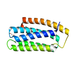
Crystal structure of Hemerythrin HHE cation binding domain-containing protein: Rv2633c homolog from Mycobacterium kansasii
Descriptor: 1,2-ETHANEDIOL, Hemerythrin HHE cation binding domain protein
Authors:Seattle Structural Genomics Center for Infectious Disease (SSGCID)
Deposit date:2019-08-22
Release date:2020-01-22
Last modified:2024-03-13
Method:X-RAY DIFFRACTION (1.75 Å)
Cite:Crystal structure of a hemerythrin-like protein from Mycobacterium kansasii and homology model of the orthologous Rv2633c protein of M. tuberculosis.
Biochem.J., 477, 2020
6XK2
DownloadVisualize
BU of 6xk2 by Molmil
Crystal structure of Ribokinase from Cryptococcus neoformans var. grubii serotype A in complex with ADP
Descriptor: 1,2-ETHANEDIOL, ADENOSINE-5'-DIPHOSPHATE, Ribokinase, ...
Authors:Seattle Structural Genomics Center for Infectious Disease (SSGCID)
Deposit date:2020-06-25
Release date:2020-07-15
Last modified:2023-10-18
Method:X-RAY DIFFRACTION (2.5 Å)
Cite:Crystal structure of Ribokinase from Cryptococcus neoformans var. grubii serotype A in complex with ADP
to be published
6XR5
DownloadVisualize
BU of 6xr5 by Molmil
Crystal Structure of Diphosphomevalonate decarboxylase (MVD1) Cryptococcus neoformans var. grubii serotype A
Descriptor: 1,2-ETHANEDIOL, ADENINE, Diphosphomevalonate decarboxylase, ...
Authors:Seattle Structural Genomics Center for Infectious Disease (SSGCID)
Deposit date:2020-07-11
Release date:2020-09-23
Last modified:2023-10-18
Method:X-RAY DIFFRACTION (1.7 Å)
Cite:Crystal Structure of Diphosphomevalonate decarboxylase (MVD1) from Cryptococcus neoformans var. grubii serotype A
to be published
6X79
DownloadVisualize
BU of 6x79 by Molmil
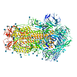
Prefusion SARS-CoV-2 S ectodomain trimer covalently stabilized in the closed conformation
Descriptor: 2-acetamido-2-deoxy-beta-D-glucopyranose, 2-acetamido-2-deoxy-beta-D-glucopyranose-(1-4)-2-acetamido-2-deoxy-beta-D-glucopyranose, Spike glycoprotein
Authors:McCallum, M, Walls, A.C, Corti, D, Veesler, D, Seattle Structural Genomics Center for Infectious Disease (SSGCID)
Deposit date:2020-05-29
Release date:2020-08-19
Last modified:2021-01-27
Method:ELECTRON MICROSCOPY (2.9 Å)
Cite:Structure-guided covalent stabilization of coronavirus spike glycoprotein trimers in the closed conformation.
Nat.Struct.Mol.Biol., 27, 2020
4W5K
DownloadVisualize
BU of 4w5k by Molmil
Structure of a mitochondrial aspartate aminotransferase from Trypanosoma brucei, K237A mutant
Descriptor: 1,2-ETHANEDIOL, Aspartate aminotransferase, mitochondrial, ...
Authors:Seattle Structural Genomics Center for Infectious Disease (SSGCID)
Deposit date:2014-08-18
Release date:2014-09-24
Last modified:2023-09-27
Method:X-RAY DIFFRACTION (1.7 Å)
Cite:Structures of aspartate aminotransferases from Trypanosoma brucei, Leishmania major and Giardia lamblia.
Acta Crystallogr.,Sect.F, 71, 2015
5K9F
DownloadVisualize
BU of 5k9f by Molmil
Crystal structure of a NIPSNAP domain protein from Burkholderia xenovorans
Descriptor: ACETATE ION, NIPSNAP domain protein, OXAMIC ACID
Authors:Seattle Structural Genomics Center for Infectious Disease (SSGCID)
Deposit date:2016-05-31
Release date:2016-06-15
Last modified:2023-09-27
Method:X-RAY DIFFRACTION (1.65 Å)
Cite:Crystal structure of a NIPSNAP domain protein from Burkholderia xenovorans
to be published
5JSC
DownloadVisualize
BU of 5jsc by Molmil
Crystal structure of a Putative acyl-CoA dehydrogenase from Burkholderia xenovorans
Descriptor: 1,2-ETHANEDIOL, CHLORIDE ION, FLAVIN-ADENINE DINUCLEOTIDE, ...
Authors:Seattle Structural Genomics Center for Infectious Disease (SSGCID)
Deposit date:2016-05-07
Release date:2016-06-08
Last modified:2023-09-27
Method:X-RAY DIFFRACTION (1.5 Å)
Cite:Crystal structure of a Putative acyl-CoA dehydrogenase from Burkholderia xenovorans
to be published
5KF0
DownloadVisualize
BU of 5kf0 by Molmil
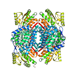
Crystal structure of an Aldedhyde dehydrogenase from Burkholderia vietnamiensis
Descriptor: (4R)-2-METHYLPENTANE-2,4-DIOL, (4S)-2-METHYL-2,4-PENTANEDIOL, ACETATE ION, ...
Authors:Seattle Structural Genomics Center for Infectious Disease (SSGCID)
Deposit date:2016-06-10
Release date:2016-11-09
Last modified:2023-09-27
Method:X-RAY DIFFRACTION (1.7 Å)
Cite:Crystal structure of an Aldedhyde dehydrogenase from Burkholderia vietnamiensis
to be published
5KIA
DownloadVisualize
BU of 5kia by Molmil
Crystal structure of L-threonine 3-dehydrogenase from Burkholderia thailandensis
Descriptor: CALCIUM ION, L-threonine 3-dehydrogenase, ZINC ION
Authors:Seattle Structural Genomics Center for Infectious Disease (SSGCID)
Deposit date:2016-06-16
Release date:2017-02-15
Last modified:2023-09-27
Method:X-RAY DIFFRACTION (2.1 Å)
Cite:Crystal structure of L-threonine 3-dehydrogenase from Burkholderia thailandensis
to be published
5KHA
DownloadVisualize
BU of 5kha by Molmil
Structure of glutamine-dependent NAD+ synthetase from Acinetobacter baumannii in complex with adenosine diphosphate (ADP)
Descriptor: 1,2-ETHANEDIOL, ADENOSINE-5'-DIPHOSPHATE, CHLORIDE ION, ...
Authors:Seattle Structural Genomics Center for Infectious Disease (SSGCID)
Deposit date:2016-06-14
Release date:2016-07-27
Last modified:2023-09-27
Method:X-RAY DIFFRACTION (2.1 Å)
Cite:Structure of glutamine-dependent NAD+ synthetase from Acinetobacter baumannii in complex with adenosine diphosphate (ADP)
to be published
5KOI
DownloadVisualize
BU of 5koi by Molmil
Crystal Structure of a Possible Enoyl-(acyl-carrier-protein) Reductase from Brucella melitensis
Descriptor: Enoyl-[acyl-carrier-protein] reductase [NADH], NICOTINAMIDE-ADENINE-DINUCLEOTIDE
Authors:Seattle Structural Genomics Center for Infectious Disease (SSGCID)
Deposit date:2016-06-30
Release date:2016-07-13
Last modified:2023-10-04
Method:X-RAY DIFFRACTION (1.7 Å)
Cite:Crystal Structure of a Possible Enoyl-(acyl-carrier-protein) Reductase from Brucella melitensis
to be published
5KWV
DownloadVisualize
BU of 5kwv by Molmil
Crystal Structure of a Pantoate-beta-alanine Ligase from Neisseria gonorrhoeae with bound AMPPNP
Descriptor: PHOSPHOAMINOPHOSPHONIC ACID-ADENYLATE ESTER, Pantothenate synthetase
Authors:Seattle Structural Genomics Center for Infectious Disease (SSGCID)
Deposit date:2016-07-19
Release date:2016-07-27
Last modified:2023-10-04
Method:X-RAY DIFFRACTION (2.25 Å)
Cite:Crystal Structure of a Pantoate-beta-alanine Ligase from Neisseria gonorrhoeae with bound AMPPNP
to be published

218853

PDB entries from 2024-04-24

PDB statisticsPDBj update infoContact PDBjnumon