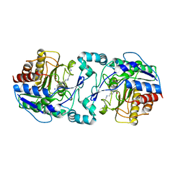
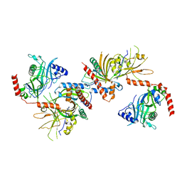
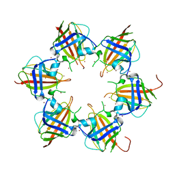
5D18
 
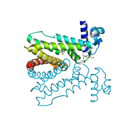 | | Crystal structure of Mycobacterium tuberculosis Rv0302, form I | | 分子名称: | 4-(2-HYDROXYETHYL)-1-PIPERAZINE ETHANESULFONIC ACID, ISOPROPYL ALCOHOL, SODIUM ION, ... | | 著者 | Chou, T.-H, Delmar, J, Su, C.-C, Yu, E. | | 登録日 | 2015-08-04 | | 公開日 | 2015-10-07 | | 最終更新日 | 2024-03-06 | | 実験手法 | X-RAY DIFFRACTION (2.04 Å) | | 主引用文献 | Crystal structure of the Mycobacterium tuberculosis transcriptional regulator Rv0302.
Protein Sci., 2015
|
|
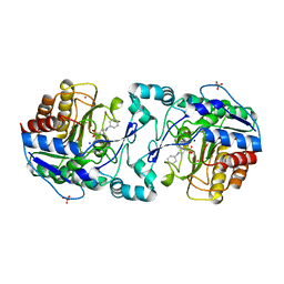
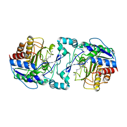
5D1R
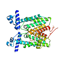 | | Crystal structure of Mycobacterium tuberculosis Rv1816 transcriptional regulator. | | 分子名称: | MAGNESIUM ION, NICKEL (II) ION, Rv1816 transcriptional regulator, ... | | 著者 | Chou, T.-H, Delmar, J, Su, C.-C, Yu, E. | | 登録日 | 2015-08-04 | | 公開日 | 2015-09-30 | | 最終更新日 | 2024-03-06 | | 実験手法 | X-RAY DIFFRACTION (2 Å) | | 主引用文献 | Structural Basis for the Regulation of the MmpL Transporters of Mycobacterium tuberculosis.
J.Biol.Chem., 290, 2015
|
|
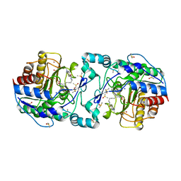
3OPO
 | |
3OW7
 | |
3K0I
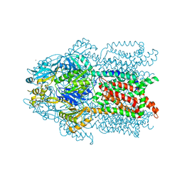 | | Crystal structure of Cu(I)CusA | | 分子名称: | COPPER (I) ION, Cation efflux system protein cusA | | 著者 | Su, C.-C. | | 登録日 | 2009-09-24 | | 公開日 | 2010-10-13 | | 最終更新日 | 2024-02-21 | | 実験手法 | X-RAY DIFFRACTION (4.116 Å) | | 主引用文献 | Crystal structure of CusA
To be Published
|
|
3K07
 | | Crystal structure of CusA | | 分子名称: | Cation efflux system protein cusA | | 著者 | Su, C.-C. | | 登録日 | 2009-09-24 | | 公開日 | 2010-09-22 | | 最終更新日 | 2024-02-21 | | 実験手法 | X-RAY DIFFRACTION (3.521 Å) | | 主引用文献 | Crystal structures of the CusA efflux pump suggest methionine-mediated metal transport.
Nature, 467, 2010
|
|
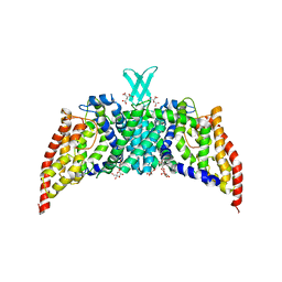
3PJZ
 | | Crystal Structure of the Potassium Transporter TrkH from Vibrio parahaemolyticus | | 分子名称: | POTASSIUM ION, Potassium uptake protein TrkH | | 著者 | Cao, Y, Jin, X, Huang, H, Levin, E.J, Zhou, M, New York Consortium on Membrane Protein Structure (NYCOMPS) | | 登録日 | 2010-11-10 | | 公開日 | 2011-01-19 | | 最終更新日 | 2017-11-08 | | 実験手法 | X-RAY DIFFRACTION (3.506 Å) | | 主引用文献 | Crystal structure of a potassium ion transporter, TrkH.
Nature, 471, 2011
|
|
3KSO
 | |
3QNQ
 | | Crystal structure of the transporter ChbC, the IIC component from the N,N'-diacetylchitobiose-specific phosphotransferase system | | 分子名称: | 2-acetamido-2-deoxy-beta-D-glucopyranose-(1-4)-2-acetamido-2-deoxy-beta-D-glucopyranose, CITRIC ACID, PTS system, ... | | 著者 | Cao, Y, Jin, X, Huang, H, Levin, E.J, Zhou, M, New York Consortium on Membrane Protein Structure (NYCOMPS) | | 登録日 | 2011-02-08 | | 公開日 | 2011-04-06 | | 最終更新日 | 2024-02-21 | | 実験手法 | X-RAY DIFFRACTION (3.295 Å) | | 主引用文献 | Crystal structure of a phosphorylation-coupled saccharide transporter.
Nature, 473, 2011
|
|
3Q9E
 | |
3Q9F
 | | Crystal Structure of APAH complexed with CAPS | | 分子名称: | 3-CYCLOHEXYL-1-PROPYLSULFONIC ACID, Acetylpolyamine amidohydrolase, PHOSPHATE ION, ... | | 著者 | Lombardi, P.M, Christianson, D.W. | | 登録日 | 2011-01-07 | | 公開日 | 2011-03-02 | | 最終更新日 | 2024-04-03 | | 実験手法 | X-RAY DIFFRACTION (2.35 Å) | | 主引用文献 | Structure of prokaryotic polyamine deacetylase reveals evolutionary functional relationships with eukaryotic histone deacetylases .
Biochemistry, 50, 2011
|
|
3Q9B
 | | Crystal Structure of APAH complexed with M344 | | 分子名称: | 4-(dimethylamino)-N-[7-(hydroxyamino)-7-oxoheptyl]benzamide, Acetylpolyamine amidohydrolase, DIMETHYL SULFOXIDE, ... | | 著者 | Lombardi, P.M, Christianson, D.W. | | 登録日 | 2011-01-07 | | 公開日 | 2011-03-02 | | 最終更新日 | 2023-09-13 | | 実験手法 | X-RAY DIFFRACTION (2.25 Å) | | 主引用文献 | Structure of prokaryotic polyamine deacetylase reveals evolutionary functional relationships with eukaryotic histone deacetylases .
Biochemistry, 50, 2011
|
|
3KSS
 | |
3KYH
 | |
3Q9C
 | |
3RBI
 | |
3RBJ
 | |
3RCC
 | |
3RBK
 | |
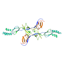
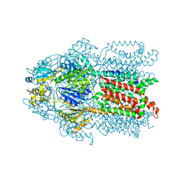
3MBW
 | | Crystal structure of the human ephrin A2 LBD and CRD domains in complex with ephrin A1 | | 分子名称: | Ephrin type-A receptor 2, Ephrin-A1, UNKNOWN ATOM OR ION, ... | | 著者 | Walker, J.R, Yermekbayeva, L, Seitova, A, Butler-Cole, C, Bountra, C, Weigelt, J, Arrowsmith, C.H, Edwards, A.M, Bochkarev, A, Dhe-Paganon, S, Structural Genomics Consortium (SGC) | | 登録日 | 2010-03-26 | | 公開日 | 2010-06-09 | | 最終更新日 | 2023-09-06 | | 実験手法 | X-RAY DIFFRACTION (2.81 Å) | | 主引用文献 | Architecture of Eph receptor clusters.
Proc.Natl.Acad.Sci.USA, 107, 2010
|
|
3NE5
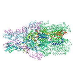 | |
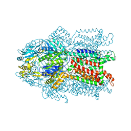
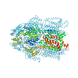
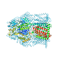
3T53
 | | Crystal structures of the extrusion state of the CusBA adaptor-transporter complex | | 分子名称: | COPPER (II) ION, Cation efflux system protein CusA, Cation efflux system protein CusB | | 著者 | Su, C.-C, Long, F, Yu, E.W. | | 登録日 | 2011-07-26 | | 公開日 | 2012-06-20 | | 最終更新日 | 2024-02-28 | | 実験手法 | X-RAY DIFFRACTION (3.37 Å) | | 主引用文献 | Charged Amino Acids (R83, E567, D617, E625, R669, and K678) of CusA Are Required for Metal Ion Transport in the Cus Efflux System.
J.Mol.Biol., 422, 2012
|
|
3T51
 | | Crystal structures of the pre-extrusion and extrusion states of the CusBA adaptor-transporter complex | | 分子名称: | COPPER (II) ION, Cation efflux system protein CusA, Cation efflux system protein CusB | | 著者 | Su, C.-C, Long, F, Yu, E.W. | | 登録日 | 2011-07-26 | | 公開日 | 2012-06-20 | | 最終更新日 | 2024-02-28 | | 実験手法 | X-RAY DIFFRACTION (3.9 Å) | | 主引用文献 | Charged Amino Acids (R83, E567, D617, E625, R669, and K678) of CusA Are Required for Metal Ion Transport in the Cus Efflux System.
J.Mol.Biol., 422, 2012
|
|
3T56
 | | Crystal structure of the pre-extrusion state of the CusBA adaptor-transporter complex | | 分子名称: | COPPER (II) ION, Cation efflux system protein CusA, Cation efflux system protein CusB | | 著者 | Su, C.-C, Long, F, Yu, E.W. | | 登録日 | 2011-07-26 | | 公開日 | 2012-06-20 | | 最終更新日 | 2024-02-28 | | 実験手法 | X-RAY DIFFRACTION (3.42 Å) | | 主引用文献 | Charged Amino Acids (R83, E567, D617, E625, R669, and K678) of CusA Are Required for Metal Ion Transport in the Cus Efflux System.
J.Mol.Biol., 422, 2012
|
|
4MT1
 | |


