7VIO
 
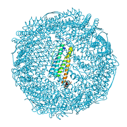 | | Crystal structure of recombinant horse spleen apo-R168H/L169C-Fr | | 分子名称: | CADMIUM ION, Ferritin light chain, GLYCEROL, ... | | 著者 | Lu, C, Peng, X, Maity, B, Ito, N, Abe, S, Wu, J, Ueno, T, Lu, D. | | 登録日 | 2021-09-27 | | 公開日 | 2022-06-08 | | 最終更新日 | 2023-11-29 | | 実験手法 | X-RAY DIFFRACTION (1.5 Å) | | 主引用文献 | Design of a gold clustering site in an engineered apo-ferritin cage.
Commun Chem, 5, 2022
|
|
7VIP
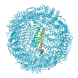 | | Crystal structure of Au(10EQ)-apo-R168H/L169C-rHLFr | | 分子名称: | CADMIUM ION, Ferritin light chain, GLYCEROL, ... | | 著者 | Lu, C, Peng, X, Maity, B, Ito, N, Abe, S, Ueno, T, Lu, D. | | 登録日 | 2021-09-27 | | 公開日 | 2022-06-08 | | 最終更新日 | 2023-11-29 | | 実験手法 | X-RAY DIFFRACTION (1.9 Å) | | 主引用文献 | Design of a gold clustering site in an engineered apo-ferritin cage.
Commun Chem, 5, 2022
|
|
7VIU
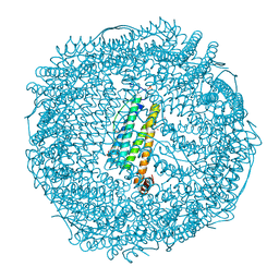 | | Crystal structure of Au(200EQ)-apo-R168C/L169C-rHLFr | | 分子名称: | CADMIUM ION, CHLORIDE ION, Ferritin light chain, ... | | 著者 | Lu, C, Peng, X, Maity, B, Ito, N, Abe, S, Ueno, T, Lu, D. | | 登録日 | 2021-09-27 | | 公開日 | 2022-06-08 | | 最終更新日 | 2023-11-29 | | 実験手法 | X-RAY DIFFRACTION (1.5 Å) | | 主引用文献 | Design of a gold clustering site in an engineered apo-ferritin cage.
Commun Chem, 5, 2022
|
|
3G36
 | | Crystal structure of the human DPY-30-like C-terminal domain | | 分子名称: | (2R,3S)-1,4-DIMERCAPTOBUTANE-2,3-DIOL, (2S,3S)-1,4-DIMERCAPTOBUTANE-2,3-DIOL, 2,3-DIHYDROXY-1,4-DITHIOBUTANE, ... | | 著者 | Wang, X, Lou, Z, Bartlam, M, Rao, Z. | | 登録日 | 2009-02-02 | | 公開日 | 2009-06-30 | | 最終更新日 | 2021-11-10 | | 実験手法 | X-RAY DIFFRACTION (1.2 Å) | | 主引用文献 | Crystal structure of the C-terminal domain of human DPY-30-like protein: A component of the histone methyltransferase complex
J.Mol.Biol., 390, 2009
|
|
1UEF
 | | Crystal Structure of Dok1 PTB Domain Complex | | 分子名称: | 13-mer peptide from Proto-oncogene tyrosine-protein kinase receptor ret, Docking protein 1 | | 著者 | Shi, N, Ye, S, Liu, Y, Zhou, W, Ding, Y, Lou, Z, Qiang, B, Yan, J, Rao, Z. | | 登録日 | 2003-05-14 | | 公開日 | 2004-05-25 | | 最終更新日 | 2023-12-27 | | 実験手法 | X-RAY DIFFRACTION (2.5 Å) | | 主引用文献 | Structural Basis for the Specific Recognition of RET by the Dok1 Phosphotyrosine Binding Domain
J.Biol.Chem., 279, 2004
|
|
1P5T
 | | Crystal Structure of Dok1 PTB Domain | | 分子名称: | Docking protein 1 | | 著者 | Shi, N, Ye, S, Liu, Y, Zhou, W, Ding, Y, Lou, Z, Qiang, B, Yuan, J, Rao, Z. | | 登録日 | 2003-04-28 | | 公開日 | 2004-02-17 | | 最終更新日 | 2011-07-13 | | 実験手法 | X-RAY DIFFRACTION (2.35 Å) | | 主引用文献 | Structural Basis for the Specific Recognition of RET by the Dok1 Phosphotyrosine Binding Domain
J.BIOL.CHEM., 279, 2004
|
|
2X7I
 | | Crystal structure of mevalonate kinase from methicillin-resistant Staphylococcus aureus MRSA252 | | 分子名称: | CHLORIDE ION, CITRIC ACID, MEVALONATE KINASE | | 著者 | Oke, M, Yan, X, Carter, L.G, Johnson, K.A, Liu, H, Mcmahon, S.A, White, M.F, Naismith, J.H. | | 登録日 | 2010-02-27 | | 公開日 | 2010-07-21 | | 最終更新日 | 2024-05-08 | | 実験手法 | X-RAY DIFFRACTION (2.2 Å) | | 主引用文献 | The Scottish Structural Proteomics Facility: Targets, Methods and Outputs.
J.Struct.Funct.Genomics, 11, 2010
|
|
2FQD
 | | Crystal Structures of E. coli Laccase CueO under different copper binding situations | | 分子名称: | Blue copper oxidase cueO, CITRIC ACID, COPPER (II) ION, ... | | 著者 | Li, X, Wei, Z, Zhang, M, Teng, M, Gong, W. | | 登録日 | 2006-01-18 | | 公開日 | 2007-01-30 | | 最終更新日 | 2024-03-13 | | 実験手法 | X-RAY DIFFRACTION (2.4 Å) | | 主引用文献 | Crystal structures of E. coli laccase CueO at different copper concentrations.
Biochem.Biophys.Res.Commun., 354, 2007
|
|
2FQE
 | | Crystal Structures of E. coli Laccase CueO under different copper binding situations | | 分子名称: | Blue copper oxidase cueO, CITRIC ACID, COPPER (II) ION, ... | | 著者 | Li, X, Wei, Z, Zhang, M, Teng, M, Gong, W. | | 登録日 | 2006-01-18 | | 公開日 | 2007-01-30 | | 最終更新日 | 2024-03-13 | | 実験手法 | X-RAY DIFFRACTION (1.92 Å) | | 主引用文献 | Crystal structures of E. coli laccase CueO at different copper concentrations.
Biochem.Biophys.Res.Commun., 354, 2007
|
|
2FQG
 | | Crystal Structures of E. coli Laccase CueO under different copper binding situations | | 分子名称: | Blue copper oxidase cueO, CITRIC ACID, COPPER (II) ION, ... | | 著者 | Li, X, Wei, Z, Zhang, M, Teng, M, Gong, W. | | 登録日 | 2006-01-18 | | 公開日 | 2007-01-30 | | 最終更新日 | 2024-03-13 | | 実験手法 | X-RAY DIFFRACTION (2.3 Å) | | 主引用文献 | Crystal structures of E. coli laccase CueO at different copper concentrations.
Biochem.Biophys.Res.Commun., 354, 2007
|
|
2FQF
 | | Crystal Structures of E. coli Laccase CueO under different copper binding situations | | 分子名称: | Blue copper oxidase cueO, CITRIC ACID, COPPER (II) ION, ... | | 著者 | Li, X, Wei, Z, Zhang, M, Teng, M, Gong, W. | | 登録日 | 2006-01-18 | | 公開日 | 2007-01-30 | | 最終更新日 | 2024-03-13 | | 実験手法 | X-RAY DIFFRACTION (2 Å) | | 主引用文献 | Crystal structures of E. coli laccase CueO at different copper concentrations.
Biochem.Biophys.Res.Commun., 354, 2007
|
|
2X5G
 | | Crystal structure of the ORF131L51M mutant from Sulfolobus islandicus rudivirus 1 | | 分子名称: | CHLORIDE ION, MALONATE ION, ORF 131 | | 著者 | Oke, M, Carter, L.G, Johnson, K.A, Liu, H, Mcmahon, S.A, Naismith, J.H, White, M.F. | | 登録日 | 2010-02-08 | | 公開日 | 2010-07-21 | | 最終更新日 | 2017-08-23 | | 実験手法 | X-RAY DIFFRACTION (2 Å) | | 主引用文献 | The Scottish Structural Proteomics Facility: Targets, Methods and Outputs.
J.Struct.Funct.Genom., 11, 2010
|
|
2X5T
 | | Crystal structure of ORF131 from Sulfolobus islandicus rudivirus 1 | | 分子名称: | MALONATE ION, ORF 131 | | 著者 | Oke, M, Carter, L.G, Johnson, K.A, Liu, H, Mcmahon, S.A, Naismith, J.H, White, M.F. | | 登録日 | 2010-02-10 | | 公開日 | 2010-07-28 | | 最終更新日 | 2023-12-20 | | 実験手法 | X-RAY DIFFRACTION (2.2 Å) | | 主引用文献 | The Scottish Structural Proteomics Facility: Targets, Methods and Outputs.
J.Struct.Funct.Genomics, 11, 2010
|
|
2X7B
 | | Crystal structure of the N-terminal acetylase Ard1 from Sulfolobus solfataricus P2 | | 分子名称: | CHLORIDE ION, COENZYME A, N-ACETYLTRANSFERASE SSO0209 | | 著者 | Oke, M, Carter, L.G, Johnson, K.A, Liu, H, Mcmahon, S.A, Mackay, D, White, M.F, Taylor, G.L, Naismith, J.H. | | 登録日 | 2010-02-25 | | 公開日 | 2010-07-21 | | 最終更新日 | 2023-12-20 | | 実験手法 | X-RAY DIFFRACTION (1.95 Å) | | 主引用文献 | The Scottish Structural Proteomics Facility: Targets, Methods and Outputs.
J.Struct.Funct.Genomics, 11, 2010
|
|
2X0O
 | | Apo structure of the Alcaligin biosynthesis protein C (AlcC) from Bordetella bronchiseptica | | 分子名称: | ALCALIGIN BIOSYNTHESIS PROTEIN, SULFATE ION | | 著者 | Johnson, K.A, Schmelz, S, Kadi, N, Mcmahon, S.A, Oke, M, Liu, H, Carter, L.G, White, M.F, Challis, G.L, Naismith, J.H. | | 登録日 | 2009-12-16 | | 公開日 | 2010-07-28 | | 最終更新日 | 2023-12-20 | | 実験手法 | X-RAY DIFFRACTION (2.4 Å) | | 主引用文献 | The Scottish Structural Proteomics Facility: Targets, Methods and Outputs.
J.Struct.Funct.Genomics, 11, 2010
|
|
2X3D
 | | Crystal Structure of SSo6206 from Sulfolobus solfataricus P2 | | 分子名称: | SSO6206 | | 著者 | Oke, M, Carter, L.G, Johnson, K.A, Liu, H, McMahon, S.A, McEwan, A.R, White, M.F, Naismith, J.H. | | 登録日 | 2010-01-24 | | 公開日 | 2010-07-28 | | 最終更新日 | 2023-12-20 | | 実験手法 | X-RAY DIFFRACTION (2.7 Å) | | 主引用文献 | The Scottish Structural Proteomics Facility: targets, methods and outputs.
J. Struct. Funct. Genomics, 11, 2010
|
|
2X4I
 | | ORF 114a from Sulfolobus islandicus rudivirus 1 | | 分子名称: | UNCHARACTERIZED PROTEIN 114 | | 著者 | Oke, M, Carter, L.G, Johnson, K.A, Liu, H, Mcmahon, S.A, Naismith, J.H, White, M.F. | | 登録日 | 2010-01-31 | | 公開日 | 2010-07-21 | | 最終更新日 | 2023-12-20 | | 実験手法 | X-RAY DIFFRACTION (2.2 Å) | | 主引用文献 | The Scottish Structural Proteomics Facility: Targets, Methods and Outputs.
J.Struct.Funct.Genomics, 11, 2010
|
|
2X3F
 | | Crystal Structure of the Methicillin-Resistant Staphylococcus aureus Sar2676, a Pantothenate Synthetase. | | 分子名称: | DIPHOSPHOMETHYLPHOSPHONIC ACID ADENOSYL ESTER, PANTHOTHENATE SYNTHETASE, SULFATE ION | | 著者 | Oke, M, Carter, L.G, Johnson, K.A, Liu, H, Mcmahon, S.A, White, M.F, Naismith, J.H. | | 登録日 | 2010-01-24 | | 公開日 | 2010-07-21 | | 最終更新日 | 2023-12-20 | | 実験手法 | X-RAY DIFFRACTION (1.95 Å) | | 主引用文献 | The Scottish Structural Proteomics Facility: Targets, Methods and Outputs.
J.Struct.Funct.Genom., 11, 2010
|
|
2X5H
 | | Crystal structure of the ORF131 L26M L51M double mutant from Sulfolobus islandicus rudivirus 1 | | 分子名称: | ORF 131, SULFATE ION | | 著者 | Oke, M, Carter, L.G, Johnson, K.A, Liu, H, Mcmahon, S.A, Naismith, J.H, White, M.F. | | 登録日 | 2010-02-08 | | 公開日 | 2010-07-21 | | 最終更新日 | 2023-12-20 | | 実験手法 | X-RAY DIFFRACTION (1.8 Å) | | 主引用文献 | The Scottish Structural Proteomics Facility: Targets, Methods and Outputs.
J.Struct.Funct.Genom., 11, 2010
|
|
2X48
 | | ORF 55 from Sulfolobus islandicus rudivirus 1 | | 分子名称: | CAG38821, PHOSPHATE ION | | 著者 | Oke, M, Carter, L, Johnson, K.A, Liu, H, Mcmahon, S, Naismith, J.H, White, M.F. | | 登録日 | 2010-01-28 | | 公開日 | 2010-07-21 | | 最終更新日 | 2024-05-08 | | 実験手法 | X-RAY DIFFRACTION (2.6 Å) | | 主引用文献 | The Scottish Structural Proteomics Facility: Targets, Methods and Outputs.
J.Struct.Funct.Genomics, 11, 2010
|
|
2X3M
 | | Crystal Structure of Hypothetical Protein ORF239 from Pyrobaculum Spherical Virus | | 分子名称: | HYPOTHETICAL PROTEIN ORF239 | | 著者 | Oke, M, Carter, L.G, Johnson, K.A, Liu, H, Mcmahon, S.A, White, M.F, Naismith, J.H. | | 登録日 | 2010-01-25 | | 公開日 | 2011-02-16 | | 最終更新日 | 2024-05-08 | | 実験手法 | X-RAY DIFFRACTION (1.45 Å) | | 主引用文献 | The Scottish Structural Proteomics Facility: Targets, Methods and Outputs.
J.Struct.Funct.Genomics, 11, 2010
|
|
2X5F
 | | Crystal structure of the methicillin-resistant Staphylococcus aureus Sar2028, an aspartate_tyrosine_phenylalanine pyridoxal-5'-phosphate dependent aminotransferase | | 分子名称: | 4-(2-HYDROXYETHYL)-1-PIPERAZINE ETHANESULFONIC ACID, ASPARTATE_TYROSINE_PHENYLALANINE PYRIDOXAL-5' PHOSPHATE-DEPENDENT AMINOTRANSFERASE, MAGNESIUM ION, ... | | 著者 | Oke, M, Carter, L.G, Johnson, K.A, Liu, H, Mcmahon, S.A, White, M.F, Naismith, J.H. | | 登録日 | 2010-02-08 | | 公開日 | 2010-07-21 | | 最終更新日 | 2024-05-08 | | 実験手法 | X-RAY DIFFRACTION (1.8 Å) | | 主引用文献 | The Scottish Structural Proteomics Facility: Targets, Methods and Outputs.
J.Struct.Funct.Genom., 11, 2010
|
|
2X4G
 | | Crystal structure of PA4631, a nucleoside-diphosphate-sugar epimerase from Pseudomonas aeruginosa | | 分子名称: | NUCLEOSIDE-DIPHOSPHATE-SUGAR EPIMERASE | | 著者 | Oke, M, Carter, L.G, Johnson, K.A, Liu, H, Mcmahon, S.A, White, M.F, Naismith, J.H. | | 登録日 | 2010-01-30 | | 公開日 | 2010-07-21 | | 最終更新日 | 2024-05-08 | | 実験手法 | X-RAY DIFFRACTION (2.65 Å) | | 主引用文献 | The Scottish Structural Proteomics Facility: Targets, Methods and Outputs.
J.Struct.Funct.Genomics, 11, 2010
|
|
2X5R
 | | Crystal Structure of the hypothetical protein ORF126 from Pyrobaculum spherical virus | | 分子名称: | HYPOTHETICAL PROTEIN ORF126, ZINC ION | | 著者 | Oke, M, Carter, L.G, Johnson, K.A, Liu, H, Mcmahon, S.A, White, M.F, Naismith, J.H. | | 登録日 | 2010-02-10 | | 公開日 | 2010-07-28 | | 最終更新日 | 2024-05-08 | | 実験手法 | X-RAY DIFFRACTION (2 Å) | | 主引用文献 | The Scottish Structural Proteomics Facility: Targets, Methods and Outputs.
J.Struct.Funct.Genomics, 11, 2010
|
|
2X4K
 | | Crystal structure of SAR1376, a putative 4-oxalocrotonate tautomerase from the methicillin-resistant Staphylococcus aureus (MRSA) | | 分子名称: | 4-OXALOCROTONATE TAUTOMERASE, ACETATE ION, PHOSPHATE ION, ... | | 著者 | Oke, M, Carter, L.G, Johnson, K.A, Liu, H, Mcmahon, S.A, White, M.F, Naismith, J.H. | | 登録日 | 2010-02-01 | | 公開日 | 2010-07-21 | | 最終更新日 | 2024-05-08 | | 実験手法 | X-RAY DIFFRACTION (1.1 Å) | | 主引用文献 | The Scottish Structural Proteomics Facility: Targets, Methods and Outputs.
J.Struct.Funct.Genomics, 11, 2010
|
|


