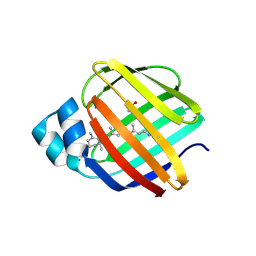
Loading
PDBj
MenuPDBj@FacebookPDBj@TwitterPDBj@YouTubewwPDB FoundationwwPDB
RCSB PDBPDBeBMRBAdv. SearchSearch help
Search by PDB author
4QGW
DownloadVisualize
BU of 4qgw by Molmil
Crystal sturcture of the R132K:R111L:L121D mutant of Cellular Retinoic Acid Binding ProteinII complexed with a synthetic ligand (Merocyanine) at 1.77 angstrom resolution
Descriptor: (2E,4E,6E)-3-methyl-6-(1,3,3-trimethyl-1,3-dihydro-2H-indol-2-ylidene)hexa-2,4-dienal, 2-[3-(2-HYDROXY-1,1-DIHYDROXYMETHYL-ETHYLAMINO)-PROPYLAMINO]-2-HYDROXYMETHYL-PROPANE-1,3-DIOL, Cellular retinoic acid-binding protein 2
Authors:Nosrati, M, Yapici, I, Geiger, J.H.
Deposit date:2014-05-26
Release date:2015-01-28
Last modified:2015-02-11
Method:X-RAY DIFFRACTION (1.77 Å)
Cite:"Turn-on" protein fluorescence: in situ formation of cyanine dyes.
J.Am.Chem.Soc., 137, 2015
4QGX
DownloadVisualize
BU of 4qgx by Molmil
Crystal structure of the R132K:R111L:L121E mutant of Cellular Retinoic Acid Binding ProteinII complexed with a synthetic ligand (Merocyanine) at 1.47 angstrom resolution
Descriptor: (2E,4E,6E)-3-methyl-6-(1,3,3-trimethyl-1,3-dihydro-2H-indol-2-ylidene)hexa-2,4-dienal, Cellular retinoic acid-binding protein 2
Authors:Nosrati, M, Yapici, I, Geiger, J.H.
Deposit date:2014-05-26
Release date:2015-01-28
Last modified:2023-09-20
Method:X-RAY DIFFRACTION (1.471 Å)
Cite:"Turn-on" protein fluorescence: in situ formation of cyanine dyes.
J.Am.Chem.Soc., 137, 2015
4QGV
DownloadVisualize
BU of 4qgv by Molmil
Crystal structure of the R132K:R111L mutant of Cellular Retinoic Acid Binding ProteinII complexed with a synthetic ligand (Merocyanine) at 1.73 angstrom resolution.
Descriptor: (2E,4E,6E)-3-methyl-6-(1,3,3-trimethyl-1,3-dihydro-2H-indol-2-ylidene)hexa-2,4-dienal, Cellular retinoic acid-binding protein 2
Authors:Nosrati, M, Yapici, I, Geiger, J.H.
Deposit date:2014-05-25
Release date:2015-01-28
Last modified:2023-09-20
Method:X-RAY DIFFRACTION (1.73 Å)
Cite:"Turn-on" protein fluorescence: in situ formation of cyanine dyes.
J.Am.Chem.Soc., 137, 2015
4QZU
DownloadVisualize
BU of 4qzu by Molmil
Crystal Structure of wild type Human Cellular Retinol Binding Protein II (hCRBPII) bound to retinol at 11 KeV beam energy
Descriptor: ACETATE ION, GLYCEROL, RETINOL, ...
Authors:Assar, Z, Geiger, J.H.
Deposit date:2014-07-28
Release date:2014-12-10
Last modified:2023-09-20
Method:X-RAY DIFFRACTION (1.5 Å)
Cite:Structures of holo wild-type human cellular retinol-binding protein II (hCRBPII) bound to retinol and retinal.
Acta Crystallogr.,Sect.D, 70, 2014
4QZT
DownloadVisualize
BU of 4qzt by Molmil
Crystal Structure of wild type Human Cellular Retinol Binding Protein II (hCRBPII) bound to retinol at 7 KeV beam energy
Descriptor: ACETATE ION, RETINOL, Retinol-binding protein 2
Authors:Assar, Z, Geiger, J.H.
Deposit date:2014-07-28
Release date:2014-12-10
Last modified:2023-09-20
Method:X-RAY DIFFRACTION (1.9 Å)
Cite:Structures of holo wild-type human cellular retinol-binding protein II (hCRBPII) bound to retinol and retinal.
Acta Crystallogr.,Sect.D, 70, 2014
4RUU
DownloadVisualize
BU of 4ruu by Molmil
Crystal structure of the Q108K:K40L mutant of human Cellular Retinol Binding ProteinII in complex with All-trans-Retinal after 24 hour incubation at 1.4 Angstrom Resolution
Descriptor: ACETATE ION, RETINAL, Retinol-binding protein 2
Authors:Nosrati, M, Geiger, J.H.
Deposit date:2014-11-21
Release date:2014-12-10
Last modified:2023-09-20
Method:X-RAY DIFFRACTION (1.4 Å)
Cite:Tuning the electronic absorption of protein-embedded all-trans-retinal.
Science, 338, 2012
4EDE
DownloadVisualize
BU of 4ede by Molmil
Crystal Structure of the Q108K:K40L:T51V:T53C:Y19W:R58W:T29L:A33W Mutant of Cellular Retinol Binding Protein Type II in Complex with All-trans-Retinal at 1.4 Angstrom Resolution
Descriptor: ACETATE ION, RETINAL, Retinol-binding protein 2
Authors:Nossoni, Z, Geiger, J.H.
Deposit date:2012-03-27
Release date:2012-12-26
Last modified:2023-09-13
Method:X-RAY DIFFRACTION (1.4 Å)
Cite:Tuning the electronic absorption of protein-embedded all-trans-retinal.
Science, 338, 2012
4EEJ
DownloadVisualize
BU of 4eej by Molmil
Crystal Structure of the Q108K:K40L:T51V:T53C:Y19W:R58W:T29L:Q4R Mutant of Cellular Retinol Binding Protein Type II in Complex with All-trans-Retinal at 1.5 Angstrom Resolution
Descriptor: ACETATE ION, RETINAL, Retinol-binding protein 2
Authors:Nossoni, Z, Geiger, J.H.
Deposit date:2012-03-28
Release date:2012-12-26
Last modified:2023-09-13
Method:X-RAY DIFFRACTION (1.501 Å)
Cite:Tuning the electronic absorption of protein-embedded all-trans-retinal.
Science, 338, 2012
4GKC
DownloadVisualize
BU of 4gkc by Molmil
Crystal structure of Q108K:K40L:T51V:T53C:R58W:T29L:Y19W:Q4A mutant of cellular retinol binding protein II complex with all-trans-retinal at 1.33
Descriptor: RETINAL, Retinol-binding protein 2
Authors:Nossoni, Z, Geiger, J.H.
Deposit date:2012-08-10
Release date:2012-12-26
Last modified:2023-09-13
Method:X-RAY DIFFRACTION (1.3 Å)
Cite:Tuning the electronic absorption of protein-embedded all-trans-retinal.
Science, 338, 2012
4I9S
DownloadVisualize
BU of 4i9s by Molmil
Crystal Structure of the R111K:R132L:Y134F:T54V:R59W Mutant of the Cellular Retinoic Acid Binding Protein Type II in Complex with All-Trans Retinal at 2.58 Angstrom Resolution
Descriptor: Cellular retinoic acid-binding protein 2, RETINAL
Authors:Nosrati, M, Geiger, J.H.
Deposit date:2012-12-05
Release date:2013-10-09
Last modified:2023-09-20
Method:X-RAY DIFFRACTION (2.58 Å)
Cite:Rational Design of a Colorimetric pH Sensor from a Soluble Retinoic Acid Chaperone.
J.Am.Chem.Soc., 135, 2013
4I9R
DownloadVisualize
BU of 4i9r by Molmil
Crystal Structure of the R111K:R132L:Y134F:T54V:R59W:A32W Mutant of the Cellular Retinoic Acid Binding Protein Type II in Complex with All-Trans Retinal at 2.6 Angstrom Resolution
Descriptor: Cellular retinoic acid-binding protein 2, RETINAL
Authors:Nosrati, M, Geiger, J.H.
Deposit date:2012-12-05
Release date:2013-10-09
Last modified:2023-09-20
Method:X-RAY DIFFRACTION (2.6 Å)
Cite:Rational Design of a Colorimetric pH Sensor from a Soluble Retinoic Acid Chaperone.
J.Am.Chem.Soc., 135, 2013
5F58
DownloadVisualize
BU of 5f58 by Molmil
Crystal structure of the Q108K:K40L:T51V:R58F mutant of human Cellular Retinol Binding Protein II in complex with All-trans-Retinal after 24 hours of incubation at 1.54 Angstrom Resolution
Descriptor: ACETATE ION, RETINAL, Retinol-binding protein 2
Authors:Nosrati, M, Nossoni, Z, Geiger, J.H.
Deposit date:2015-12-04
Release date:2016-12-07
Last modified:2023-09-27
Method:X-RAY DIFFRACTION (1.54 Å)
Cite:Crystal structure of the Q108K:K40L:T51V:R58F mutant of human Cellular Retinol Binding Protein II in complex with All-trans-Retinal after 24 hours of incubation at 1.54 Angstrom Resolution
To Be Published
5F6B
DownloadVisualize
BU of 5f6b by Molmil
Crystal structure of the Q108K:K40L:T51V:R58Y:Y19W mutant of human Cellular Retinol Binding Protein II in complex with All-trans-Retinal at 1.3 Angstrom Resolution
Descriptor: ACETATE ION, RETINAL, Retinol-binding protein 2
Authors:Nosrati, M, Nossoni, Z, Geiger, J.H.
Deposit date:2015-12-05
Release date:2016-12-07
Last modified:2023-09-27
Method:X-RAY DIFFRACTION (1.31 Å)
Cite:Crystal structure of the Q108K:K40L:T51V:R58Y:Y19W mutant of human Cellular Retinol Binding ProteinII in complex with All-trans-Retinal at 1.31 Angstrom Resolution
To Be Published
5FAZ
DownloadVisualize
BU of 5faz by Molmil
Crystal structure of the Q108K:K40L:T51V mutant of human Cellular Retinol Binding Protein II in complex with All-trans-Retinal after 24 hours of incubation at 1.4 Angstrom Resolution
Descriptor: ACETATE ION, RETINAL, Retinol-binding protein 2
Authors:Nosrati, M, Nossoni, Z, Geiger, J.H.
Deposit date:2015-12-13
Release date:2016-12-14
Last modified:2023-09-27
Method:X-RAY DIFFRACTION (1.4 Å)
Cite:Crystal structure of the Q108K:K40L:T51V mutant of human Cellular Retinol Binding Protein II in complex with All-trans-Retinal after 24 hours of incubation at 1.54 Angstrom Resolution
To Be Published
5FEN
DownloadVisualize
BU of 5fen by Molmil
Crystal structure of the Q108K:K40L:T53C mutant of human Cellular Retinol Binding Protein II in complex with All-trans-Retinal after 24 hours of incubation at 1.55 Angstrom Resolution
Descriptor: ACETATE ION, RETINAL, Retinol-binding protein 2
Authors:Nossoni, Z, Nosrati, M, Geiger, J.H.
Deposit date:2015-12-17
Release date:2016-12-21
Last modified:2023-09-27
Method:X-RAY DIFFRACTION (1.55 Å)
Cite:Crystal structure of the Q108K:K40L:T53C mutant of human Cellular Retinol Binding Protein II in complex with All-trans-Retinal after 24 hours of incubation at 1.55 Angstrom Resolution
To Be Published
5FFH
DownloadVisualize
BU of 5ffh by Molmil
Crystal structure of the Q108K:K40L:T51V:R58W:Y19W mutant of human Cellular Retinol Binding Protein II in complex with All-trans-Retinal at 1.68 Angstrom Resolution
Descriptor: ACETATE ION, RETINAL, Retinol-binding protein 2
Authors:Nossoni, Z, Nosrati, M, Geiger, J.H.
Deposit date:2015-12-18
Release date:2016-12-21
Last modified:2023-09-27
Method:X-RAY DIFFRACTION (1.68 Å)
Cite:Crystal structure of the Q108K:K40L:T51V:R58W:Y19W mutant of human Cellular Retinol Binding ProteinII in complex with All-trans-Retinal at 1.68 Angstrom Resolution
To Be Published
5F7G
DownloadVisualize
BU of 5f7g by Molmil
Crystal structure of the Q108K:K40L:T51V:R58Y:Y19W:Q38L mutant of human Cellular Retinol Binding Protein II in complex with All-trans-Retinal at 1.48 Angstrom Resolution
Descriptor: ACETATE ION, RETINAL, Retinol-binding protein 2
Authors:Nosrati, M, Nossoni, Z, Geiger, J.H.
Deposit date:2015-12-08
Release date:2016-12-14
Last modified:2023-09-27
Method:X-RAY DIFFRACTION (1.48 Å)
Cite:Crystal structure of the Q108K:K40L:T51V:R58F mutant of human Cellular Retinol Binding Protein II in complex with All-trans-Retinal after 24 hours of incubation at 1.54 Angstrom Resolution
To Be Published
2FRS
DownloadVisualize
BU of 2frs by Molmil
Crystal structure of the f15w mutant of apo-cellular retinoic acid binding protein type ii at 1.51 angstroms resolution
Descriptor: Cellular retinoic acid-binding protein 2, SODIUM ION
Authors:Vaezeslami, S, Geiger, J.H.
Deposit date:2006-01-20
Release date:2006-09-19
Last modified:2024-02-14
Method:X-RAY DIFFRACTION (1.51 Å)
Cite:The structure of Apo-wild-type cellular retinoic acid binding protein II at 1.4 A and its relationship to ligand binding and nuclear translocation.
J.Mol.Biol., 363, 2006
2FS6
DownloadVisualize
BU of 2fs6 by Molmil
Crystal Structure of Apo-Cellular Retinoic Acid Binding Protein Type II At 1.35 Angstroms Resolution
Descriptor: ACETATE ION, CHLORIDE ION, Cellular retinoic acid-binding protein 2, ...
Authors:Vaezeslami, S, Geiger, J.H.
Deposit date:2006-01-21
Release date:2006-09-19
Last modified:2024-02-14
Method:X-RAY DIFFRACTION (1.35 Å)
Cite:The structure of Apo-wild-type cellular retinoic acid binding protein II at 1.4 A and its relationship to ligand binding and nuclear translocation.
J.Mol.Biol., 363, 2006
2FR3
DownloadVisualize
BU of 2fr3 by Molmil
Crystal Structure of Cellular Retinoic Acid Binding Protein Type II in Complex with All-Trans-Retinoic Acid at 1.48 Angstroms Resolution
Descriptor: ACETATE ION, Cellular retinoic acid binding protein 2, RETINOIC ACID
Authors:Vaezeslami, S, Geiger, J.H.
Deposit date:2006-01-18
Release date:2006-09-19
Last modified:2024-02-14
Method:X-RAY DIFFRACTION (1.48 Å)
Cite:The structure of Apo-wild-type cellular retinoic acid binding protein II at 1.4 A and its relationship to ligand binding and nuclear translocation.
J.Mol.Biol., 363, 2006
2FS7
DownloadVisualize
BU of 2fs7 by Molmil
Crystal Structure of Apo-Cellular Retinoic Acid Binding Protein Type II At 1.55 Angstroms Resolution
Descriptor: ACETATE ION, CHLORIDE ION, Cellular retinoic acid-binding protein 2
Authors:Vaezeslami, S, Geiger, J.H.
Deposit date:2006-01-21
Release date:2006-09-19
Last modified:2024-02-14
Method:X-RAY DIFFRACTION (1.55 Å)
Cite:The structure of Apo-wild-type cellular retinoic acid binding protein II at 1.4 A and its relationship to ligand binding and nuclear translocation.
J.Mol.Biol., 363, 2006
2G78
DownloadVisualize
BU of 2g78 by Molmil
Crystal Structure of the R132K:Y134F Mutant of Cellular Retinoic Acid Binding Protein Type II in Complex with All-Trans-Retinoic Acid at 1.70 Angstroms Resolution
Descriptor: Cellular retinoic acid-binding protein 2, RETINOIC ACID, SODIUM ION
Authors:Vaezeslami, S, Geiger, J.H.
Deposit date:2006-02-27
Release date:2007-04-17
Last modified:2024-02-14
Method:X-RAY DIFFRACTION (1.7 Å)
Cite:Protein design: reengineering cellular retinoic acid binding protein II into a rhodopsin protein mimic.
J.Am.Chem.Soc., 129, 2007
2G7B
DownloadVisualize
BU of 2g7b by Molmil
Crystal Structure of the R132K:R111L:L121E mutant of Cellular Retinoic Acid Binding Protein Type II In Complex With All-Trans-Retinal At 1.18 Angstroms Resolution
Descriptor: ALL-TRANS AXEROPHTHENE, Cellular retinoic acid-binding protein 2, SODIUM ION
Authors:Vaezeslami, S, Geiger, J.H.
Deposit date:2006-02-27
Release date:2007-04-17
Last modified:2021-10-20
Method:X-RAY DIFFRACTION (1.18 Å)
Cite:Protein design: reengineering cellular retinoic acid binding protein II into a rhodopsin protein mimic.
J.Am.Chem.Soc., 129, 2007
2G79
DownloadVisualize
BU of 2g79 by Molmil
Crystal Structure of the R132K:Y134F Mutant of Cellular Retinoic Acid Binding Protein Type II in Complex with All-Trans-Retinal at 1.69 Angstroms Resolution
Descriptor: Cellular retinoic acid-binding protein 2, RETINAL, SODIUM ION, ...
Authors:Vaezeslami, S, Geiger, J.H.
Deposit date:2006-02-27
Release date:2007-04-17
Last modified:2024-02-14
Method:X-RAY DIFFRACTION (1.69 Å)
Cite:Protein design: reengineering cellular retinoic acid binding protein II into a rhodopsin protein mimic.
J.Am.Chem.Soc., 129, 2007
6E5E
DownloadVisualize
BU of 6e5e by Molmil
Crystal structure of the apo domain-swapped dimer Q108K:T51D mutant of human cellular retinol binding protein II
Descriptor: ACETATE ION, Retinol-binding protein 2
Authors:Ghanbarpour, A, Geiger, J.
Deposit date:2018-07-20
Release date:2019-10-16
Last modified:2023-10-11
Method:X-RAY DIFFRACTION (1.696 Å)
Cite:Engineering the hCRBPII Domain-Swapped Dimer into a New Class of Protein Switches.
J.Am.Chem.Soc., 141, 2019

218853

PDB entries from 2024-04-24

PDB statisticsPDBj update infoContact PDBjnumon