1E1K
 
 | |
1DZZ
 | |
1E1M
 | |
1E1L
 | |
1E4Y
 | |
1EB4
 | |
1QJP
 | |
1QJ8
 | |
1BH3
 | |
1E4V
 | |
1AKE
 | |
2BVG
 | |
2BVF
 | |
2BVL
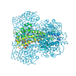
 | | Crystal structure of the catalytic domain of toxin B from Clostridium difficile in complex with UDP, Glc and manganese ion | | Descriptor: | HEXATANTALUM DODECABROMIDE, MANGANESE (II) ION, SULFATE ION, ... | | Authors: | Reinert, D.J, Jank, T, Aktories, K, Schulz, G.E. | | Deposit date: | 2005-06-30 | | Release date: | 2005-08-03 | | Last modified: | 2024-05-08 | | Method: | X-RAY DIFFRACTION (2.2 Å) | | Cite: | Structural Basis for the Function of Clostridium Difficile Toxin B.
J.Mol.Biol., 351, 2005
|
|
2CGJ
 | |
2CGK
 | |
2CB4
 | | Crystal structure of the catalytic domain of the mosquitocidal toxin from Bacillus sphaericus, mutant E197Q | | Descriptor: | MOSQUITOCIDAL TOXIN | | Authors: | Reinert, D.J, Carpusca, I, Aktories, K, Schulz, G.E. | | Deposit date: | 2005-12-29 | | Release date: | 2006-02-22 | | Last modified: | 2011-07-13 | | Method: | X-RAY DIFFRACTION (2.5 Å) | | Cite: | Structure of the Mosquitocidal Toxin from Bacillus Sphaericus.
J.Mol.Biol., 357, 2006
|
|
2CGL
 | |
2CB6
 | | Crystal structure of the catalytic domain of the mosquitocidal toxin from Bacillus sphaericus, mutant E195Q | | Descriptor: | MOSQUITOCIDAL TOXIN | | Authors: | Reinert, D.J, Carpusca, I, Aktories, K, Schulz, G.E. | | Deposit date: | 2005-12-29 | | Release date: | 2006-02-22 | | Last modified: | 2023-12-13 | | Method: | X-RAY DIFFRACTION (3 Å) | | Cite: | Structure of the Mosquitocidal Toxin from Bacillus Sphaericus.
J.Mol.Biol., 357, 2006
|
|
2BVH
 | |
2BVM
 | | Crystal structure of the catalytic domain of toxin B from Clostridium difficile in complex with UDP, Glc and manganese ion | | Descriptor: | MANGANESE (II) ION, SULFATE ION, TOXIN B, ... | | Authors: | Reinert, D.J, Jank, T, Aktories, K, Schulz, G.E. | | Deposit date: | 2005-06-30 | | Release date: | 2005-08-03 | | Last modified: | 2023-12-13 | | Method: | X-RAY DIFFRACTION (2.55 Å) | | Cite: | Structural Basis for the Function of Clostridium Difficile Toxin B.
J.Mol.Biol., 351, 2005
|
|
2BIW
 | | Crystal structure of apocarotenoid cleavage oxygenase from Synechocystis, native enzyme | | Descriptor: | (3R)-3-HYDROXY-8'-APOCAROTENOL, APOCAROTENOID-CLEAVING OXYGENASE, FE (III) ION | | Authors: | Kloer, D.P, Ruch, S, Al-Babili, S, Beyer, P, Schulz, G.E. | | Deposit date: | 2005-01-26 | | Release date: | 2005-04-14 | | Last modified: | 2024-05-01 | | Method: | X-RAY DIFFRACTION (2.39 Å) | | Cite: | The Structure of a Retinal-Forming Carotenoid Oxygenase
Science, 308, 2005
|
|
2BIX
 | | Crystal structure of apocarotenoid cleavage oxygenase from Synechocystis, Fe-free apoenzyme | | Descriptor: | (HYDROXYETHYLOXY)TRI(ETHYLOXY)OCTANE, APOCAROTENOID-CLEAVING OXYGENASE, GLYCEROL | | Authors: | Kloer, D.P, Ruch, S, Al-Babili, S, Beyer, P, Schulz, G.E. | | Deposit date: | 2005-01-26 | | Release date: | 2005-04-14 | | Last modified: | 2024-05-08 | | Method: | X-RAY DIFFRACTION (2.68 Å) | | Cite: | The Structure of a Retinal-Forming Carotenoid Oxygenase
Science, 308, 2005
|
|
2AG1
 | |
2AG0
 | |


