Loading
PDBj
MenuPDBj@FacebookPDBj@TwitterPDBj@YouTubewwPDB FoundationwwPDB
RCSB PDBPDBeBMRBAdv. SearchSearch help
Search by PDB author
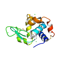
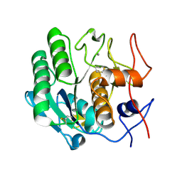
8DCT
DownloadVisualize
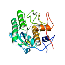
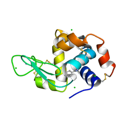
BU of 8dct by Molmil
Lysozyme cluster 3 dual apo structure
Descriptor: 2-acetamido-2-deoxy-alpha-D-glucopyranose, BENZAMIDINE, CHLORIDE ION, ...
Authors:Soares, A.S, Yamada, Y, Jakoncic, J, Schneider, D.K, Bernstein, H.J.
Deposit date:2022-06-17
Release date:2022-08-17
Last modified:2023-10-18
Method:X-RAY DIFFRACTION (2 Å)
Cite:Serial crystallography with multi-stage merging of thousands of images.
Acta Crystallogr.,Sect.F, 78, 2022
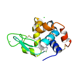
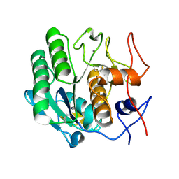
8DCW
DownloadVisualize
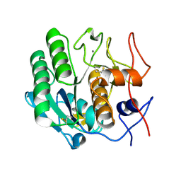
BU of 8dcw by Molmil
Lysozyme cluster 0062 (NAG and benzamidine ligands)
Descriptor: 2-acetamido-2-deoxy-alpha-D-glucopyranose, BENZAMIDINE, CHLORIDE ION, ...
Authors:Soares, A.S, Yamada, Y, Jakoncic, J, Schneider, D.K, Bernstein, H.J.
Deposit date:2022-06-17
Release date:2022-08-17
Last modified:2023-10-18
Method:X-RAY DIFFRACTION (2 Å)
Cite:Serial crystallography with multi-stage merging of thousands of images.
Acta Crystallogr.,Sect.F, 78, 2022
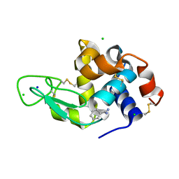
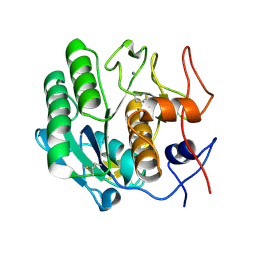
8DCU
DownloadVisualize
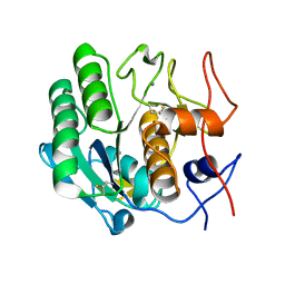
BU of 8dcu by Molmil
Lysozyme cluster 0028 (benzamidine ligand)
Descriptor: 2-acetamido-2-deoxy-alpha-D-glucopyranose, BENZAMIDINE, CHLORIDE ION, ...
Authors:Soares, A.S, Yamada, Y, Jakoncic, J, Schneider, D.K, Bernstein, H.J.
Deposit date:2022-06-17
Release date:2022-08-17
Last modified:2024-10-30
Method:X-RAY DIFFRACTION (2 Å)
Cite:Serial crystallography with multi-stage merging of thousands of images.
Acta Crystallogr.,Sect.F, 78, 2022
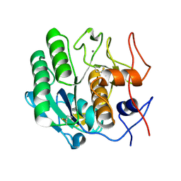
8DCV
DownloadVisualize
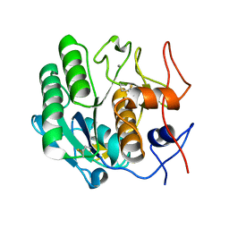
BU of 8dcv by Molmil
Lysozyme cluster 0043, NAG ligand
Descriptor: 2-acetamido-2-deoxy-alpha-D-glucopyranose, BENZAMIDINE, CHLORIDE ION, ...
Authors:Soares, A.S, Yamada, Y, Jakoncic, J, Schneider, D.K, Bernstein, H.J.
Deposit date:2022-06-17
Release date:2022-08-17
Last modified:2024-10-30
Method:X-RAY DIFFRACTION (2 Å)
Cite:Serial crystallography with multi-stage merging of thousands of images.
Acta Crystallogr.,Sect.F, 78, 2022
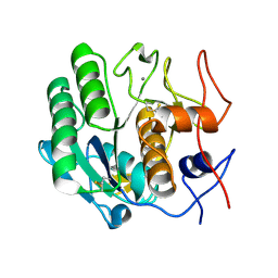
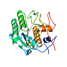
3DE6
DownloadVisualize
BU of 3de6 by Molmil
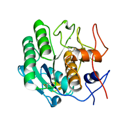
Proteinase K by Classical hanging drop method after the third step of high X-Ray dose on ESRF ID23-1 beamline
Descriptor: CALCIUM ION, Proteinase K
Authors:Pechkova, E, Tripathi, S.K, Nicolini, C.
Deposit date:2008-06-08
Release date:2009-06-09
Last modified:2024-10-16
Method:X-RAY DIFFRACTION (2.2 Å)
Cite:Radiation stability of proteinase K crystals grown by LB nanotemplate method
J.Struct.Biol., 168, 2009
3DE3
DownloadVisualize
BU of 3de3 by Molmil
Proteinase K by Classical hanging drop method before high X-Ray dose on ESRF ID23-1 beamline
Descriptor: CALCIUM ION, Proteinase K
Authors:Pechkova, E, Tripathi, S.K, Nicolini, C.
Deposit date:2008-06-08
Release date:2009-06-09
Last modified:2011-07-13
Method:X-RAY DIFFRACTION (1.43 Å)
Cite:Radiation stability of proteinase K crystals grown by LB nanotemplate method
J.Struct.Biol., 168, 2009
3DE5
DownloadVisualize
BU of 3de5 by Molmil
roteinase K by Classical hanging drop method after the second step of high X-Ray dose on ESRF ID23-1 beamline
Descriptor: CALCIUM ION, Proteinase K
Authors:Pechkova, E, Tripathi, S.K, Nicolini, C.
Deposit date:2008-06-08
Release date:2009-06-09
Last modified:2024-10-30
Method:X-RAY DIFFRACTION (2.1 Å)
Cite:Radiation stability of proteinase K crystals grown by LB nanotemplate method
J.Struct.Biol., 168, 2009
3DDZ
DownloadVisualize
BU of 3ddz by Molmil
Proteinase K by LB nanotemplate method after the first step of high X-Ray dose on ESRF ID23-1 beamline
Descriptor: CALCIUM ION, Proteinase K
Authors:Pechkova, E, Tripathi, S.K, Nicolini, C.
Deposit date:2008-06-07
Release date:2009-06-09
Last modified:2023-11-01
Method:X-RAY DIFFRACTION (1.7 Å)
Cite:Radiation stability of proteinase K crystals grown by LB nanotemplate method
J.Struct.Biol., 168, 2009
3DE1
DownloadVisualize
BU of 3de1 by Molmil
Proteinase K by LB nanotemplate method after the third step of high X-Ray dose on ESRF ID23-1 beamline
Descriptor: CALCIUM ION, Proteinase K
Authors:Pechkova, E, Tripathi, S.K, Nicolini, C.
Deposit date:2008-06-07
Release date:2009-06-09
Last modified:2024-10-09
Method:X-RAY DIFFRACTION (2 Å)
Cite:Radiation stability of proteinase K crystals grown by LB nanotemplate method
J.Struct.Biol., 168, 2009
3D9Q
DownloadVisualize
BU of 3d9q by Molmil
Proteinase K by LB nanotemplate method before high X-Ray dose on ESRF ID23-1 beamline
Descriptor: CALCIUM ION, Proteinase K
Authors:Pechkova, E, Tripathi, S.K, Nicolini, C.
Deposit date:2008-05-27
Release date:2009-06-02
Last modified:2023-11-01
Method:X-RAY DIFFRACTION (1.43 Å)
Cite:Radiation stability of proteinase K crystals grown by LB nanotemplate method
J.Struct.Biol., 168, 2009
3DE0
DownloadVisualize
BU of 3de0 by Molmil
Proteinase K by LB nanotemplate method after the second step of high X-Ray dose on ESRF ID23-1 beamline
Descriptor: CALCIUM ION, Proteinase K
Authors:Pechkova, E, Tripathi, S.K, Nicolini, C.
Deposit date:2008-06-07
Release date:2009-06-09
Last modified:2023-11-01
Method:X-RAY DIFFRACTION (1.9 Å)
Cite:Radiation stability of proteinase K crystals grown by LB nanotemplate method
J.Struct.Biol., 168, 2009
3DE2
DownloadVisualize
BU of 3de2 by Molmil
Proteinase K by LB nanotemplate method after the fourth step of high X-Ray dose on ESRF ID23-1 beamline
Descriptor: CALCIUM ION, Proteinase K
Authors:Pechkova, E, Tripathi, S.K, Nicolini, C.
Deposit date:2008-06-07
Release date:2009-06-09
Last modified:2011-07-13
Method:X-RAY DIFFRACTION (2.1 Å)
Cite:Radiation stability of proteinase K crystals grown by LB nanotemplate method
J.Struct.Biol., 168, 2009
3DE7
DownloadVisualize
BU of 3de7 by Molmil
Proteinase K by Classical hanging drop method after the fourth step of high X-Ray dose on ESRF ID23-1 beamline
Descriptor: CALCIUM ION, Proteinase K
Authors:Pechkova, E, Tripathi, S.K, Nicolini, C.
Deposit date:2008-06-08
Release date:2009-06-09
Last modified:2011-07-13
Method:X-RAY DIFFRACTION (2.3 Å)
Cite:Radiation stability of proteinase K crystals grown by LB nanotemplate method
J.Struct.Biol., 168, 2009
3DE4
DownloadVisualize
BU of 3de4 by Molmil
Proteinase K by Classical hanging drop method after the first step of high X-Ray dose on ESRF ID23-1 beamline
Descriptor: CALCIUM ION, Proteinase K
Authors:Pechkova, E, Tripathi, S.K, Nicolini, C.
Deposit date:2008-06-08
Release date:2009-06-09
Last modified:2024-10-16
Method:X-RAY DIFFRACTION (1.8 Å)
Cite:Radiation stability of proteinase K crystals grown by LB nanotemplate method
J.Struct.Biol., 168, 2009
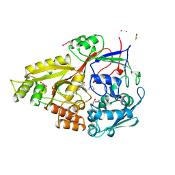
2OLB
DownloadVisualize
BU of 2olb by Molmil
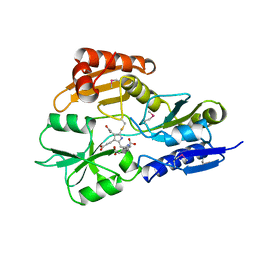
OLIGOPEPTIDE BINDING PROTEIN (OPPA) COMPLEXED WITH TRI-LYSINE
Descriptor: ACETATE ION, OLIGO-PEPTIDE BINDING PROTEIN, TRIPEPTIDE LYS-LYS-LYS, ...
Authors:Tame, J, Wilkinson, A.J.
Deposit date:1995-09-10
Release date:1996-01-29
Last modified:2024-10-23
Method:X-RAY DIFFRACTION (1.4 Å)
Cite:The crystal structures of the oligopeptide-binding protein OppA complexed with tripeptide and tetrapeptide ligands.
Structure, 3, 1995
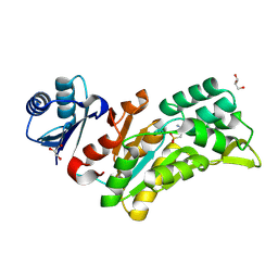
8Q4S
DownloadVisualize
BU of 8q4s by Molmil
Crystal structure of phosphoserine phosphatase (SerB) from Brucella melitensis in complex with AP4 and magnesium.
Descriptor: (2S)-2-amino-4-phosphonobutanoic acid, GLYCEROL, MAGNESIUM ION, ...
Authors:Scaillet, T, Wouters, J.
Deposit date:2023-08-07
Release date:2024-08-21
Method:X-RAY DIFFRACTION (2.08 Å)
Cite:Crystal structure of phosphoserine phosphatase (SerB) from Brucella melitensis in complex with AP4 and magnesium.
To Be Published
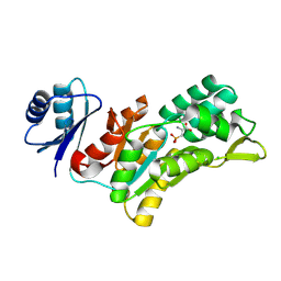
8QOB
DownloadVisualize
BU of 8qob by Molmil
Crystal structure of phosphoserine phosphatase (SerB) from Brucella melitensis in complex with AP3 and magnesium
Descriptor: (2~{R})-2-azanyl-3-phosphono-propanoic acid, CHLORIDE ION, MAGNESIUM ION, ...
Authors:Scaillet, T, Wouters, J.
Deposit date:2023-09-28
Release date:2024-10-09
Method:X-RAY DIFFRACTION (2.74 Å)
Cite:Crystal structure of phosphoserine phosphatase (SerB) from Brucella melitensis in complex with AP3 and magnesium
To Be Published
8BHD
DownloadVisualize
BU of 8bhd by Molmil
N-terminal domain of Plasmodium berghei glutamyl-tRNA synthetase (Tbxo4 derivative crystal structure)
Descriptor: GLYCEROL, Glutamate--tRNA ligase, SULFATE ION, ...
Authors:Benas, P, Jaramillo Ponce, J.R, Legrand, P, Frugier, M, Sauter, C.
Deposit date:2022-10-31
Release date:2023-01-25
Last modified:2024-06-19
Method:X-RAY DIFFRACTION (3.17 Å)
Cite:Solution X-ray scattering highlights discrepancies in Plasmodium multi-aminoacyl-tRNA synthetase complexes.
Protein Sci., 32, 2023
3DVS
DownloadVisualize
BU of 3dvs by Molmil
Proteinase K by LB nanotmplate method after the second step of high dose on ESRF ID14-2 beamline
Descriptor: CALCIUM ION, Proteinase K
Authors:Pechkova, E, Tripathi, S.K, Nicolini, C.
Deposit date:2008-07-20
Release date:2009-07-07
Last modified:2023-11-01
Method:X-RAY DIFFRACTION (1.02 Å)
Cite:Radiation damage in protein structural characterization by Synchrotron Radiation: State of the art and Nanotechnology-based perspective
To be Published
8AMS
DownloadVisualize
BU of 8ams by Molmil
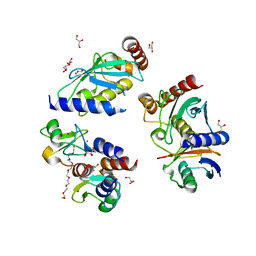
Complex of human TRIM2 RING domain, UBCH5C, and Ubiquitin
Descriptor: 3,6,9,12,15,18,21-HEPTAOXATRICOSANE-1,23-DIOL, GLYCEROL, Polyubiquitin-C, ...
Authors:Perez-Borrajero, C, Kotova, I, Murciano, B, Hennig, J.
Deposit date:2022-08-04
Release date:2023-11-15
Method:X-RAY DIFFRACTION (2.4 Å)
Cite:Structural and biophysical studies of TRIM2 and TRIM3
To Be Published
1AH5
DownloadVisualize
BU of 1ah5 by Molmil
REDUCED FORM SELENOMETHIONINE-LABELLED HYDROXYMETHYLBILANE SYNTHASE DETERMINED BY MAD
Descriptor: 3-[5-{[3-(2-carboxyethyl)-4-(carboxymethyl)-5-methyl-1H-pyrrol-2-yl]methyl}-4-(carboxymethyl)-1H-pyrrol-3-yl]propanoic acid, HYDROXYMETHYLBILANE SYNTHASE
Authors:Helliwell, J.R, Nieh, Y.P, Harrop, S.J, Cassetta, A.
Deposit date:1997-04-13
Release date:1997-10-15
Last modified:2013-09-18
Method:X-RAY DIFFRACTION (2.4 Å)
Cite:Determination of the structure of seleno-methionine-labelled hydroxymethylbilane synthase in its active form by multi-wavelength anomalous dispersion.
Acta Crystallogr.,Sect.D, 55, 1999
1REQ
DownloadVisualize
BU of 1req by Molmil
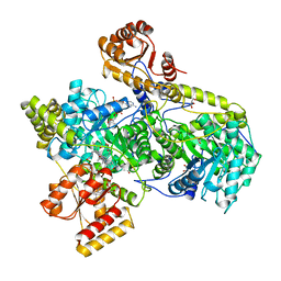
METHYLMALONYL-COA MUTASE
Descriptor: COBALAMIN, DESULFO-COENZYME A, GLYCEROL, ...
Authors:Evans, P.R, Mancia, F.
Deposit date:1996-01-19
Release date:1997-01-27
Last modified:2024-02-14
Method:X-RAY DIFFRACTION (2 Å)
Cite:How coenzyme B12 radicals are generated: the crystal structure of methylmalonyl-coenzyme A mutase at 2 A resolution.
Structure, 4, 1996
3REQ
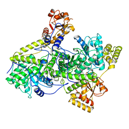
DownloadVisualize
BU of 3req by Molmil
METHYLMALONYL-COA MUTASE, SUBSTRATE-FREE STATE (POOR QUALITY STRUCTURE)
Descriptor: ADENOSINE, COBALAMIN, METHYLMALONYL-COA MUTASE
Authors:Evans, P.R, Mancia, F.
Deposit date:1997-12-04
Release date:1998-03-18
Last modified:2024-05-22
Method:X-RAY DIFFRACTION (2.7 Å)
Cite:Conformational changes on substrate binding to methylmalonyl CoA mutase and new insights into the free radical mechanism.
Structure, 6, 1998
1QIG
DownloadVisualize
BU of 1qig by Molmil
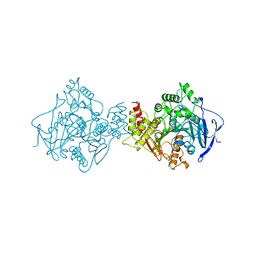
SPECIFIC CHEMICAL AND STRUCTURAL DAMAGE AT NINE TIME POINTS (POINT D) CAUSED BY INTENSE SYNCHROTRON RADIATION TO TORPEDO CALIFORNICA ACETYLCHOLINESTERASE
Descriptor: ACETYLCHOLINESTERASE
Authors:Kryger, G, Weik, M, Ravelli, R.B.G.
Deposit date:1999-06-14
Release date:2000-01-28
Last modified:2023-08-16
Method:X-RAY DIFFRACTION (2.3 Å)
Cite:Specific chemical and structural damage to proteins produced by synchrotron radiation.
Proc.Natl.Acad.Sci.USA, 97, 2000
1QIF
DownloadVisualize
BU of 1qif by Molmil
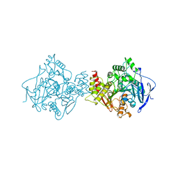
SPECIFIC CHEMICAL AND STRUCTURAL DAMAGE AT NINE TIME POINTS (POINT C) CAUSED BY INTENSE SYNCHROTRON RADIATION TO TORPEDO CALIFORNICA ACETYLCHOLINESTERASE
Descriptor: ACETYLCHOLINESTERASE
Authors:Kryger, G, Weik, M, Ravelli, R.B.G.
Deposit date:1999-06-14
Release date:2000-01-28
Last modified:2023-08-16
Method:X-RAY DIFFRACTION (2.1 Å)
Cite:Specific chemical and structural damage to proteins produced by synchrotron radiation.
Proc.Natl.Acad.Sci.USA, 97, 2000

226707

PDB entries from 2024-10-30

PDB statisticsPDBj update infoContact PDBjnumon