6MQY
 
 | |
6MOV
 | |
6MKV
 | |
6MQX
 | |
6MQI
 | |
6MQZ
 | |
6MOR
 | |
6MQW
 | |
6MOX
 | |
6MLB
 | |
6MR0
 | |
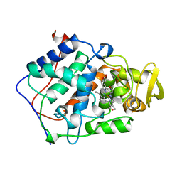
1BEK
 | |
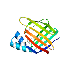
1BEP
 | | EFFECT OF UNNATURAL HEME SUBSTITUTION ON KINETICS OF ELECTRON TRANSFER IN CYTOCHROME C PEROXIDASE | | Descriptor: | YEAST CYTOCHROME C PEROXIDASE, [7-ETHENYL-12-FORMYL-3,8,13,17-TERTRAMETHYL-21H,23H-PORPHINE-2,18-DIPROPANOATO(2)-N21,N22,N23,N24]IRON | | Authors: | Miller, M, Kraut, J. | | Deposit date: | 1998-05-16 | | Release date: | 1998-10-21 | | Last modified: | 2024-02-07 | | Method: | X-RAY DIFFRACTION (2.2 Å) | | Cite: | Effect of Unnatural Heme Substitution on Kinetics of Electron Transfer in Cytochrome C Peroxidase
To be Published
|
|
4YGH
 | |
4YH0
 | |
4YDA
 | |
4YDB
 | |
4YCE
 | |
4YFR
 | |
4YKM
 | |
4YFQ
 | |
4YBP
 | |
4YGZ
 | |
4YKO
 | |
4YFP
 | |


