Loading
PDBj
MenuPDBj@FacebookPDBj@TwitterPDBj@YouTubewwPDB FoundationwwPDB
RCSB PDBPDBeBMRBAdv. SearchSearch help
Search by PDB author
8F6T
DownloadVisualize
BU of 8f6t by Molmil
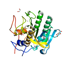
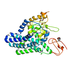
Cryo-EM structure of alkane 1-monooxygenase AlkB-AlkG complex from Fontimonas thermophila
Descriptor: Alkane 1-monooxygenase, DODECANE, FE (III) ION
Authors:Chai, J, Guo, G, McSweeney, S, Shanklin, J, Liu, Q.
Deposit date:2022-11-17
Release date:2023-04-05
Last modified:2024-05-22
Method:ELECTRON MICROSCOPY (2.76 Å)
Cite:Structural basis for enzymatic terminal C-H bond functionalization of alkanes.
Nat.Struct.Mol.Biol., 30, 2023
2C6R
DownloadVisualize
BU of 2c6r by Molmil
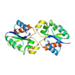
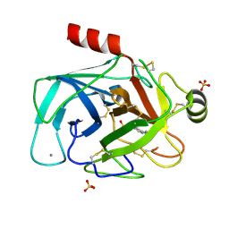
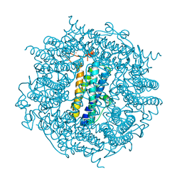
FE-SOAKED CRYSTAL STRUCTURE OF THE DPS92 FROM DEINOCOCCUS RADIODURANS
Descriptor: CHLORIDE ION, DNA-BINDING STRESS RESPONSE PROTEIN, DPS FAMILY, ...
Authors:Cuypers, M.G, Romao, C.V, Mitchell, E, Mcsweeney, S.
Deposit date:2005-11-11
Release date:2007-02-20
Last modified:2024-05-08
Method:X-RAY DIFFRACTION (2.1 Å)
Cite:The Crystal Structure of the Dps2 from Deinococcus Radiodurans Reveals an Unusual Pore Profile with a Non-Specific Metal Binding Site.
J.Mol.Biol., 371, 2007
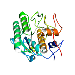
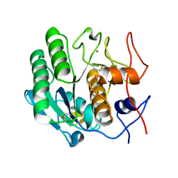
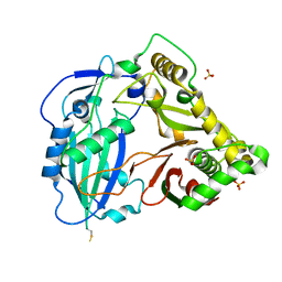
4YTA
DownloadVisualize
BU of 4yta by Molmil
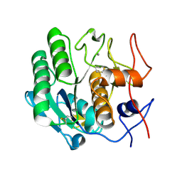
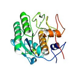
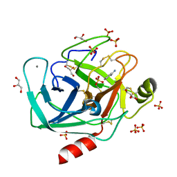
BOND LENGTH ANALYSIS OF ASP, GLU AND HIS RESIDUES IN TRYPSIN AT 1.2A RESOLUTION
Descriptor: BENZAMIDINE, CALCIUM ION, Cationic trypsin, ...
Authors:Fisher, S.J, Helliwell, J.R, Blakeley, M.P, Cianci, M, McSweeny, S.
Deposit date:2015-03-17
Release date:2015-05-27
Last modified:2024-10-16
Method:X-RAY DIFFRACTION (1.2 Å)
Cite:Protonation-state determination in proteins using high-resolution X-ray crystallography: effects of resolution and completeness.
Acta Crystallogr. D Biol. Crystallogr., 68, 2012
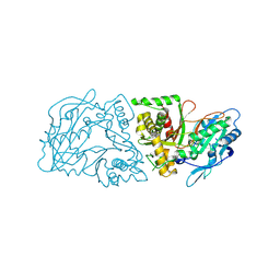
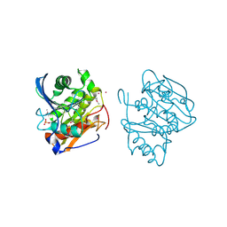
1DJL
DownloadVisualize
BU of 1djl by Molmil
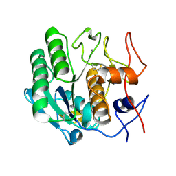
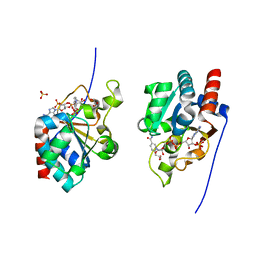
THE CRYSTAL STRUCTURE OF HUMAN TRANSHYDROGENASE DOMAIN III WITH BOUND NADP
Descriptor: GLYCEROL, NADP NICOTINAMIDE-ADENINE-DINUCLEOTIDE PHOSPHATE, SULFATE ION, ...
Authors:White, S.A, Peak, S.J, Cotton, N.P, Jackson, J.B.
Deposit date:1999-12-03
Release date:2000-12-06
Last modified:2024-02-07
Method:X-RAY DIFFRACTION (2 Å)
Cite:The high-resolution structure of the NADP(H)-binding component (dIII) of proton-translocating transhydrogenase from human heart mitochondria.
Structure Fold.Des., 8, 2000
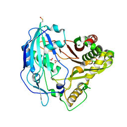
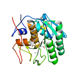
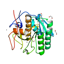
8G83
DownloadVisualize
BU of 8g83 by Molmil
Structure of NAD+ consuming protein Acinetobacter baumannii TIR domain
Descriptor: NAD(+) hydrolase AbTIR
Authors:Klontz, E.H, Wang, Y, Glendening, G, Carr, J, Tsibouris, T, Buddula, S, Nallar, S, Soares, A, Snyder, G.A.
Deposit date:2023-02-17
Release date:2023-10-11
Last modified:2023-11-15
Method:X-RAY DIFFRACTION (3.03 Å)
Cite:The structure of NAD + consuming protein Acinetobacter baumannii TIR domain shows unique kinetics and conformations.
J.Biol.Chem., 299, 2023
3DE1
DownloadVisualize
BU of 3de1 by Molmil
Proteinase K by LB nanotemplate method after the third step of high X-Ray dose on ESRF ID23-1 beamline
Descriptor: CALCIUM ION, Proteinase K
Authors:Pechkova, E, Tripathi, S.K, Nicolini, C.
Deposit date:2008-06-07
Release date:2009-06-09
Last modified:2024-10-09
Method:X-RAY DIFFRACTION (2 Å)
Cite:Radiation stability of proteinase K crystals grown by LB nanotemplate method
J.Struct.Biol., 168, 2009
3DDZ
DownloadVisualize
BU of 3ddz by Molmil
Proteinase K by LB nanotemplate method after the first step of high X-Ray dose on ESRF ID23-1 beamline
Descriptor: CALCIUM ION, Proteinase K
Authors:Pechkova, E, Tripathi, S.K, Nicolini, C.
Deposit date:2008-06-07
Release date:2009-06-09
Last modified:2023-11-01
Method:X-RAY DIFFRACTION (1.7 Å)
Cite:Radiation stability of proteinase K crystals grown by LB nanotemplate method
J.Struct.Biol., 168, 2009
3D9Q
DownloadVisualize
BU of 3d9q by Molmil
Proteinase K by LB nanotemplate method before high X-Ray dose on ESRF ID23-1 beamline
Descriptor: CALCIUM ION, Proteinase K
Authors:Pechkova, E, Tripathi, S.K, Nicolini, C.
Deposit date:2008-05-27
Release date:2009-06-02
Last modified:2023-11-01
Method:X-RAY DIFFRACTION (1.43 Å)
Cite:Radiation stability of proteinase K crystals grown by LB nanotemplate method
J.Struct.Biol., 168, 2009
3DE2
DownloadVisualize
BU of 3de2 by Molmil
Proteinase K by LB nanotemplate method after the fourth step of high X-Ray dose on ESRF ID23-1 beamline
Descriptor: CALCIUM ION, Proteinase K
Authors:Pechkova, E, Tripathi, S.K, Nicolini, C.
Deposit date:2008-06-07
Release date:2009-06-09
Last modified:2011-07-13
Method:X-RAY DIFFRACTION (2.1 Å)
Cite:Radiation stability of proteinase K crystals grown by LB nanotemplate method
J.Struct.Biol., 168, 2009
3DE0
DownloadVisualize
BU of 3de0 by Molmil
Proteinase K by LB nanotemplate method after the second step of high X-Ray dose on ESRF ID23-1 beamline
Descriptor: CALCIUM ION, Proteinase K
Authors:Pechkova, E, Tripathi, S.K, Nicolini, C.
Deposit date:2008-06-07
Release date:2009-06-09
Last modified:2023-11-01
Method:X-RAY DIFFRACTION (1.9 Å)
Cite:Radiation stability of proteinase K crystals grown by LB nanotemplate method
J.Struct.Biol., 168, 2009
3DE6
DownloadVisualize
BU of 3de6 by Molmil
Proteinase K by Classical hanging drop method after the third step of high X-Ray dose on ESRF ID23-1 beamline
Descriptor: CALCIUM ION, Proteinase K
Authors:Pechkova, E, Tripathi, S.K, Nicolini, C.
Deposit date:2008-06-08
Release date:2009-06-09
Last modified:2024-10-16
Method:X-RAY DIFFRACTION (2.2 Å)
Cite:Radiation stability of proteinase K crystals grown by LB nanotemplate method
J.Struct.Biol., 168, 2009
3DE3
DownloadVisualize
BU of 3de3 by Molmil
Proteinase K by Classical hanging drop method before high X-Ray dose on ESRF ID23-1 beamline
Descriptor: CALCIUM ION, Proteinase K
Authors:Pechkova, E, Tripathi, S.K, Nicolini, C.
Deposit date:2008-06-08
Release date:2009-06-09
Last modified:2011-07-13
Method:X-RAY DIFFRACTION (1.43 Å)
Cite:Radiation stability of proteinase K crystals grown by LB nanotemplate method
J.Struct.Biol., 168, 2009
3DE5
DownloadVisualize
BU of 3de5 by Molmil
roteinase K by Classical hanging drop method after the second step of high X-Ray dose on ESRF ID23-1 beamline
Descriptor: CALCIUM ION, Proteinase K
Authors:Pechkova, E, Tripathi, S.K, Nicolini, C.
Deposit date:2008-06-08
Release date:2009-06-09
Last modified:2011-07-13
Method:X-RAY DIFFRACTION (2.1 Å)
Cite:Radiation stability of proteinase K crystals grown by LB nanotemplate method
J.Struct.Biol., 168, 2009
3DE7
DownloadVisualize
BU of 3de7 by Molmil
Proteinase K by Classical hanging drop method after the fourth step of high X-Ray dose on ESRF ID23-1 beamline
Descriptor: CALCIUM ION, Proteinase K
Authors:Pechkova, E, Tripathi, S.K, Nicolini, C.
Deposit date:2008-06-08
Release date:2009-06-09
Last modified:2011-07-13
Method:X-RAY DIFFRACTION (2.3 Å)
Cite:Radiation stability of proteinase K crystals grown by LB nanotemplate method
J.Struct.Biol., 168, 2009
3DE4
DownloadVisualize
BU of 3de4 by Molmil
Proteinase K by Classical hanging drop method after the first step of high X-Ray dose on ESRF ID23-1 beamline
Descriptor: CALCIUM ION, Proteinase K
Authors:Pechkova, E, Tripathi, S.K, Nicolini, C.
Deposit date:2008-06-08
Release date:2009-06-09
Last modified:2024-10-16
Method:X-RAY DIFFRACTION (1.8 Å)
Cite:Radiation stability of proteinase K crystals grown by LB nanotemplate method
J.Struct.Biol., 168, 2009
3DVS
DownloadVisualize
BU of 3dvs by Molmil
Proteinase K by LB nanotmplate method after the second step of high dose on ESRF ID14-2 beamline
Descriptor: CALCIUM ION, Proteinase K
Authors:Pechkova, E, Tripathi, S.K, Nicolini, C.
Deposit date:2008-07-20
Release date:2009-07-07
Last modified:2023-11-01
Method:X-RAY DIFFRACTION (1.02 Å)
Cite:Radiation damage in protein structural characterization by Synchrotron Radiation: State of the art and Nanotechnology-based perspective
To be Published
4G2M
DownloadVisualize
BU of 4g2m by Molmil
Structure of a Lys-HCT mutant from Coffea canephora (Crystal form 2)
Descriptor: Hydroxycinnamoyl-CoA shikimate/quinate hydroxycinnamoyltransferase
Authors:Lallemand, L.A, McCarthy, J.G, McCarthy, A.A.
Deposit date:2012-07-12
Release date:2012-08-01
Last modified:2012-11-21
Method:X-RAY DIFFRACTION (2.5 Å)
Cite:A structural basis for the biosynthesis of the major chlorogenic acids found in coffee.
Plant Physiol., 160, 2012
4G22
DownloadVisualize
BU of 4g22 by Molmil
Structure of a Lys-HCT mutant from Coffea canephora (Crystal form 1)
Descriptor: CHLORIDE ION, GLYCEROL, Hydroxycinnamoyl-CoA shikimate/quinate hydroxycinnamoyltransferase
Authors:McCarthy, A.A, Lallemand, L.A, McCarthy, J.G.
Deposit date:2012-07-11
Release date:2012-08-01
Last modified:2012-11-21
Method:X-RAY DIFFRACTION (1.7 Å)
Cite:A structural basis for the biosynthesis of the major chlorogenic acids found in coffee.
Plant Physiol., 160, 2012
4G0B
DownloadVisualize
BU of 4g0b by Molmil
Structure of native HCT from Coffea canephora
Descriptor: Hydroxycinnamoyl-CoA shikimate/quinate hydroxycinnamoyltransferase, SULFATE ION
Authors:Lallemand, L.A, McCarthy, J.G, McCarthy, A.A.
Deposit date:2012-07-09
Release date:2012-08-01
Last modified:2023-09-13
Method:X-RAY DIFFRACTION (3 Å)
Cite:A structural basis for the biosynthesis of the major chlorogenic acids found in coffee.
Plant Physiol., 160, 2012
5FXM
DownloadVisualize
BU of 5fxm by Molmil
Structure of FAE solved by SAD from data collected by Direct Data Collection (DDC) using the ESRF RoboDiff goniometer
Descriptor: CADMIUM ION, ENDO-1,4-BETA-XYLANASE Y
Authors:Bowler, M.W, Nurizzo, D.
Deposit date:2016-03-02
Release date:2016-03-16
Last modified:2017-08-23
Method:X-RAY DIFFRACTION (1.99 Å)
Cite:Robodiff: Combining a Sample Changer and Goniometer for Highly Automated Macromolecular Crystallography Experiments.
Acta Crystallogr.,Sect.D, 72, 2016
5WJH
DownloadVisualize
BU of 5wjh by Molmil
Using sound pulses to solve the crystal harvesting bottleneck
Descriptor: (CARBAMOYLMETHYL-CARBOXYMETHYL-AMINO)-ACETIC ACID, CALCIUM ION, Proteinase K
Authors:Soares, A.S, Brennan, H.M, McCarthy, L, Leroy, L.
Deposit date:2017-07-22
Release date:2017-08-02
Last modified:2023-10-04
Method:X-RAY DIFFRACTION (1.63 Å)
Cite:Using sound pulses to solve the crystal-harvesting bottleneck.
Acta Crystallogr D Struct Biol, 74, 2018
8BHD
DownloadVisualize
BU of 8bhd by Molmil
N-terminal domain of Plasmodium berghei glutamyl-tRNA synthetase (Tbxo4 derivative crystal structure)
Descriptor: GLYCEROL, Glutamate--tRNA ligase, SULFATE ION, ...
Authors:Benas, P, Jaramillo Ponce, J.R, Legrand, P, Frugier, M, Sauter, C.
Deposit date:2022-10-31
Release date:2023-01-25
Last modified:2024-06-19
Method:X-RAY DIFFRACTION (3.17 Å)
Cite:Solution X-ray scattering highlights discrepancies in Plasmodium multi-aminoacyl-tRNA synthetase complexes.
Protein Sci., 32, 2023
5WHW
DownloadVisualize
BU of 5whw by Molmil
Using sound pulses to solve the crystal harvesting bottleneck
Descriptor: 1,2-ETHANEDIOL, BICINE, CALCIUM ION, ...
Authors:Soares, A.S, Brennan, H.M, Natarajan, R, McCarthy, L, Leroy, L.
Deposit date:2017-07-18
Release date:2017-08-02
Last modified:2023-10-04
Method:X-RAY DIFFRACTION (1.71 Å)
Cite:Using sound pulses to solve the crystal-harvesting bottleneck.
Acta Crystallogr D Struct Biol, 74, 2018
5WJG
DownloadVisualize
BU of 5wjg by Molmil
Using sound pulses to solve the crystal harvesting bottleneck
Descriptor: 1,2-ETHANEDIOL, CALCIUM ION, L(+)-TARTARIC ACID, ...
Authors:Soares, A.S, Brennan, H.M, McCarthy, L, Leroy, L.
Deposit date:2017-07-22
Release date:2017-08-02
Last modified:2023-10-04
Method:X-RAY DIFFRACTION (1.5 Å)
Cite:Using sound pulses to solve the crystal-harvesting bottleneck.
Acta Crystallogr D Struct Biol, 74, 2018
5FXL
DownloadVisualize
BU of 5fxl by Molmil
Structure of trypsin solved by MR from data collected by Direct Data Collection (DDC) using the ESRF RoboDiff goniometer
Descriptor: BENZAMIDINE, CALCIUM ION, CATIONIC TRYPSIN, ...
Authors:Bowler, M.W, Nurizzo, D.
Deposit date:2016-03-02
Release date:2016-03-16
Last modified:2024-01-10
Method:X-RAY DIFFRACTION (1.78 Å)
Cite:Robodiff: Combining a Sample Changer and Goniometer for Highly Automated Macromolecular Crystallography Experiments.
Acta Crystallogr.,Sect.D, 72, 2016

226262

PDB entries from 2024-10-16

PDB statisticsPDBj update infoContact PDBjnumon