Loading
PDBj
MenuPDBj@FacebookPDBj@X(formerly Twitter)PDBj@BlueSkyPDBj@YouTubewwPDB FoundationwwPDB
RCSB PDBPDBeBMRBAdv. SearchSearch help
Search by PDB author
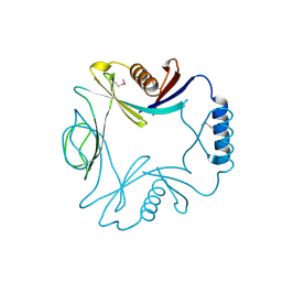
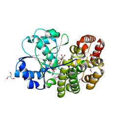
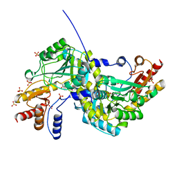
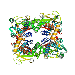
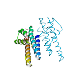
1O5H
DownloadVisualize
BU of 1o5h by Molmil
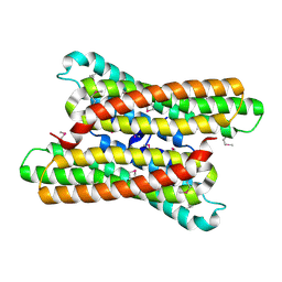
Crystal structure of formiminotetrahydrofolate cyclodeaminase (TM1560) from Thermotoga maritima at 2.80 A resolution
Descriptor: Formiminotetrahydrofolate cyclodeaminase
Authors:Joint Center for Structural Genomics (JCSG)
Deposit date:2003-09-17
Release date:2003-09-30
Last modified:2024-10-30
Method:X-RAY DIFFRACTION (2.8 Å)
Cite:Crystal structure of a formiminotetrahydrofolate cyclodeaminase (TM1560) from Thermotoga maritima at 2.80 A resolution reveals a new fold
Proteins, 58, 2005
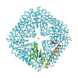
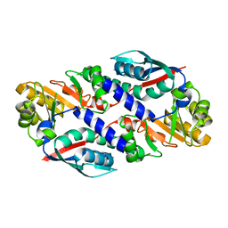
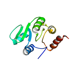
1O3U
DownloadVisualize
BU of 1o3u by Molmil
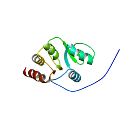
Crystal structure of an hepn domain protein (tm0613) from thermotoga maritima at 1.75 A resolution
Descriptor: conserved hypothetical protein TM0613
Authors:Joint Center for Structural Genomics (JCSG)
Deposit date:2003-03-28
Release date:2003-04-22
Last modified:2024-10-30
Method:X-RAY DIFFRACTION (1.75 Å)
Cite:Crystal structure of an HEPN domain protein (TM0613) from Thermotoga maritima at 1.75 A resolution.
PROTEINS, 54, 2004
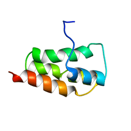
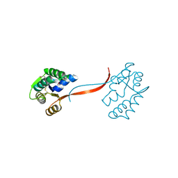
1O22
DownloadVisualize
BU of 1o22 by Molmil
Crystal structure of an orphan protein (TM0875) from Thermotoga maritima at 2.00 A resolution
Descriptor: orphan protein TM0875
Authors:Joint Center for Structural Genomics (JCSG)
Deposit date:2003-02-15
Release date:2003-04-01
Last modified:2024-10-30
Method:X-RAY DIFFRACTION (2 Å)
Cite:Crystal structure of an orphan protein (TM0875) from Thermotoga maritima at 2.00-A resolution reveals a new fold.
Proteins, 56, 2004
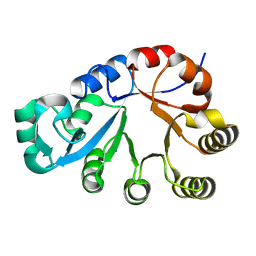
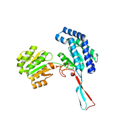
1O4V
DownloadVisualize
BU of 1o4v by Molmil
Crystal structure of the catalytic subunit of a phosphoribosylaminoimidazole mutase (tm0446) from thermotoga maritima at 1.77 A resolution
Descriptor: SULFATE ION, phosphoribosylaminoimidazole mutase PurE
Authors:Joint Center for Structural Genomics (JCSG)
Deposit date:2003-07-11
Release date:2003-07-22
Last modified:2023-09-20
Method:X-RAY DIFFRACTION (1.77 Å)
Cite:Crystal structure of a phosphoribosylaminoimidazole mutase PurE (TM0446) from Thermotoga maritima at 1.77 A resolution
Proteins, 55, 2004
2KYS
DownloadVisualize
BU of 2kys by Molmil
NMR Structure of the SARS Coronavirus Nonstructural Protein Nsp7 in Solution at pH 6.5
Descriptor: Non-structural protein 7
Authors:Johnson, M.A, Jaudzems, K, Wilson, I.A, Wuthrich, K, Joint Center for Structural Genomics (JCSG)
Deposit date:2010-06-07
Release date:2010-06-16
Last modified:2024-05-15
Method:SOLUTION NMR
Cite:NMR Structure of the SARS-CoV Nonstructural Protein 7 in Solution at pH 6.5.
J.Mol.Biol., 402, 2010
1O1Z
DownloadVisualize
BU of 1o1z by Molmil
Crystal structure of glycerophosphodiester phosphodiesterase (GDPD) (TM1621) from Thermotoga maritima at 1.60 A resolution
Descriptor: SODIUM ION, glycerophosphodiester phosphodiesterase
Authors:Joint Center for Structural Genomics (JCSG)
Deposit date:2003-02-12
Release date:2003-04-01
Last modified:2024-05-22
Method:X-RAY DIFFRACTION (1.6 Å)
Cite:Crystal structure of a glycerophosphodiester phosphodiesterase (GDPD) from Thermotoga maritima (TM1621) at 1.60 A resolution.
Proteins, 56, 2004
1O2D
DownloadVisualize
BU of 1o2d by Molmil
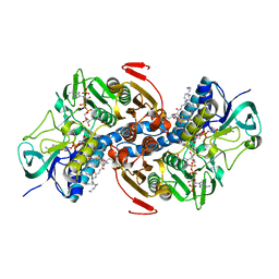
Crystal structure of Alcohol dehydrogenase, iron-containing (TM0920) from Thermotoga maritima at 1.30 A resolution
Descriptor: 2-AMINO-2-HYDROXYMETHYL-PROPANE-1,3-DIOL, Alcohol dehydrogenase, iron-containing, ...
Authors:Joint Center for Structural Genomics (JCSG)
Deposit date:2003-02-27
Release date:2003-06-10
Last modified:2023-01-25
Method:X-RAY DIFFRACTION (1.3 Å)
Cite:Crystal structure of an iron-containing 1,3-propanediol dehydrogenase (TM0920) from Thermotoga maritima at 1.3 A resolution
Proteins, 54, 2004
1O4U
DownloadVisualize
BU of 1o4u by Molmil
Crystal structure of a nicotinate nucleotide pyrophosphorylase (tm1645) from thermotoga maritima at 2.50 A resolution
Descriptor: Type II quinolic acid phosphoribosyltransferase
Authors:Joint Center for Structural Genomics (JCSG)
Deposit date:2003-07-09
Release date:2003-07-22
Last modified:2023-09-20
Method:X-RAY DIFFRACTION (2.5 Å)
Cite:Crystal structure of a type II quinolic acid phosphoribosyltransferase (TM1645) from Thermotoga maritima at 2.50 A resolution
Proteins, 55, 2004
1O4W
DownloadVisualize
BU of 1o4w by Molmil
CRYSTAL STRUCTURE OF a PIN (PILT N-TERMINUS) DOMAIN CONTAINING PROTEIN (AF0591) FROM ARCHAEOGLOBUS FULGIDUS AT 1.90 A RESOLUTION
Descriptor: PIN (PilT N-terminus) domain
Authors:Joint Center for Structural Genomics (JCSG)
Deposit date:2003-07-16
Release date:2003-07-29
Last modified:2024-11-06
Method:X-RAY DIFFRACTION (1.9 Å)
Cite:Crystal structure of a PIN (PilT N-terminus) domain (AF0591) from Archaeoglobus fulgidus at 1.90 A resolution
Proteins, 56, 2004
1O20
DownloadVisualize
BU of 1o20 by Molmil
Crystal structure of Gamma-glutamyl phosphate reductase (TM0293) from Thermotoga maritima at 2.00 A resolution
Descriptor: Gamma-glutamyl phosphate reductase
Authors:Joint Center for Structural Genomics (JCSG)
Deposit date:2003-02-12
Release date:2003-04-01
Last modified:2024-11-13
Method:X-RAY DIFFRACTION (2 Å)
Cite:Crystal structure of gamma-glutamyl phosphate reductase (TM0293) from Thermotoga maritima at 2.0 A resolution.
Proteins, 54, 2004
1O4S
DownloadVisualize
BU of 1o4s by Molmil
Crystal structure of Aspartate aminotransferase (TM1255) from Thermotoga maritima at 1.90 A resolution
Descriptor: Aspartate aminotransferase, PYRIDOXAL-5'-PHOSPHATE, SULFATE ION
Authors:Joint Center for Structural Genomics (JCSG)
Deposit date:2003-06-26
Release date:2003-07-08
Last modified:2023-09-20
Method:X-RAY DIFFRACTION (1.9 Å)
Cite:Crystal structure of an aspartate aminotransferase (TM1255) from Thermotoga maritima at 1.90 A resolution
Proteins, 55, 2004
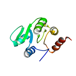
2JZD
DownloadVisualize
BU of 2jzd by Molmil
NMR structure of the domain 527-651 of the SARS-CoV nonstructural protein nsp3
Descriptor: Replicase polyprotein 1ab
Authors:Chatterjee, A, Johnson, M.A, Serrano, P, Pedrini, B, Joseph, J, Saikatendu, K, Neuman, B, Stevens, R.C, Wilson, I.A, Buchmeier, M.J, Kuhn, P, Wuthrich, K, Joint Center for Structural Genomics (JCSG)
Deposit date:2008-01-04
Release date:2008-02-05
Last modified:2024-05-29
Method:SOLUTION NMR
Cite:Nuclear magnetic resonance structure shows that the severe acute respiratory syndrome coronavirus-unique domain contains a macrodomain fold.
J.Virol., 83, 2009
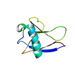
2HSX
DownloadVisualize
BU of 2hsx by Molmil
NMR Structure of the nonstructural protein 1 (nsp1) from the SARS coronavirus
Descriptor: Leader protein; p65 homolog; NSP1 (EC 3.4.22.-)
Authors:Almeida, M.S, Herrmann, T, Geralt, M, Johnson, M.A, Saikatendu, K, Joseph, J, Subramanian, R.C, Neuman, B.W, Buchmeier, M.J, Stevens, R.C, Kuhn, P, Wilson, I.A, Wuthrich, K, Joint Center for Structural Genomics (JCSG)
Deposit date:2006-07-24
Release date:2007-02-06
Last modified:2024-05-29
Method:SOLUTION NMR
Cite:Novel beta-barrel fold in the nuclear magnetic resonance structure of the replicase nonstructural protein 1 from the severe acute respiratory syndrome coronavirus.
J.Virol., 81, 2007
2JZE
DownloadVisualize
BU of 2jze by Molmil
NMR structure of the domain 527-651 of the SARS-CoV nonstructural protein nsp3, single conformer closest to the mean coordinates of an ensemble of twenty energy minimized conformers
Descriptor: Replicase polyprotein 1ab
Authors:Chatterjee, A, Johnson, M.A, Serrano, P, Pedrini, B, Joseph, J, Saikatendu, K, Neuman, B, Stevens, R.C, Wilson, I.A, Buchmeier, M.J, Kuhn, P, Wuthrich, K, Joint Center for Structural Genomics (JCSG)
Deposit date:2008-01-04
Release date:2008-02-05
Last modified:2024-05-29
Method:SOLUTION NMR
Cite:Nuclear magnetic resonance structure shows that the severe acute respiratory syndrome coronavirus-unique domain contains a macrodomain fold.
J.Virol., 83, 2009
1O58
DownloadVisualize
BU of 1o58 by Molmil
Crystal structure of O-acetylserine sulfhydrylase (TM0665) from Thermotoga maritima at 1.80 A resolution
Descriptor: O-acetylserine sulfhydrylase, PHOSPHATE ION
Authors:Joint Center for Structural Genomics (JCSG)
Deposit date:2003-08-20
Release date:2003-09-02
Last modified:2024-05-22
Method:X-RAY DIFFRACTION (1.8 Å)
Cite:Crystal structure of O-acetylserine sulfhydrylase (TM0665) from Thermotoga maritima at 1.8 A resolution
Proteins, 56, 2004
1O5U
DownloadVisualize
BU of 1o5u by Molmil
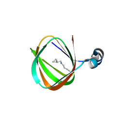
Crystal structure of a duf861 family protein (tm1112) from thermotoga maritima at 1.83 A resolution
Descriptor: UNKNOWN LIGAND, novel Thermotoga maritima enzyme TM1112
Authors:Joint Center for Structural Genomics (JCSG)
Deposit date:2003-10-06
Release date:2003-11-11
Last modified:2024-11-13
Method:X-RAY DIFFRACTION (1.83 Å)
Cite:Crystal structure of a novel Thermotoga maritima enzyme (TM1112) from the cupin family at 1.83 A resolution
Proteins, 56, 2004
2JZF
DownloadVisualize
BU of 2jzf by Molmil
NMR Conformer closest to the mean coordinates of the domain 513-651 of the SARS-CoV nonstructural protein nsp3
Descriptor: Replicase polyprotein 1ab
Authors:Chatterjee, A, Johnson, M.A, Serrano, P, Pedrini, B, Joseph, J, Saikatendu, K, Neuman, B, Stevens, R.C, Wilson, I.A, Buchmeier, M.J, Kuhn, P, Wuthrich, K, Joint Center for Structural Genomics (JCSG)
Deposit date:2008-01-04
Release date:2008-02-05
Last modified:2024-05-29
Method:SOLUTION NMR
Cite:Nuclear magnetic resonance structure shows that the severe acute respiratory syndrome coronavirus-unique domain contains a macrodomain fold.
J.Virol., 83, 2009
1TYP
DownloadVisualize
BU of 1typ by Molmil
SUBSTRATE INTERACTIONS BETWEEN TRYPANOTHIONE REDUCTASE AND N1-GLUTATHIONYLSPERMIDINE DISULPHIDE AT 0.28-NM RESOLUTION
Descriptor: FLAVIN-ADENINE DINUCLEOTIDE, GLUTATHIONE, NADP NICOTINAMIDE-ADENINE-DINUCLEOTIDE PHOSPHATE, ...
Authors:Bailey, S, Hunter, W.N.
Deposit date:1992-06-29
Release date:1994-01-31
Last modified:2024-06-05
Method:X-RAY DIFFRACTION (2.8 Å)
Cite:Substrate interactions between trypanothione reductase and N1-glutathionylspermidine disulphide at 0.28-nm resolution.
Eur.J.Biochem., 213, 1993
1EO8
DownloadVisualize
BU of 1eo8 by Molmil
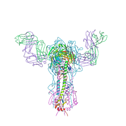
INFLUENZA VIRUS HEMAGGLUTININ COMPLEXED WITH A NEUTRALIZING ANTIBODY
Descriptor: 2-acetamido-2-deoxy-beta-D-glucopyranose, 2-acetamido-2-deoxy-beta-D-glucopyranose-(1-4)-2-acetamido-2-deoxy-beta-D-glucopyranose, ANTIBODY (HEAVY CHAIN), ...
Authors:Fleury, D, Gigant, B, Daniels, R.S, Skehel, J.J, Knossow, M, Bizebard, T.
Deposit date:2000-03-22
Release date:2000-04-12
Last modified:2020-07-29
Method:X-RAY DIFFRACTION (2.8 Å)
Cite:Structural evidence for recognition of a single epitope by two distinct antibodies.
Proteins, 40, 2000
1KB5
DownloadVisualize
BU of 1kb5 by Molmil
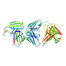
MURINE T-CELL RECEPTOR VARIABLE DOMAIN/FAB COMPLEX
Descriptor: ANTIBODY DESIRE-1, KB5-C20 T-CELL ANTIGEN RECEPTOR
Authors:Housset, D, Mazza, G, Gregoire, C, Piras, C, Malissen, B, Fontecilla-Camps, J.C.
Deposit date:1997-04-06
Release date:1998-04-08
Last modified:2023-08-09
Method:X-RAY DIFFRACTION (2.5 Å)
Cite:The three-dimensional structure of a T-cell antigen receptor V alpha V beta heterodimer reveals a novel arrangement of the V beta domain.
EMBO J., 16, 1997
1KVF
DownloadVisualize
BU of 1kvf by Molmil
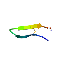
EMP-18 Erythropoietin Receptor Agonist Peptide
Descriptor: PROTEIN: EMP-18 Receptor Agonist
Authors:Skelton, N.J, Russell, S, de Sauvage, F, Cochran, A.G.
Deposit date:2002-01-25
Release date:2002-03-06
Last modified:2024-11-13
Method:SOLUTION NMR
Cite:Amino acid determinants of beta-hairpin conformation in erythropoeitin receptor agonist peptides derived from a phage display library
J.Mol.Biol., 316, 2002
1KVG
DownloadVisualize
BU of 1kvg by Molmil
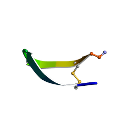
EPO-3 beta Hairpin Peptide
Descriptor: Protein: EPO-3 Receptor Agonist
Authors:Skelton, N.J, Russell, S, de Sauvage, F, Cochran, A.G.
Deposit date:2002-01-25
Release date:2002-03-06
Last modified:2022-02-23
Method:SOLUTION NMR
Cite:Amino Acid Determinants of beta-Hairpin Conformation in Erythropoeitin Receptor Agonist Peptides Derived from a Phage Display Library
J.Mol.Biol., 316, 2002
1NAM
DownloadVisualize
BU of 1nam by Molmil
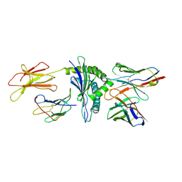
MURINE ALLOREACTIVE SCFV TCR-PEPTIDE-MHC CLASS I MOLECULE COMPLEX
Descriptor: 2-acetamido-2-deoxy-beta-D-glucopyranose-(1-4)-2-acetamido-2-deoxy-beta-D-glucopyranose, BM3.3 T Cell Receptor alpha-Chain, BM3.3 T Cell Receptor beta-Chain, ...
Authors:Reiser, J.-B, Darnault, C, Gregoire, C, Mosser, T, Mazza, G, Kearnay, A, van der Merwe, P.A, Fontecilla-Camps, J.C, Housset, D, Malissen, B.
Deposit date:2002-11-28
Release date:2003-03-11
Last modified:2024-11-06
Method:X-RAY DIFFRACTION (2.7 Å)
Cite:CDR3 loop flexibility contributes to the degeneracy of TCR recognition
Nat.Immunol., 4, 2003
1QB6
DownloadVisualize
BU of 1qb6 by Molmil
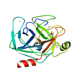
BOVINE TRYPSIN 3,3'-[3,5-DIFLUORO-4-METHYL-2, 6-PYRIDINEDIYLBIS(OXY)]BIS(BENZENECARBOXIMIDAMIDE) (ZK-805623) COMPLEX
Descriptor: 3,3'-[3,5-DIFLUORO-4-METHYL-2,6-PYRIDYLENEBIS(OXY)]-BIS(BENZENECARBOXIMIDAMIDE), CALCIUM ION, POTASSIUM ION, ...
Authors:Whitlow, M.
Deposit date:1999-04-29
Release date:2000-04-29
Last modified:2024-11-13
Method:X-RAY DIFFRACTION (1.8 Å)
Cite:Crystallographic analysis of potent and selective factor Xa inhibitors complexed to bovine trypsin.
Acta Crystallogr.,Sect.D, 55, 1999
1QBO
DownloadVisualize
BU of 1qbo by Molmil
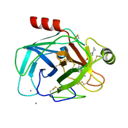
BOVINE TRYPSIN 7-[[6-[[1-(1-IMINOETHYL)PIPERIDIN-4-YL]OXY]-2-METHYL-BENZIMIDAZOL-1-YL]METHYL]NAPHTHALENE-2-CARBOXIMIDAMID ZK-806711 INHIBITOR COMPLEX
Descriptor: 7-[[6-[[1-(1-IMINOETHYL)PIPERIDIN-4-YL]OXY]-2-METHYL-BENZIMIDAZOL-1-YL]METHYL]NAPHTHALENE-2-CARBOXIMIDAMID, CALCIUM ION, PROTEIN (TRYPSIN)
Authors:Whitlow, M.
Deposit date:1999-04-26
Release date:2000-05-03
Last modified:2024-10-16
Method:X-RAY DIFFRACTION (1.8 Å)
Cite:Crystallographic analysis of potent and selective factor Xa inhibitors complexed to bovine trypsin.
Acta Crystallogr.,Sect.D, 55, 1999

227344

PDB entries from 2024-11-13

PDB statisticsPDBj update infoContact PDBjnumon