Loading
PDBj
MenuPDBj@FacebookPDBj@TwitterPDBj@YouTubewwPDB FoundationwwPDB
RCSB PDBPDBeBMRBAdv. SearchSearch help
Search by PDB author
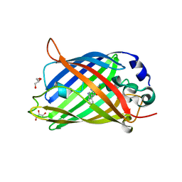
3LVD
DownloadVisualize
BU of 3lvd by Molmil
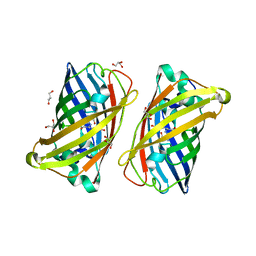
Crystal structure of GFP-like protein aceGFP_G222E (A. coerulescens). UV-photoconverted green form.
Descriptor: GLYCEROL, Green fluorescent protein
Authors:Pletneva, N.V, Pletnev, V.Z, Pletnev, S.V.
Deposit date:2010-02-19
Release date:2010-03-09
Last modified:2024-10-09
Method:X-RAY DIFFRACTION (1.751 Å)
Cite:Structural evidence for a dehydrated intermediate in green fluorescent protein chromophore biosynthesis.
J.Biol.Chem., 285, 2010
4RYW
DownloadVisualize
BU of 4ryw by Molmil
Crystal structure of the photoconverted green fluorescent protein NowGFP_conv (the variant of cyan Cerulean) at pH 7.0
Descriptor: GLYCEROL, NowGFP_conv
Authors:Pletnev, V.Z, Pletneva, N.V, Pletnev, S.V.
Deposit date:2014-12-17
Release date:2015-09-02
Last modified:2023-12-06
Method:X-RAY DIFFRACTION (2.5 Å)
Cite:Structure of the green fluorescent protein NowGFP with an anionic tryptophan-based chromophore.
Acta Crystallogr.,Sect.D, 71, 2015
3LVC
DownloadVisualize
BU of 3lvc by Molmil
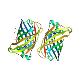
Crystal structure of GFP-like protein aceGFP_G222E (A. coerulescens). Colorless form.
Descriptor: GLYCEROL, Green fluorescent protein
Authors:Pletneva, N.V, Pletnev, V.Z, Pletnev, S.V.
Deposit date:2010-02-19
Release date:2010-03-09
Last modified:2023-11-15
Method:X-RAY DIFFRACTION (1.14 Å)
Cite:Structural evidence for a dehydrated intermediate in green fluorescent protein chromophore biosynthesis.
J.Biol.Chem., 285, 2010
3LVA
DownloadVisualize
BU of 3lva by Molmil
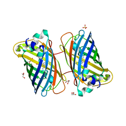
Crystal structure of colorless GFP-like protein from Aequorea coerulescens
Descriptor: GLYCEROL, Green fluorescent protein, SULFATE ION
Authors:Pletneva, N.V, Pletnev, V.Z, Pletnev, S.V.
Deposit date:2010-02-19
Release date:2010-03-09
Last modified:2023-11-15
Method:X-RAY DIFFRACTION (1.5 Å)
Cite:Structural evidence for a dehydrated intermediate in green fluorescent protein chromophore biosynthesis.
J.Biol.Chem., 285, 2010
1P58
DownloadVisualize
BU of 1p58 by Molmil
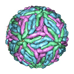
Complex Organization of Dengue Virus Membrane Proteins as Revealed by 9.5 Angstrom Cryo-EM reconstruction
Descriptor: Envelope protein M, Major envelope protein E
Authors:Zhang, W, Chipman, P.R, Corver, J, Johnson, P.R, Zhang, Y, Mukhopadhyay, S, Baker, T.S, Strauss, J.H, Rossmann, M.G, Kuhn, R.J.
Deposit date:2003-04-25
Release date:2003-11-04
Last modified:2024-02-14
Method:ELECTRON MICROSCOPY (9.5 Å)
Cite:Visualization of membrane protein domains by cryo-electron microscopy of dengue virus
Nat.Struct.Biol., 10, 2003
<12


226262

PDB entries from 2024-10-16

PDB statisticsPDBj update infoContact PDBjnumon