Loading
PDBj
MenuPDBj@FacebookPDBj@TwitterPDBj@YouTubewwPDB FoundationwwPDB
RCSB PDBPDBeBMRBAdv. SearchSearch help
Search by PDB author
7T1X
DownloadVisualize
BU of 7t1x by Molmil
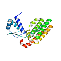
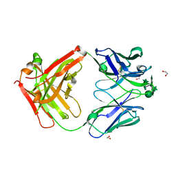
Crystal structure of human Fab A194-01 in complex with its synthetic heptasaccharide Ara6-Man epitope (BSI110888)
Descriptor: 1,2-ETHANEDIOL, Fab A194-01 heavy chain, Fab A194-01 light chain, ...
Authors:Seattle Structural Genomics Center for Infectious Disease (SSGCID)
Deposit date:2021-12-02
Release date:2022-12-07
Last modified:2024-04-03
Method:X-RAY DIFFRACTION (2.6 Å)
Cite:Crystal structure of human Fab A194-01 in complex with its synthetic heptasaccharide Ara6-Man epitope (BSI110888)
to be published
7SSQ
DownloadVisualize
BU of 7ssq by Molmil
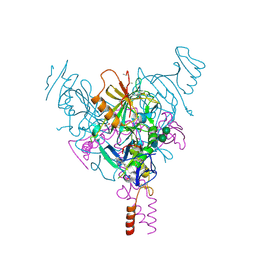
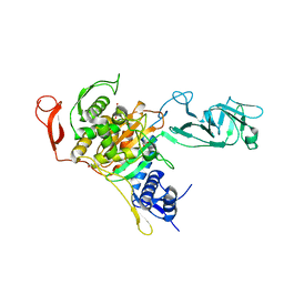
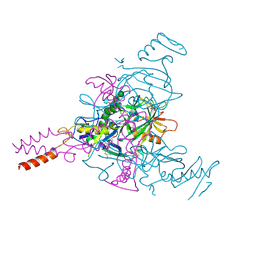
Crystal Structure of Ebola zaire Envelope glycoprotein GP in complex with compound ARN0075231
Descriptor: 1,2-ETHANEDIOL, 2-acetamido-2-deoxy-beta-D-glucopyranose, 2-acetamido-2-deoxy-beta-D-glucopyranose-(1-2)-alpha-D-mannopyranose-(1-6)-[alpha-D-mannopyranose-(1-3)]beta-D-mannopyranose-(1-4)-2-acetamido-2-deoxy-beta-D-glucopyranose-(1-4)-2-acetamido-2-deoxy-beta-D-glucopyranose, ...
Authors:Seattle Structural Genomics Center for Infectious Disease, Seattle Structural Genomics Center for Infectious Disease (SSGCID)
Deposit date:2021-11-11
Release date:2023-08-09
Method:X-RAY DIFFRACTION (2.25 Å)
Cite:Crystal Structure of Ebola zaire Envelope glycoprotein GP in complex with compound ARN0075231
to be published
7SSR
DownloadVisualize
BU of 7ssr by Molmil
Crystal Structure of Ebola zaire Envelope glycoprotein GP in complex with compound ARN0075093
Descriptor: (1R,2s,3S,5s,7s)-N-[(1r,4r)-4-(aminomethyl)cyclohexyl]-5-phenyladamantane-2-carboxamide, 1,2-ETHANEDIOL, 2-acetamido-2-deoxy-beta-D-glucopyranose, ...
Authors:Seattle Structural Genomics Center for Infectious Disease, Seattle Structural Genomics Center for Infectious Disease (SSGCID)
Deposit date:2021-11-11
Release date:2023-08-09
Method:X-RAY DIFFRACTION (2.5 Å)
Cite:Crystal Structure of Ebola zaire Envelope glycoprotein GP in complex with compound ARN0075093
to be published
7TI7
DownloadVisualize
BU of 7ti7 by Molmil
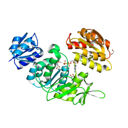
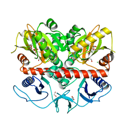
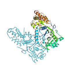
Crystal Structure of UDP-N-acetylmuramoylalanine-D-glutamate ligase from Acinetobacter baumannii AB5075-UW in complex with ADP
Descriptor: 1,2-ETHANEDIOL, ADENOSINE-5'-DIPHOSPHATE, CALCIUM ION, ...
Authors:Seattle Structural Genomics Center for Infectious Disease (SSGCID)
Deposit date:2022-01-12
Release date:2022-01-26
Last modified:2023-11-15
Method:X-RAY DIFFRACTION (1.5 Å)
Cite:Crystal Structure of UDP-N-acetylmuramoylalanine-D-glutamate ligase from Acinetobacter baumannii AB5075-UW in complex with ADP
to be published
7U5Y
DownloadVisualize
BU of 7u5y by Molmil
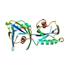
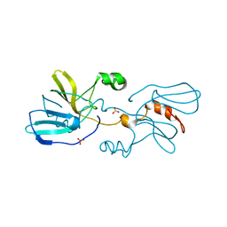
Crystal structure of ribulose-phosphate 3-epimerase from Pseudomonas aeruginosa
Descriptor: Ribulose-phosphate 3-epimerase, ZINC ION
Authors:Seattle Structural Genomics Center for Infectious Disease (SSGCID)
Deposit date:2022-03-02
Release date:2022-03-09
Last modified:2023-10-18
Method:X-RAY DIFFRACTION (2.55 Å)
Cite:Crystal structure of ribulose-phosphate 3-epimerase from Pseudomonas aeruginosa
To be Published
3LUZ
DownloadVisualize
BU of 3luz by Molmil
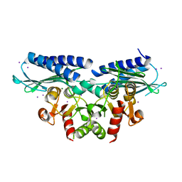
Crystal structure of extragenic suppressor protein suhB from Bartonella henselae, via combined iodide SAD molecular replacement
Descriptor: Extragenic suppressor protein suhB, IODIDE ION, MAGNESIUM ION
Authors:Seattle Structural Genomics Center for Infectious Disease (SSGCID)
Deposit date:2010-02-18
Release date:2010-03-02
Last modified:2023-09-06
Method:X-RAY DIFFRACTION (2.05 Å)
Cite:SAD phasing using iodide ions in a high-throughput structural genomics environment.
J Struct Funct Genomics, 12, 2011
7V0H
DownloadVisualize
BU of 7v0h by Molmil
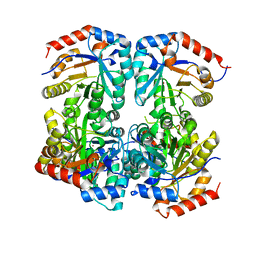
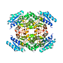
Crystal Structure of Putative glucose 1-dehydrogenase from Burkholderia cenocepacia in complex with NADP and a potential reaction product
Descriptor: (2R)-2-(hydroxymethyl)pentanedioic acid, CALCIUM ION, GLYCEROL, ...
Authors:Seattle Structural Genomics Center for Infectious Disease, Seattle Structural Genomics Center for Infectious Disease (SSGCID)
Deposit date:2022-05-10
Release date:2022-06-08
Last modified:2023-10-18
Method:X-RAY DIFFRACTION (1.55 Å)
Cite:Crystal Structure of Putative glucose 1-dehydrogenase from Burkholderia cenocepacia in complex with NADP and a potential reaction product
to be published
3PM6
DownloadVisualize
BU of 3pm6 by Molmil
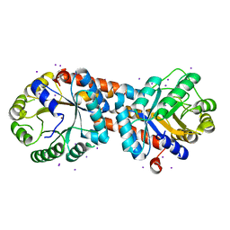
Crystal structure of a putative fructose-1,6-biphosphate aldolase from Coccidioides immitis solved by combined SAD MR
Descriptor: IODIDE ION, Putative fructose-bisphosphate aldolase, ZINC ION
Authors:Seattle Structural Genomics Center for Infectious Disease (SSGCID)
Deposit date:2010-11-16
Release date:2010-12-15
Last modified:2023-09-06
Method:X-RAY DIFFRACTION (2.2 Å)
Cite:SAD phasing using iodide ions in a high-throughput structural genomics environment.
J Struct Funct Genomics, 12, 2011
7U5F
DownloadVisualize
BU of 7u5f by Molmil
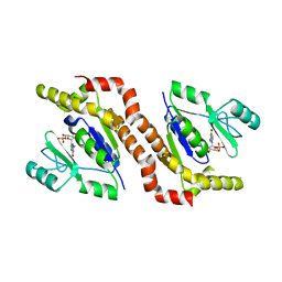
Crystal structure of guanylate kinase from Pseudomonas aeruginosa PAO1 in complex with GMP
Descriptor: GUANOSINE-5'-MONOPHOSPHATE, Guanylate kinase, MAGNESIUM ION
Authors:Seattle Structural Genomics Center for Infectious Disease (SSGCID)
Deposit date:2022-03-02
Release date:2022-03-09
Last modified:2023-10-18
Method:X-RAY DIFFRACTION (2 Å)
Cite:Crystal structure of guanylate kinase from Pseudomonas aeruginosa PAO1 in complex with GMP
To be Published
7U35
DownloadVisualize
BU of 7u35 by Molmil
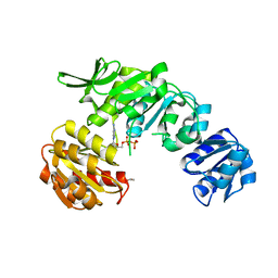
Crystal Structure of UDP-N-acetylmuramoylalanine--D-glutamate ligase (MurD) from Pseudomonas aeruginosa PAO1 in complex with ADP
Descriptor: 1,2-ETHANEDIOL, ADENOSINE-5'-DIPHOSPHATE, UDP-N-acetylmuramoylalanine--D-glutamate ligase
Authors:Seattle Structural Genomics Center for Infectious Disease, Seattle Structural Genomics Center for Infectious Disease (SSGCID)
Deposit date:2022-02-25
Release date:2022-03-09
Last modified:2023-10-18
Method:X-RAY DIFFRACTION (1.95 Å)
Cite:Crystal Structure of UDP-N-acetylmuramoylalanine--D-glutamate ligase (MurD) from Pseudomonas aeruginosa PAO1 in complex with ADP
to be published
7U5Q
DownloadVisualize
BU of 7u5q by Molmil
Crystal structure of transcriptional regulator, GntR family, from Brucella melitensis
Descriptor: BROMIDE ION, Transcriptional regulator, GntR family protein, ...
Authors:Seattle Structural Genomics Center for Infectious Disease (SSGCID)
Deposit date:2022-03-02
Release date:2022-03-09
Last modified:2024-04-03
Method:X-RAY DIFFRACTION (3 Å)
Cite:Crystal structure of transcriptional regulator, GntR family, from Brucella melitensis
To be Published
7U4H
DownloadVisualize
BU of 7u4h by Molmil
Crystal Structure of Penicillin-binding protein 1A (Pbp1a) from Chlamydia trachomatis
Descriptor: 1,2-ETHANEDIOL, 4-(2-HYDROXYETHYL)-1-PIPERAZINE ETHANESULFONIC ACID, CHLORIDE ION, ...
Authors:Seattle Structural Genomics Center for Infectious Disease (SSGCID)
Deposit date:2022-02-28
Release date:2022-03-09
Last modified:2023-10-18
Method:X-RAY DIFFRACTION (3.1 Å)
Cite:Crystal Structure of Penicillin-binding protein 1A (Pbp1a) from Chlamydia trachomatis
to be published
7U56
DownloadVisualize
BU of 7u56 by Molmil
Crystal Structure of D-alanine--D-alanine ligase from Klebsiella pneumoniae subsp. pneumoniae in complex with AMP
Descriptor: 1,2-ETHANEDIOL, ADENOSINE MONOPHOSPHATE, CHLORIDE ION, ...
Authors:Seattle Structural Genomics Center for Infectious Disease (SSGCID)
Deposit date:2022-03-01
Release date:2022-03-09
Last modified:2023-10-18
Method:X-RAY DIFFRACTION (1.85 Å)
Cite:Crystal Structure of D-alanine--D-alanine ligase from Klebsiella pneumoniae subsp. pneumoniae in complex with AMP
to be published
4KYX
DownloadVisualize
BU of 4kyx by Molmil
Crystal structure of ADP-ribose pyrophosphatase MutT from Rickettsia felis
Descriptor: ADP-ribose pyrophosphatase MutT
Authors:Seattle Structural Genomics Center for Infectious Disease (SSGCID)
Deposit date:2013-05-29
Release date:2013-08-07
Last modified:2024-02-28
Method:X-RAY DIFFRACTION (1.8 Å)
Cite:Crystal structure of ADP-ribose pyrophosphatase MutT from Rickettsia felis
To be Published
7ULH
DownloadVisualize
BU of 7ulh by Molmil
Crystal Structure of a Short chain dehydrogenase from Mycobacterium avium 104
Descriptor: Short chain dehydrogenase
Authors:Seattle Structural Genomics Center for Infectious Disease, Seattle Structural Genomics Center for Infectious Disease (SSGCID)
Deposit date:2022-04-04
Release date:2022-04-13
Last modified:2023-10-18
Method:X-RAY DIFFRACTION (2.6 Å)
Cite:Crystal Structure of Crystal Structure of a Short chain dehydrogenase from Mycobacterium avium 104
to be published
7ULZ
DownloadVisualize
BU of 7ulz by Molmil
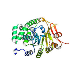
Crystal Structure of Methionine-tRNA ligase / Methionyl-tRNA synthetase (MetRS) from Pseudomonas aeruginosa PAO1
Descriptor: METHIONINE, Methionine--tRNA ligase, SULFATE ION
Authors:Seattle Structural Genomics Center for Infectious Disease (SSGCID)
Deposit date:2022-04-05
Release date:2022-04-27
Last modified:2023-10-18
Method:X-RAY DIFFRACTION (2.8 Å)
Cite:Crystal Structure of Methionine-tRNA ligase / Methionyl-tRNA synthetase (MetRS) from Pseudomonas aeruginosa PAO1
to be published
4LW8
DownloadVisualize
BU of 4lw8 by Molmil
Crystal structure of a putative epimerase from Burkholderia cenocepacia J2315
Descriptor: 4-(2-HYDROXYETHYL)-1-PIPERAZINE ETHANESULFONIC ACID, Putative epimerase
Authors:Seattle Structural Genomics Center for Infectious Disease (SSGCID)
Deposit date:2013-07-26
Release date:2013-09-04
Last modified:2023-09-20
Method:X-RAY DIFFRACTION (2.1 Å)
Cite:Crystal structure of a putative epimerase from Burkholderia cenocepacia J2315
TO BE PUBLISHED
3KW3
DownloadVisualize
BU of 3kw3 by Molmil
Crystal structure of alanine racemase from Bartonella henselae with covalently bound pyridoxal phosphate
Descriptor: Alanine racemase
Authors:Seattle Structural Genomics Center for Infectious Disease (SSGCID)
Deposit date:2009-11-30
Release date:2009-12-22
Last modified:2023-11-22
Method:X-RAY DIFFRACTION (2.04 Å)
Cite:SAD phasing using iodide ions in a high-throughput structural genomics environment.
J.Struct.Funct.Genom., 12, 2011
6UDF
DownloadVisualize
BU of 6udf by Molmil
Crystal structure of Enoyl-[acyl-carrier-protein] reductase [NADH] (InhA) from Mycobacterium kansasii
Descriptor: Enoyl-[acyl-carrier-protein] reductase [NADH], FORMIC ACID
Authors:Seattle Structural Genomics Center for Infectious Disease (SSGCID)
Deposit date:2019-09-19
Release date:2019-10-16
Last modified:2023-10-11
Method:X-RAY DIFFRACTION (1.95 Å)
Cite:Crystal stucture of Enoyl-[acyl-carrier-protein] reductase [NADH] (InhA) from Mycobacterium kansasii
to be published
6ULO
DownloadVisualize
BU of 6ulo by Molmil
Structure of an N-terminally truncated uncharacterized protein from Leptospira interrogans serogroup Icterohaemorrhagiae serovar Copenhageni (strain Fiocruz L1-130)
Descriptor: CHLORIDE ION, Uncharacterized protein
Authors:Seattle Structural Genomics Center for Infectious Disease (SSGCID)
Deposit date:2019-10-08
Release date:2019-10-16
Last modified:2024-03-13
Method:X-RAY DIFFRACTION (1.3 Å)
Cite:Structure of an N-terminally truncated uncharacterized protein from Leptospira interrogans serogroup Icterohaemorrhagiae serovar Copenhageni (strain Fiocruz L1-130)
to be published
6TYJ
DownloadVisualize
BU of 6tyj by Molmil
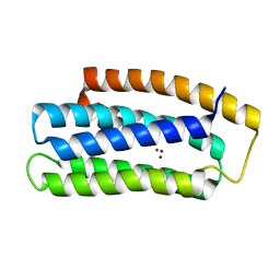
Crystal structure of zinc-bound Hemerythrin HHE cation binding domain-containing protein (soak): Rv2633c homolog from Mycobacterium kansasii
Descriptor: Hemerythrin HHE cation binding domain protein, ZINC ION
Authors:Seattle Structural Genomics Center for Infectious Disease (SSGCID)
Deposit date:2019-08-09
Release date:2020-01-22
Last modified:2023-10-11
Method:X-RAY DIFFRACTION (1.6 Å)
Cite:Crystal structure of zinc-bound Hemerythrin HHE cation binding domain-containing protein (soak): Rv2633c homolog from Mycobacterium kansasii
to be published
6UDG
DownloadVisualize
BU of 6udg by Molmil
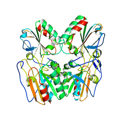
Crystal structure of a Probable thiol peroxidase from Elizabethkingia anophelis NUHP1
Descriptor: Thiol peroxidase
Authors:Seattle Structural Genomics Center for Infectious Disease (SSGCID)
Deposit date:2019-09-19
Release date:2019-10-16
Last modified:2024-04-03
Method:X-RAY DIFFRACTION (2.65 Å)
Cite:Crystal structure of a Probable thiol peroxidase from Elizabethkingia anophelis NUHP1
TO BE PUBLISHED
6UM4
DownloadVisualize
BU of 6um4 by Molmil
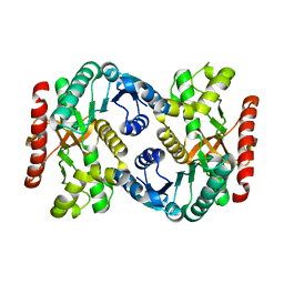
Crystal structure of malate dehydrogenase from Naegleria fowleri ATCC 30863
Descriptor: 1,2-ETHANEDIOL, Malate dehydrogenase
Authors:Seattle Structural Genomics Center for Infectious Disease (SSGCID)
Deposit date:2019-10-09
Release date:2019-10-23
Last modified:2024-04-03
Method:X-RAY DIFFRACTION (2.05 Å)
Cite:Crystal structure of malate dehydrogenase from Naegleria fowleri ATCC 30863
TO BE PUBLISHED
6UJD
DownloadVisualize
BU of 6ujd by Molmil
Crystal structure of Cysteine-tRNA ligase from Elizabethkingia sp.
Descriptor: 1,2-ETHANEDIOL, 1-METHOXY-2-[2-(2-METHOXY-ETHOXY]-ETHANE, CHLORIDE ION, ...
Authors:Seattle Structural Genomics Center for Infectious Disease (SSGCID)
Deposit date:2019-10-02
Release date:2019-10-16
Last modified:2023-10-11
Method:X-RAY DIFFRACTION (2.6 Å)
Cite:Crystal structure of Cysteine-tRNA ligase from Elizabethkingia sp.
TO BE PUBLISHED
6V45
DownloadVisualize
BU of 6v45 by Molmil
Crystal structure of a Probable carnitine operon oxidoreductase caia from Brucella melitensis
Descriptor: Probable carnitine operon oxidoreductase caia, SULFATE ION
Authors:Seattle Structural Genomics Center for Infectious Disease (SSGCID)
Deposit date:2019-11-27
Release date:2019-12-11
Last modified:2024-04-03
Method:X-RAY DIFFRACTION (2.6 Å)
Cite:Crystal structure of a Probable carnitine operon oxidoreductase caia from Brucella melitensis
to be published

222926

PDB entries from 2024-07-24

PDB statisticsPDBj update infoContact PDBjnumon