Loading
PDBj
MenuPDBj@FacebookPDBj@TwitterPDBj@YouTubewwPDB FoundationwwPDB
RCSB PDBPDBeBMRBAdv. SearchSearch help
Search by PDB author
2ERV
DownloadVisualize
BU of 2erv by Molmil
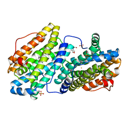
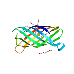
Crystal structure of the outer membrane enzyme PagL
Descriptor: CALCIUM ION, PENTAETHYLENE GLYCOL MONODECYL ETHER, hypothetical protein Paer03002360
Authors:Rutten, L, Geurtsen, J, Lambert, W, Smolenaers, J.J, Bonvin, A.M, van der Ley, P, Egmond, M.R, Gros, P, Tommassen, J.
Deposit date:2005-10-25
Release date:2006-04-11
Last modified:2023-08-23
Method:X-RAY DIFFRACTION (2 Å)
Cite:Crystal structure and catalytic mechanism of the LPS 3-O-deacylase PagL from Pseudomonas aeruginosa.
Proc.Natl.Acad.Sci.USA, 103, 2006
6QO8
DownloadVisualize
BU of 6qo8 by Molmil
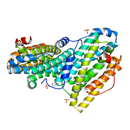
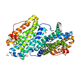
Crystal structure of ribonucleotide reductase NrdF from Bacillus anthracis anaerobically soaked with ferrous ions
Descriptor: 2-[BIS-(2-HYDROXY-ETHYL)-AMINO]-2-HYDROXYMETHYL-PROPANE-1,3-DIOL, FE (II) ION, Ribonucleoside-diphosphate reductase subunit beta, ...
Authors:Grave, K, Hogbom, M.
Deposit date:2019-02-12
Release date:2019-08-21
Last modified:2024-01-24
Method:X-RAY DIFFRACTION (1.31921768 Å)
Cite:Redox-induced structural changes in the di-iron and di-manganese forms of Bacillus anthracis ribonucleotide reductase subunit NrdF suggest a mechanism for gating of radical access.
J.Biol.Inorg.Chem., 24, 2019
6QO9
DownloadVisualize
BU of 6qo9 by Molmil
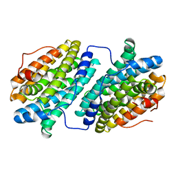
Crystal structure of ribonucleotide reductase NrdF from Bacillus anthracis soaked with manganese ions
Descriptor: 2-[BIS-(2-HYDROXY-ETHYL)-AMINO]-2-HYDROXYMETHYL-PROPANE-1,3-DIOL, MANGANESE (II) ION, Ribonucleoside-diphosphate reductase subunit beta, ...
Authors:Grave, K, Hogbom, M.
Deposit date:2019-02-12
Release date:2019-08-21
Last modified:2024-01-24
Method:X-RAY DIFFRACTION (1.299 Å)
Cite:Redox-induced structural changes in the di-iron and di-manganese forms of Bacillus anthracis ribonucleotide reductase subunit NrdF suggest a mechanism for gating of radical access.
J.Biol.Inorg.Chem., 24, 2019
6QO7
DownloadVisualize
BU of 6qo7 by Molmil
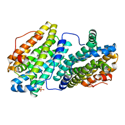
Crystal structure of ribonucleotide reductase NrdF from Bacillus anthracis aerobically soaked with ferrous ions (photo-reduced)
Descriptor: CHLORIDE ION, FE (II) ION, Ribonucleoside-diphosphate reductase subunit beta
Authors:Grave, K, Hogbom, M.
Deposit date:2019-02-12
Release date:2019-08-21
Last modified:2024-05-15
Method:X-RAY DIFFRACTION (1.628 Å)
Cite:Redox-induced structural changes in the di-iron and di-manganese forms of Bacillus anthracis ribonucleotide reductase subunit NrdF suggest a mechanism for gating of radical access.
J.Biol.Inorg.Chem., 24, 2019
6QO5
DownloadVisualize
BU of 6qo5 by Molmil
Crystal structure of apo (metal-free) ribonucleotide reductase NrdF from Bacillus anthracis
Descriptor: CHLORIDE ION, Ribonucleoside-diphosphate reductase subunit beta, SULFATE ION
Authors:Grave, K, Hogbom, M.
Deposit date:2019-02-12
Release date:2019-08-21
Last modified:2024-01-24
Method:X-RAY DIFFRACTION (1.511 Å)
Cite:Redox-induced structural changes in the di-iron and di-manganese forms of Bacillus anthracis ribonucleotide reductase subunit NrdF suggest a mechanism for gating of radical access.
J.Biol.Inorg.Chem., 24, 2019
6QOB
DownloadVisualize
BU of 6qob by Molmil
Crystal structure of ribonucleotide reductase NrdF from Bacillus anthracis with partially oxidised di-iron metallocofactor
Descriptor: 2-[BIS-(2-HYDROXY-ETHYL)-AMINO]-2-HYDROXYMETHYL-PROPANE-1,3-DIOL, FE (III) ION, Ribonucleoside-diphosphate reductase subunit beta, ...
Authors:Grave, K, Hogbom, M.
Deposit date:2019-02-12
Release date:2019-08-21
Last modified:2024-01-24
Method:X-RAY DIFFRACTION (1.457 Å)
Cite:Redox-induced structural changes in the di-iron and di-manganese forms of Bacillus anthracis ribonucleotide reductase subunit NrdF suggest a mechanism for gating of radical access.
J.Biol.Inorg.Chem., 24, 2019

226707

數據於2024-10-30公開中

PDB statisticsPDBj update infoContact PDBjnumon